Looks like no one added any tags here yet for you.
What are Biomacromolecules?
Biomacromolecules are large molecules composed of smaller units.
What is a polymer?
Polymers are a large molecule composed of a chain of smaller repeating subunits called monomers.
Define monomers.
Monomers are the smaller building blocks of a polymer.
List the four biomacromolecules and their corresponding monomers.
Lipids: Glycerol and Fatty Acid
Polysaccharide: Monosaccharide
Nucleic Acid: Nucleotide
Polypeptide/Protein: Amino Acid
Describe proteins.
Proteins, also known as polypeptides, are one of 4 biomacromolecules. They are large complex structures which are crucial to functioning of all living organisms. Proteins are extremely functionally diverse.
Define proteome.
The proteome of a cell refers to all the proteins that are found within a cell or that can be expressed by the cell.
Describe the chemical structure of an amino acid.
The chemical structure of an amino acid is composed of a central carbon atom, a carboxyl group, an amino group, an R group and a hydrogen atom. There are 20 different amino acids each with its own unique R group also known as side chain or variable. The R group is the only thing that differentiates amino acids.
Explain how amino acids are polymerised.
Amino acids are polymerised at the ribosome in the process of translation. A hydroxyl group (hydrogen + oxygen) is taken off the carboxyl group of one amino acid and forms a covalent bond with a hydrogen atom from an amino group of another amino acid, forming a water molecule (H2O). The amino acid residues are then joined together in a covalent bond called a peptide bond.
What is the difference between peptides and polypeptides?
Short chains (20 or less amino acids) are called peptides. Long chains (100 or more amino acids) are called polypeptides. The reaction is known as condensation polymerisation and the bonds are referred to as dehydration bonds or condensation bonds.
Why is protein folding important?
To function correctly, proteins must be folded into their correct shape. The four levels of structure describe how polypeptide chains fold to form this functional structure, with each level being more complex.
What is the primary structure of a protein?
Primary Structure refers to the order or sequence of amino acids in a polypeptide chain which is determined by the dna sequence of the gene, this determines the folding, hence the function of the protein.
Describe the secondary structure of proteins.
When amino acids form hydrogen bonds with each other, the polypeptide chain folds and coils forming local structures such as alpha helices and beta pleated sheets. Remember that secondary structure is only a region of the polypeptide chain.
What is an alpha helix structure?
An alpha helix is a right-handed spiralling of the polypeptide chain when there is repulsion between equally sided R groups. This helix structure is held by hydrogen bonds between hydrogen in each amino group and oxygen in the carboxyl group.
Define the tertiary structure of a protein.
The tertiary structure is the overall shape of the entire protein (not just a section or region). It is the shape of the outer surface of the folded protein. The tertiary structure is the most important because the shape of a protein to a large extent determines its function. They are affected by:
disulfide bonding
ionic bonds
hydrogen bonds
hydrophobic interactions
How are polypeptides folded into their 3D shape?
Polypeptides are folded into their 3D shape by other proteins in the cell called chaperone proteins which assist in folding into their correct 3D structures without becoming part of the final structure. They are then held in position by hydrogen bonds and disulphide bonds between cysteine amino acids.
How do amino acid side chains contribute to protein structure?
Hydrophobic non-polar amino acid side chains (R-group) usually point interior. Hydrophilic polar side chains usually point exterior. Hydrophilic polar side chains also help suspend the protein in the watery environment, so they don't sink.
What is the quaternary structure of proteins?
Some proteins have a quaternary structure which means that it has more than one polypeptide chain, each with its own primary, secondary and tertiary structure. These polypeptides are held together by hydrogen bonds and attraction between polar amino acids and amino acid side chains.
What is the difference between a polypeptide and a protein?
A polypeptide is a single chain of amino acid residue while a protein is a functional unit made up of one or more polypeptides.
What are prosthetic groups in proteins?
Many proteins have non-amino acid groups (prosthetic) added to them. For example, a protein with a carbohydrate group added is called a glycoprotein. A lipid prosthetic attached to a protein would be called a lipoprotein. A protein with a prosthetic can be called a conjugated protein.
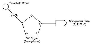
What are nucleotides and what are their components?
Nucleotides are the monomers that make up nucleic acids (DNA and RNA). They are composed of:
A pentose (5 carbon) sugar
A nitrogenous base
One or more phosphate groups
What are the five different nitrogen bases and how are they categorized?
PURINES:
Guanine (G)
Adenine (A)
PYRIMIDINES:
Cytosine (C)
Thymine (T)
Uracil (U)
How are carbons numbered in the pentose sugar and what are the three most important carbons?
The carbons are numbered in clockwise direction from 1' (one prime) to 5' (five prime). The three most important carbons are:
1': attaches to the nitrogenous base
3': attaches to the phosphate of the adjacent nucleotide joining them together
5': attaches the phosphate group to the sugar on the same nucleotide
What are polynucleotide chains?
They form when many nucleotides join together by strong covalent bonds called phosphodiester bonds between the phosphate group of one nucleotide and the 3'-hydroxyl (-OH) group of the sugar in the next nucleotide.
Form via condensation reaction between sugar and phosphate group
Nucleotides can ONLY be joined by carbon 3'
The linkage of phosphate groups and sugars is commonly referred to as the sugar-phosphate backbone of nucleic acids
3’ and 5’ are significant in contributing to the directional nature of nucleic acids
What's the difference between ribose and deoxyribose?
Ribose is found in RNA, deoxyribose is found in DNA
Ribose has a hydroxyl group (hydrogen + oxygen) on the 2' carbon
Deoxyribose lacks oxygen on the 2' carbon (hence "deoxy")
Deoxyribose won't pair with Uracil and ribose won't pair with Thymine
What is RNA and what are its three main types and their functions?
RNA is a single strand of nucleotides found in different parts of the cell, primarily involved in protein synthesis. IT IS NON CODING DNA
mRNA (messenger RNA): mRNA carries a transcript of the DNA template strand to the ribosome and therefore determines the order amino acids are assembled into a polypeptide chain at the ribosome.
tRNA (transfer RNA): Carries its corresponding amino acid to the mRNA codon using its anticodon, which complementary ensuring the correct amino acid is added to the growing protein chain.
rRNA (ribosomal RNA): Forms part of the ribosome, where it helps facilitate translation by positioning tRNAs and catalyzing the formation of peptide bonds between amino acids
How does RNA polymerization work?
RNA polymerization works as follows:
RNA is built by an enzyme called DNA polymerase
RNA polymerase moves along the DNA template strand in a 5'-3' direction, adding new nucleotides to 3' end
Uses triphosphate nucleotides:
ATP (Adenosine Triphosphate)
GTP (Guanosine Triphosphate)
CTP (Cytosine Triphosphate)
UTP (Uracil Triphosphate)
RNA polymerase breaks of two phosphates which releases the right amount of energy to join the nucleotide residue to the polynucleotide chain
This Is condensation polymerisation reaction except instead of a water molecule being produced when working with amino acids, a disulfide molecule is produced
What are the key characteristics of DNA?
Polymer of nucleotides composed of 2 polynucleotide chains running antiparallel
One strand goes 5' to 3', the other 3' to 5'
Composed of phosphate group, deoxyribose sugar, and nitrogenous bases (A, C, T, G)
Base pairing rules:
Adenine (A) pairs with Thymine (T) using 2 hydrogen bonds
Guanine (G) pairs with Cytosine (C) using 3 hydrogen bonds
DNA compresses and stores itself effectively by forming a double helix
In nuclear DNA this Helix structure coils around proteins called histones which then condenses to form chromosomes
central dogma of molecular biology
The ‘central dogma of molecular biology’ describes the flow of genetic information in a cell. It explains that information can be passed from nucleic acid to nucleic acid or protein, but once it has passed into a protein it cannot get out again.
Gene:
Genes are sections of DNA which carry the code required to synthesise proteins
Genetic code
Protein synthesis relies on genetic code which is a series of rules that dictate how information is translated and transcribed into functional proteins
Genetic code relies on the grouping of adjacent nucleotides into groups of 3
In DNA these groups are called triplets
When triplets are transcribed into mRNA they are known as a Codon
Each codon codes for a specific amino acid in the final polypeptide chain
There are specific codons which instruct the cell to start or stop protein synthesis
Promoter
The promoter region is an upstream (5' end) binding site (a sequence of DNA) for RNA polymerase that is responsible for initiating gene expression-transcription. When RNA polymerase binds to the promoter region of a gene, it allows for the transcription of that particular gene. Therefore, the promoter region effectively denotes the starting position and direction of transcription. In eukaryotes, the promoter region is often the sequence of bases 'TATAAA', commonly known as the TATA box.
Introns
Introns are regions of the template strand DNA that do not contribute to the final protein (non-coding regions) as they are removed during RNA processing. Importantly, only eukaryotic genes contain introns - prokaryotic genes do not contain introns.
Exons
Exons are regions of coding DNA, which are transcribed and translated into the final protein. These can be found in both eukaryotes and prokaryotes.
Termination sequence
The termination sequence represents a sequence of DNA that signals for the end of transcription or the release of the mrna transcription in premrna
Operator
The operator region serves as the binding site for repressor proteins, which can then inhibit gene expression. This region is typically only found in prokaryotic genes, as eukaryotes have different regions for regulating gene expression.
are operators found in prokaryotic genes
yes
What is gene expression and what are its three stages?
Gene expression is the process of reading the information stored within a gene to create a functional resource typically a protein. There are three stages to this process:
Transcription: The process of where a DNA strand is used as a template to create pre-mRNA
RNA processing: Modifying the pre – mRNA molecules to produce mRNA
Translation: The process which involves decoding mRNA strands into a polypeptide chain
What is transcription and why can't DNA leave the nucleus?
Transcription is the first stage of gene expression and involves the creation of a pre-mRNA molecule by converting genetic information in DNA to pre -mRNA. DNA cannot leave the cell's nucleus which is why mRNA is created so that it can leave the nucleus and transport the code for proteins.
What occurs in Stage 1 (INITIATION) of transcription?
To begin transcription, specific proteins called transcription factors bind to the promoter region to initiate transcription. With the help of transcription factors, RNA polymerase binds to the promoter region of the DNA. This results in hydrogen bonds to break meaning that the DNA double helix strands begin to unwind and unzip. RNA polymerase is then able to start transcription.
What happens in Stage 2 (ELONGATION) of transcription?
RNA polymerase moves along the template strand of DNA, reading the nucleotide sequence 3' → 5' and uses free-floating complementary RNA nucleotides to produce a new single stranded RNA molecule known as pre-mRNA. The pre-mRNA molecule is synthesised in a 5' to 3' direction. This pre-mRNA strand has a complementary nucleotide sequence to the DNA template strand. The strand of DNA that is not read by RNA polymerase is called the coding strand. The coding strand is identical to the pre-mRNA strand (except the pre-mRNA includes uracil instead of thymine).
What happens in Stage 3 (TERMINATION) of transcription?
Transcription ends when RNA polymerase reaches the termination sequence of a gene, signalling the end of transcription. RNA polymerase then detaches, releasing the pre-mRNA molecule. The DNA molecule winds up again into a double helix.
What happens during RNA Processing?
After transcription pre-mRNA must undergo RNA processing before becoming mRNA and going into the ribosome. In RNA processing a Methyl – Guanine cap (Methyl – G cap) is added to the 5' end in the upstream end of pre-mRNA . A chain of adenosine nucleotides (poly – A- tail) is added to the 3' end to stabilise the mRNA molecule preventing it from degrading and allowing it to bind to the ribosome during translation.
What is splicing and alternative splicing?
Splicing: In the process of splicing, a complex molecule called a spliceosome removes the introns and rejoins the remaining exons together.
Alternative Splicing: Sometimes, exons are removed during splicing, meaning a single pre-mRNA strand can generate multiple mRNA molecules, depending on which exons are retained or spliced out. This allows a single gene to produce different proteins.
Where does translation begin and what is its purpose?
After transcription and RNA processing, mRNA molecules exit the nucleus through a nuclear pore. They travel to a ribosome either in the cytosol or attached to a rough endoplasmic reticulum. In this stage mRNA is decoded and translated into a sequence of amino acids forming a polypeptide chain.
What happens in Stage 1 of translation?
The 5' end of the mRNA molecule binds to the ribosome, which scans until it recognizes the start codon. A tRNA molecule with the complementary anticodon temporarily binds to the codon within the ribosome and delivers the corresponding amino acid
What happens in Stage 2 of translation?
The mRNA chain is fed through the ribosome so that the next codon is matched to its complementary tRNA anticodon. The tRNA molecule then delivers the corresponding amino acid which binds to the previous amino acid via condensation reaction - allows for formation of peptide bonds. The tRNA molecule then leaves the ribosome and is free to pick up another amino acid.
What happens in Stage 3 of translation?
This process continues until the ribosome reaches the stop codon. This signals the end of translation. The polypeptide chain is then released by the ribosome into the cytosol or E.R. Following translation, the mRNA molecule can be reused to produce more polypeptides.
dna vs rn copying
copying dna is replication, copying rna is transcription
when does the ribsosome start to work
when the small and large subunit bind together then the ribosome starts to work
Why do we need gene regulation?
Creating proteins is extremely energy expensive and with a finite energy supply we need to limit the amount of energy our cells use
What is gene regulation used for?
To control the expression of genes by switching transcription on or off
What is a Structural Gene?
A segment of DNA which codes for proteins that play a role in the structure or function of a cell or organism
What is a Regulatory Gene?
segment of DNA responsible for producing proteins that control the expression of other genes
What is an operon?
In certain organisms such as prokaryotes, multiple structural genes that share a common purpose can often be arranged into groups, so their expression is efficiently controlled by a single promoter and operator in what is known as an operon.
What is the function of the trp operon?
The trp operon contains a series of genes that are involved in the production of the amino acid tryptophan which can be then used in protein synthesis
What are the structural genes in the trp operon?
trpE, trpD, trpC, trpB, trpA
what does trp R do
the promoter region have transcription factors (repressor proteins) which binds to the rna polymerase. the repressor proteins are made by trp R .
What controls these structural genes in the trp operon?
These genes are controlled by a common promoter and operator
What happens in repression when tryptophan levels are high?
When there are high levels of tryptophan are present, they bind to the repressor protein making it go from inactive to active
What happens to the repressor protein when tryptophan binds to it?
This induces conformational change in the repressor protein (changes the 3D shape of the protein) resulting in an increase in affinity between the operator region and the repressor protein
How does the repressor protein prevent transcription?
This allows the repressor protein to bind to the operator, blocking RNA polymerase from binding to the promoter region.
when the repressor is floating around it called inactive repressor
What happens when tryptophan levels are low?
Overtime tryptophan will detach from the repressor protein when levels are low decreasing the affinity between the operator region and the repressor protein to allow for the operon structural genes to be expressed allowing for the production of tryptophan.
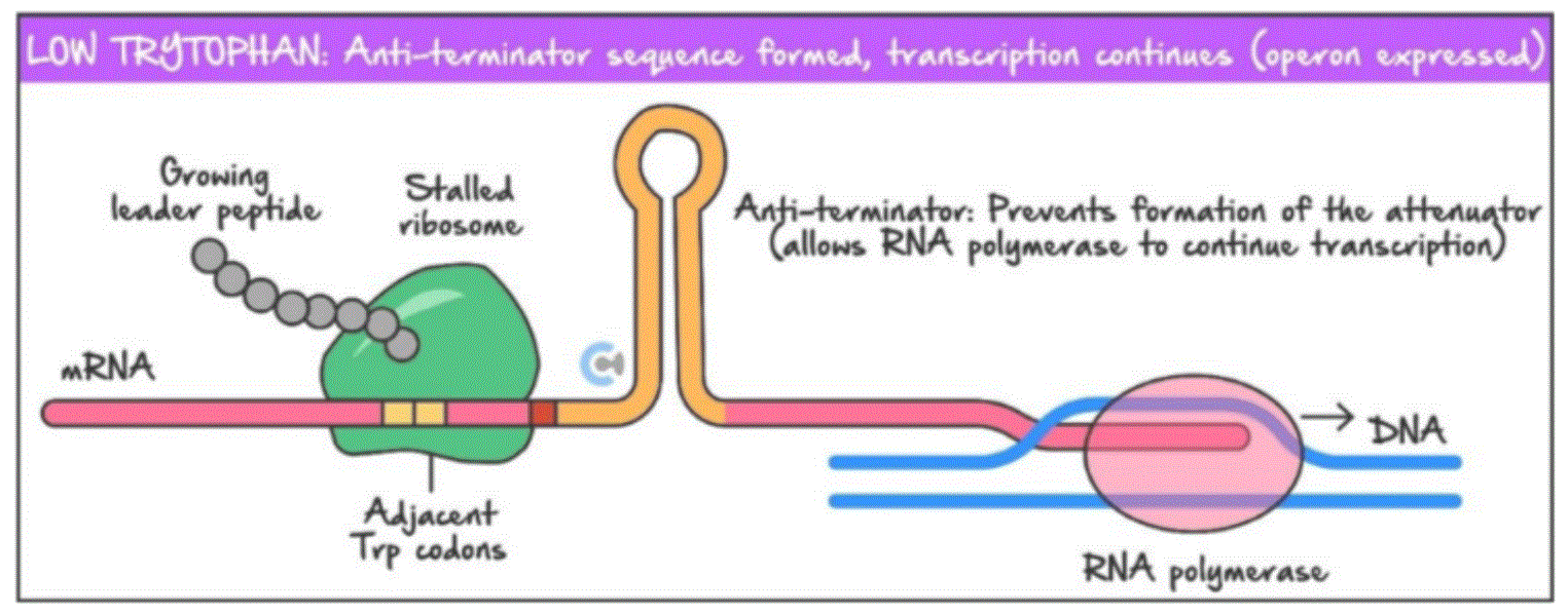
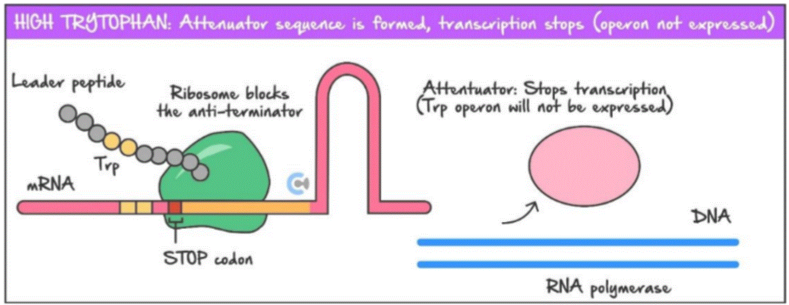
What is attenuation?
In prokaryotes transcription and translation occur at the same time, attenuation serves the purpose of stopping tryptophan production after transcription and translation have already begun.
What is the leader sequence?
Between the operator and the trp structural genes is a leader sequence containing two trp codons and a stop codon, when translating mRNA molecules the position of the ribosome on the leader sequence dictates the formation of two hairpin loops, which can either allow for the trp genes to be transcribed, or prevent transcription of the trp genes.
What happens during attenuation when tryptophan levels are low?
When tryptophan is low in both tRNA molecules and the cytosol the ribosome will be stalled at the two trp codons on the leader sequence of the mRNA.
This will result in the formation of the anti-terminator loop allow the trp structural genes to be transcribed by RNA polymerase and translated by a ribosome
Thus increasing tryptophan levels
What happens during attenuation when tryptophan levels are high?
When tryptophan levels are not abundant in the cytosol but are high in tRNA molecules, the ribosome will move past the two trp codons and reach a stop codon on the leader sequence of the mRNA strand
This will block the anti-terminator loop from forming, resulting in the formation of the attenuator loop in the leader sequence.
This will cause the attenuator region at the end of the leader sequence to detach, causing the mRNA strand to detach, stopping transcription as the trp operon is not expressed (RNA polymerase can no longer continue transcribing the mRNA strand)
Exocytosis
Exocytosis is the process by which contents of a vesicle are released into the extracellular environment
It is a form of bulk transport which is a form of active transport and there requires energy to operate
3 stages of exocytosis:
1) A vesicle is transported to the membrane
2) The membrane of the vesicle fuses with the plasma membrane
3) The products are released into the extracellular environment
1. Rough Endoplasmic Reticulum (RER) – The Protein Factory 🏭
📌 What happens here?
Proteins that must be exported out of the cell begin their synthesis at ribosomes attached to the membrane of the rough endoplasmic reticulum (RER). These membrane-bound ribosomes translate mRNA into polypeptides, which are then translocated into the lumen of the RER for further processing and modification.
These proteins enter the lumen (inner space) of the RER, where they are modified as they move towards golgi appartus.
🔹 Modifications in the RER:
✅ Protein folding
✅ Early glycosylation (N-linked
✅ Disulfide bond formation –
✅ Quality control
2. Golgi Apparatus – The Post Office 📦
The Golgi apparatus is a stack of flattened membrane sacs called cisternae. Its function is to modify and package proteins for export from the cell via vesicles ready for secretion from the cell.
Proteins are delivered to the cis side of the Golgi apparatus from the rough endoplasmic reticulum. As they move through the Golgi apparatus, proteins are modified by resident enzymes which add or remove sugars, or add phosphate or sulphate groups. Different modifications take place in the cis, medial, and trans compartments.
Modifications are necessary to target the proteins to their intended destination – like shipping labels.
3. Exocytosis – The Final Shipment 🚀
📌 What happens here?
Secretory vesicles move toward the plasma membrane.
The vesicle fuses with the membrane, releasing the protein outside the cell.
This process is used for:
✅ Secreting hormones
✅ Releasing neurotransmitters
✅ Delivering enzymes or antibodies
How Cargo Proteins Bind and Direct Vesicles for Exocytosis 🚀
1⃣ Vesicle Formation at the Golgi (Trans Face)
Cargo proteins (the proteins to be exported) bind to specific receptor proteins on the membrane of the vesicle.
This binding occurs because the cargo protein has a complementary shape to the receptor, ensuring only the correct proteins are packaged.
Once the vesicle is formed, it buds off from the Golgi apparatus and travels toward the plasma membrane.
2⃣ Receptor Recognition & Targeting 🎯
When the vesicle reaches the plasma membrane, the membrane-bound receptor proteins recognize specific docking sites on the membrane.
The receptor undergoes a conformational (shape) change, which helps position the vesicle correctly.
This ensures the vesicle attaches only to the right part of the membrane and doesn’t fuse randomly.
3⃣ Shape Change & Membrane Fusion 🔄
The binding of the receptor to the membrane docking site induces further shape changes in both the vesicle and membrane proteins.
This brings the vesicle very close to the membrane, causing the lipid bilayers to merge.
As a result, the cargo proteins are released outside the cell (exocytosis).
What are endonucleases?
Endonucleases refer to a broad range of enzymes responsible for cutting strands of DNA
When are endonucleases known as restriction endonucleases?
When these enzymes target recognition sites, they are known as restriction endonucleases
How do restriction endonucleases cut DNA?
Restriction Endonucleases cut DNA by splitting the phosphodiester bond holding the nucleotides together"
Why do bacteria produce restriction endonucleases?
Restriction endonucleases are often found in bacteria where they are produced as a defence mechanism for invading viral DNA
What is the typical length of a recognition site for a restriction endonuclease?
The recognition site of a restriction endonuclease is usually 4-6 nucleotides long"
What is special about the sequence of nucleotides in a recognition site?
The sequence of nucleotides is typically a palindrome meaning the 5' to 3' sequence of a template strand is the same as the 5' to 3' of the coding strand
What types of ends can endonucleases create?
Endonucleases either create sticky or blunt ends
What are blunt end nucleases?
Blunt ends nucleases cut DNA in the middle of a specfic recognition site with a straight cut
What are sticky end nucleases?
Sticky end nucleases are restriction enzymes that cut DNA in a staggered manner at a specific restriction site, leaving overhanging, unpaired nucleotides at the ends.
Why are they called sticky ends?
They are called sticky ends because the two overhanging nucleotides are complementary and are attracted to stick to each other via hydrogen bonding
What are ligases?
Ligases are enzymes that join two fragments of DNA or RNA together, acting like molecule glue"
How do ligases join DNA fragments?
This occurs by catalysing the formation of phosphodiester bonds between both fragments to merge them together"
What are the two types of ligase enzymes?
There are two types of ligase enzyme: DNA ligase to join together DNA and RNA ligase to join together RNA"
How do ligases compare to endonucleases?
Ligase are like the opposite of endonucleases with the exception that ligases are not as specific
What types of DNA ends can ligases join?
Meaning that ligase can join together any blunt or sticky ends"
What do polymerases do?
Polymerases synthesise polymer chains from monomer building blocks"
What are the two types of polymerase used for gene manipulation?
There are two types of polymerase used for gene manipulation:
DNA polymerase: Used in replication or amplification of DNA
RNA polymerase: Used in transcription of genes"
Why do polymerases require a primer?
Since nucleotides can only be added to the 3' end polymerase require a primer to attach to the start of the template strand"
What is a primer?
A primer is a short single strand of nucleotides that are complementary to the template strand"
How does polymerase synthesize DNA once attached to a primer?
Once attached to the primer, the polymerase enzyme can read and synthesis a complementary strand in a 5' to 3' direction"
What is PCR and what is it used for?
"PCR is a technique used to amplify DNA by making multiple copies
What are three ways scientists can analyze DNA after PCR?
Paternity Testing
Forensic Testing of bodily fluids
Analysing gene fragments for genetic diseases
What does it mean to amplify DNA?
"Amplify: to increase the quantity of a molecule by making many copies"
What is denaturation in the context of PCR?
"Denature: (denaturation is the name of the process) the disruption of a molecules structure by an external factor such as heat."
What is Taq polymerase and what does it do?
Taq polymerase ( MUST name this DNA polymerase. Note how it is spelt!) heat resistant DNA polymerase enzyme isolated from thermophilic bacteria. It amplifies a single stranded DNA molecule by attaching complementary DNA nucleotides.
What does it mean to elongate DNA?
Elongate: to synthesise a longer polynucleotide"
What does annealing mean in PCR?
"Anneal: the joining of two molecules for example two complementary DNA strands during the cooling phase of PCR. ( involves the PRIMERS)"
What is a forward primer and what does it do?
forward primer binds to the 3'end of the template strand and reads the DNA in the same direction of the RNA polymerase