Chapter 4, Cell Injury, Aging, & Death
 Call Kai
 Learn
Learn Practice Test
 Spaced Repetition
 Match
Match Flashcards
 Knowt Play
1/25

There's no tags or description
Looks like no tags are added yet.
Name | Mastery | Learn | Test | Matching | Spaced | Call with Kai | Chat |
|---|
No analytics yet
Send a link to your students to track their progress
26 Terms
Reversible cell injury
mild/short lived injury
Hydropic swelling
cellular swelling due to accumulation of water intracellular accumulations
excessive amounts of fat
accumulation of abnormal substances produced by cell & pigments and particles cell cannot digest
(reversible cell injury)
Cellular adaptation
persistent sublethal injury
atrophy
hypertrophy
hyperplasia
metaplasia
dysplasia
anaplasia
neoplasia
Atrophy
cells shrink and reduce differentiated functions
Causes: Disuse, enervation, ischemia, nutrient deficiency, interruption of endocrine signals, persistent cell injury

Hypertrophy
increase in cell mass, augmented functional capacity
Causes: Response to increased physiologic or pathophysiologic demands

Hyperplasia
increased number of cells + functional capacity
Increased physiologic demands and persistent cell injury
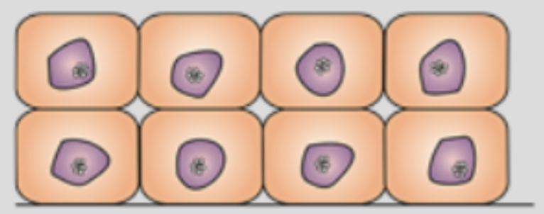
Metaplasia
replacement of one differentiated cell type with another (reversible)
Causes: adaptation to persistent injury with replacement of a cell type better suited for situation

Dysplasia
disorganized appearance of cells because of abnormal variations in size, shape and arrangement
Causes: adaptive effort gone wrong
VERY high chance to go cancerous

Anaplasia
cellular deviance characterized by a loss of differentiation and structural organization. Anaplastic cells lose their specialized functions, vary wildly in size and shape, and are a hallmark feature of aggressive, malignant tumors.

Neoplasia
"new growth"; an abnormal, unregulated, and autonomous proliferation of cells that continues even after the original stimulus is removed. Neoplasms can be benign (non-cancerous and localized) or malignant (cancerous and capable of invading surrounding tissue or metastasizing)

Irreversible cell injury
Necrosis
Apoptosis
Necrosis
cell death resulting from inflammation/consequence of ischemia or toxins. characterized by cell membrane rupture, spilling of contents into surrounding fluid, and subsequent inflammation
Has 4 types:
Coagulative
Liquefactive
Fat necrosis
Caseous
Coagulative
the most common type; begins with ischemia and ends in tissue degradation (ex. dry gangrene)
Liquefactive
occurs when dead cells dissolve quickly due to lysosomal enzymes, often forming an abscess or cyst (ex. wet gangrene)
Fat necrosis
death of adipose tissue resulting from trauma or pancreatitis, appearing as chalky white areas
Caseous
characteristic of lung damage from tuberculosis; leaves tissue looking soft and clumpy like cheese
Apoptosis
programmed cellular suicide triggered by internal signaling pathways. Response to injury that does not directly kill the cell. Does not cause inflammation and not always pathological
Ischemia
interruption of blood flow to an area; it is the most common cause of tissue hypoxia and overall cell injury
Ischemia stages
Blockage in artery
Decreased oxygenation
Decreased cellular metabolism
Vital organs becoming hypoxic
ATP production inhibited
Failure of sodium pump causing cells to enlarge and rupture
Ischemia-Reprefusion Injury
additional cellular damage that occurs after blood supply is restored, driven by calcium overload, formation of reactive oxygen species (free radicals), and localized inflammation
Hypoxia
insufficient oxygen reaching tissues, which deprives cells of the energy required to maintain basic physiological function
_____ _____ is caused by ischemia.
Tissue hypoxia
Bacteria
single-celled microorganisms that primarily cause cell injury externally by producing toxins or competing for nutrients. MOST DAMAGE CELLS FROM THE OUTSIDE
Viruses
obligate intracellular agents that must enter inside host cells to replicate, causing cell injury directly from the inside. ENTER A CELL TO DAMAGE FROM THE INSIDE
Immune responses do more damage than the direct effects of the _______ _____.
Infectious agent
Somatic death
the death of the entire organism, characterized by the absence of respiration and heartbeat. Key postmortem changes include rigor mortis (stiffening of muscles within 6 hours) and postmortem autolysis (widespread tissue deterioration within 24 to 48 hours)