Human Anatomy Bone Development
 Call Kai
 Learn
Learn Practice Test
 Spaced Repetition
 Match
Match Flashcards
 Knowt Play
1/29

There's no tags or description
Looks like no tags are added yet.
Name | Mastery | Learn | Test | Matching | Spaced | Call with Kai | Chat |
|---|
No analytics yet
Send a link to your students to track their progress
30 Terms
Skeletal System
Protects and supports organs, framework of muscles for movement, storage of minerals, creation of new blood cells (hematopoeisis), energy metabolism. Organs include bones, joints, and cartilage.
Support
Hard framework. Support weight of body.
Movement
Skeletal muscle attach to bone by tendon. Bones act as lever.
Protection
Surround and protect inner organs
Mineral storage
Reservoir of minerals (calcium, phosphate). Stored minerals released into bloodstream and distributed to body as needed.
Blood cell formation & energy storage
Contain red marrow for making blood cells (hematopoeisis). Yellow marrow for stroage of fat (energy source).
Energy metabolism
Osteoblasts secrete hormone (osteocalcin) that aids in blood sugar regulation and energy metabolism.Osteoclacin stimulates pancreatic secretions (insulin) to reduce blood sugar and allow sugar to enter into cells. Also influences fat cells to store less fat.
Types of Bone
Long bones (femur), Flat bones (sternum), Irregular bones (vertebrae), Short bones (talus)
Long Bone
Most bones. Longer than wide.
Diaphysis = shaft.
Proximal epiphysis = “top” round end.
Distal epiphysis = “bottom” round end.
Each epiphysis covered by thin layer of articular (hyaline) cartilage to form joint.
Epiphyseal plate = “growth plate” disc of hyaline cartilage in center of shaft that grows to lengthen the bone in childhood.
Medullar cavity = very center of diaphysis, holds yellow bone marrow
Periosteum = connective tissue membrane. Covers outside of bone except where articular cartilage is present. Dense irregular CT forms superficial layer to resist tension while bending. Deep layer surrounds compact bone, contains osteoblasts/clasts for remodeling. Many nerves & blood vessels.
Endosteum = thinner CT that covers internal spongy bone, marrow/medullar cavity, and lines central canal. Contain osteoblasts/clasts.

Short Bone
Roughly cube-shaped. Wrist and ankle, within tendons (patella). Reduce tension, pull on tendon, friction, abrasion, tearing in high impact areas.
Flat bones
Thin flattened, somewhat curved usually. Cranial bones, ribs, sternum, scapula.
Irregular bone
various shapes. Vertebrae, hip bones.
Osteoprogenitor cells
undifferentiated stem cells originating in mesenchyme in bone marrow, then differentiate into osteoblasts. Found in inner (deep) layer of periosteum and the endosteum.
Osteoblasts
Produce and secrete organic components (ground substance & collagen fibers) of bone matrix. Found in deep layer of periosteum and the endosteum.
Osteocytes
Osteoblasts that have become “trapped” in matrix. Maintain bone health through sensory and regulatory actions. Body found in lacunae (solid matrix), spider legs found in canaliculi.
Osteoclasts
Derived from white blood cells (monocytes) that leave the bloodstream and enter bone tissue. Resorbtion of bone. Secrete HCl to break down bone by dissolving minerals of matrix(bone). Secrete lysosomal enzymes to digest the organic components (collagen, ground substance). Found on bone surfaces where resorbtion and remodeling is needed (old, injured, unneeded sites).
Remodeling
New bone formed when we are active. Old bone broken down and absorbed when not needed/injured.
Osteoclasts break down, osteoblasts build. Osteocytes = mastermind.
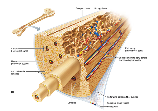
Compact Bone
External layer of bone. Weight-bearing support, protection, structural strength.
Osteon (Haversian System) = parallel to main compression stresses. “weight-bearing pillars”. Contain: lamellae, central canal, perforating canals, canaliculi,
Lamella = layer of matrix compact bone “rings of tree”. Has collagen fibers and minerals (hydroxyapatite). Strength and compression, stress, twisting resistance.
Central canal (Haversian canal) = contains blood vessels and nerves
Perforating canals (Volkmann’s canals) = Connect blood and nerve supply of periosteum and central canal to marrow cavity.
Canaliculi = connect lacunae to one another and the capillaries in central canals. Supply osteon its nutrients through osteocyte connections and remove waste.
Spongy bone
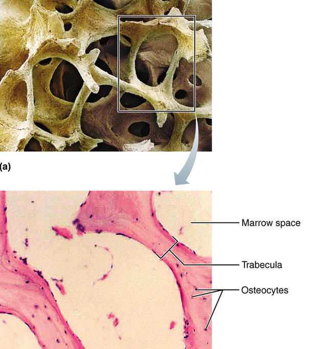
Inner layer of bone. For shock absorption and lighten weight.
Trabeculae: needle-like or flat pieces that form honeycomb network. Open space between trabeculae is filled with bone marrow. Contain several layers of lemellae containing osteocytes.
Osteocytes = receive nutrients from capillaries of endosteum via canaliculi.
Blood vessels
Nutrient foramen is hole in center of diaphysis where nutrient artery and nutrient vein enter perpendicular to bone length. Epiphyseal artery and vein do same for epiphyses.
Capillaries enter through periosteum. Flow to central canals of osteons through Volkmann’s canals. Flow through spongy bone through Volkmann’s canals. Flow through spongy bone to medullar cavity.
Organic components of bone
Flexibility, resists stretching and tearing. Collagen fibers. Ground substance (water, proteoglycans, glycoproteins). Called osteiod.
Inorganic components of bone
Hardness. Mineral salts that harden. Hydrated calcium phosphate (hydroxyapatite).
Intramembranous Ossification
“dermal ossification”. Process of dermal bones developing directly from mesenchyme (embryonic dermis, mesoderm) w/o first being modeled by cartilage.
Produces flat and irregular bones.
Most skull bones and clavicle.
Begins in week 8 of development.
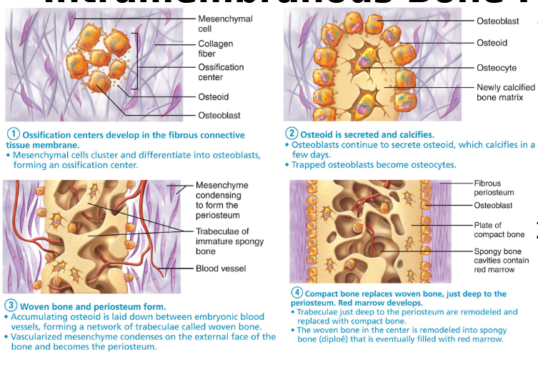
Mesenchymal cells w/in fibrous CT membrane → osteoprogenitor cells → osteoblasts. Forms ossification center.
osteoblasts secrete osteiod (organic part of matrix) → osteiod mineralized/hardened/calcified (by addition of inorganic minerals: calcium, phosphate) → osteoblasts become trapped in hard matrix → osteocytes
New bone tissue forms between embryonic blood vessels, creating trabeculae made of woven bone tissue (immature bone).
Mesenchyme surrounding the outer immature woven bone condenses to become periosteum.
Compact bone replaces woven bone (trabeculae) just underneath periosteum.
Spongy bone replaces woven bone in center and filled w/ red bone marrow.
Endochondral Ossification
Bone formed using hyaline cartilage as model.
Usually producing long bones.
From skull down, except clavicle.
2nd month of development until early adulthood.
Bone exists first as hyaline cartilage. Perichondrium of cartilage is invaded by blood vessels to now become periosteum.
Osteoblast in periosteum lay down bone collar (osteiod) around the diaphysis of model.
At same time, chondrocytes in ossification center enlarge to signal surrounding cartilage matrix to calcify (harden). Trapped chondrocytes die and leave cavities. Cartilage matrix begins to deteriorate. Cartilage at ends of bone are unaffected, continue to grow and elongate bone.
Periosteal bud (contain nutrient artery & vein, osteoprogenitor cells, osteoclasts, bone marrow forming cells) invades cavities in ossification center. Osteoclasts help breakdown cartilage matrix. Osteoprogenitors differentiate into osteoblasts and place osteoid around remaining cartilage matrix. Forms spongy bone. Bone tissue from periosteum continues to surround diaphysis.
Cartilage in center continues calcifying and being replaced by spongy bone, lengthening bone. Cartilage at epiphyses continue to grow rapidly elongating bone. Osteoclasts breakdown spongy bone in center to make medullar cavity. Secondary ossification centers in epiphyses are invaded by epiphyseal vessels. Same process at center begins in epiphysis ends.
Cartilage remains only at epiphyses surfaces (articular cartilage) and epiphyseal plates.

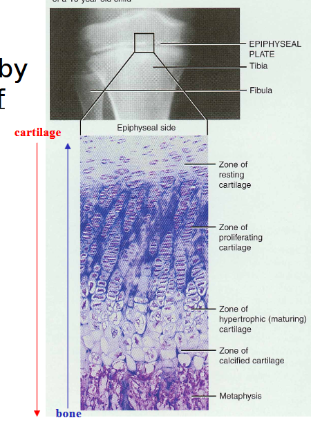
Epiphyseal Plates
Growth plates.
Proliferation zone. Chondroblasts proliferate quickly, pushing epiphyses further from diaphysis.
Hypertrophic zone. Older cartilage cells enlarge and signal matrix to calcify.
Calcification zone. Calcified matrix forms and chondrocytes die near diaphysis side. Calcified cartilage exists close to diaphysis.
Ossification zone. Osteoclasts destroy the calcified cartilage and osteoblasts replace with bone tissue.
Become fully ossifed at 18-21. Growth stops.
Increasing bone width
Osteoblasts in periosteum add bone tissue to circumferential lamellae to widen bone at external diaphysis surface.
Osteoclasts from endosteum remove bone tissue from internal diaphysis wall.
Hormones and Growth
Pituitary gland release growth hormone, thyroxine (thyroid gland), testosterone stimulate and regulate growth of epiphyseal plate. Androgen and estrogen (sex hormones) first promote growth in puberty, then promote end of growth at adulthood.
Fracture Repair
Hematome. Blood vessels break, clots form to create hematoma.
Fibrocartilaginous callus. New blood vessels grow in the clot. Fibers and cartilage are brought. Callus made of fibrocartilage tissue and hyaline cartilage.
Bony callus. Woven bone forms in callus (endochondral ossification). Osteoblasts laying matrix to replace cartilage.
Bone remodeling. Excess bone in medullar cavity and outside of bone is removed. Compact bone replaces callus to reconstruct shaft walls.

Calcitonin & Remodeling
inhibits osteoclasts. Decreases bone breakdown and calcium level in blood.
Released by thyroid when blood calcium too high. Promotes absorption of calcium into bone
Parathyroid hormone & Remodeling
Stimulate osteoclast activity and increase breakdown of bone. Released by parathyroid gland when calcium in blood too low. Promotes absorption of calcium into kidney, intestine, etc.