Correlative Imaging - Radiography
 Call Kai
 Learn
Learn Practice Test
 Spaced Repetition
 Match
Match Flashcards
 Knowt Play
1/21

There's no tags or description
Looks like no tags are added yet.
Name | Mastery | Learn | Test | Matching | Spaced | Call with Kai | Chat |
|---|
No analytics yet
Send a link to your students to track their progress
22 Terms
Discuss the historical development of Radiography.
November 8, 1895 - Wilhelm Conrad Roentgen observed a photographic screen emitting light (fluorescing) & called it x-rays
Hand-held fluoroscopes - helped made by Thomas Edison
1896 - realization that x-rays can be harmful
How x-rays are made
X-rays are produced by fast-moving electrons in an x-ray tube
X-ray tube contains an anode and cathode
Cathode electrons are released - thermionic emission
Properties of x-rays
Electromagnetic waves of very short wavelength and high frequency
Can penetrate matter
Affect a photographic emulsion in the same way that light does
Cause fluorescence (emotion of light) in certain materials
Cause ionization (gaining or losing an electron)
Unaffected by electric and magnetic fields
Cannot easily be reflected or refracted
May be diffracted by passage through crystals
Primary Radiation
Exists in x-ray tube
Absorbed or attenuated in patient
Remnant Radiation
Radiation that passes through and exits the patient
Used to produce radiographic images
Appearance of different tissues on radiographs
Metal: full attenuation of x-ray beam; resultant image is white.
Bone: nearly full attenuation of x-ray beam; resultant image is white to shades of gray.
Water and soft tissues: partial attenuation of x-ray beam; resultant image is gray.
Fat: almost no attenuation of x-ray beam; resultant image is very dark gray to black.
Air: no attenuation of x-ray beam; resultant image is black.
Density
The density of the structure (the amount of blackness on the image) may be affected by the superimposition of bones, muscles, or other soft tissue organs
The "denser" a structure is, the more it attenuates the x-ray beam
Contrast
Refers to the density difference between two adjacent structures
Recorded Detail
Refers to the spatial resolution of the image
Distortion
Refers to the accuracy of the shape of the structure represented in the image
Contrast Agents
Contrast media are solutions or gases introduced into the body to provide contrast on a radiograph between an organ and its surrounding tissue
Positive agents and negative agents decrease opacity (radiolucent). Positive and negative contrast media may be used at the same time for double contrast studies such as the barium enema. The barium coats the large intestine and provides density while the air acts to inflate and separate the haustral folds.
Positive Contrast Agents
Increase opacity (radiopaque) of a structure
Negative Contrast Agents
Decrease opacity (radiolucent) of a structure
Equipment used in interventional radiography
Fluoroscopy
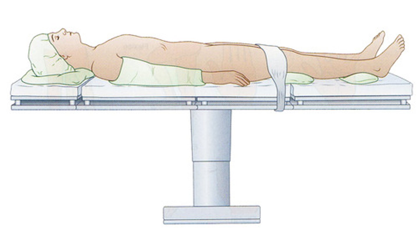
Supine
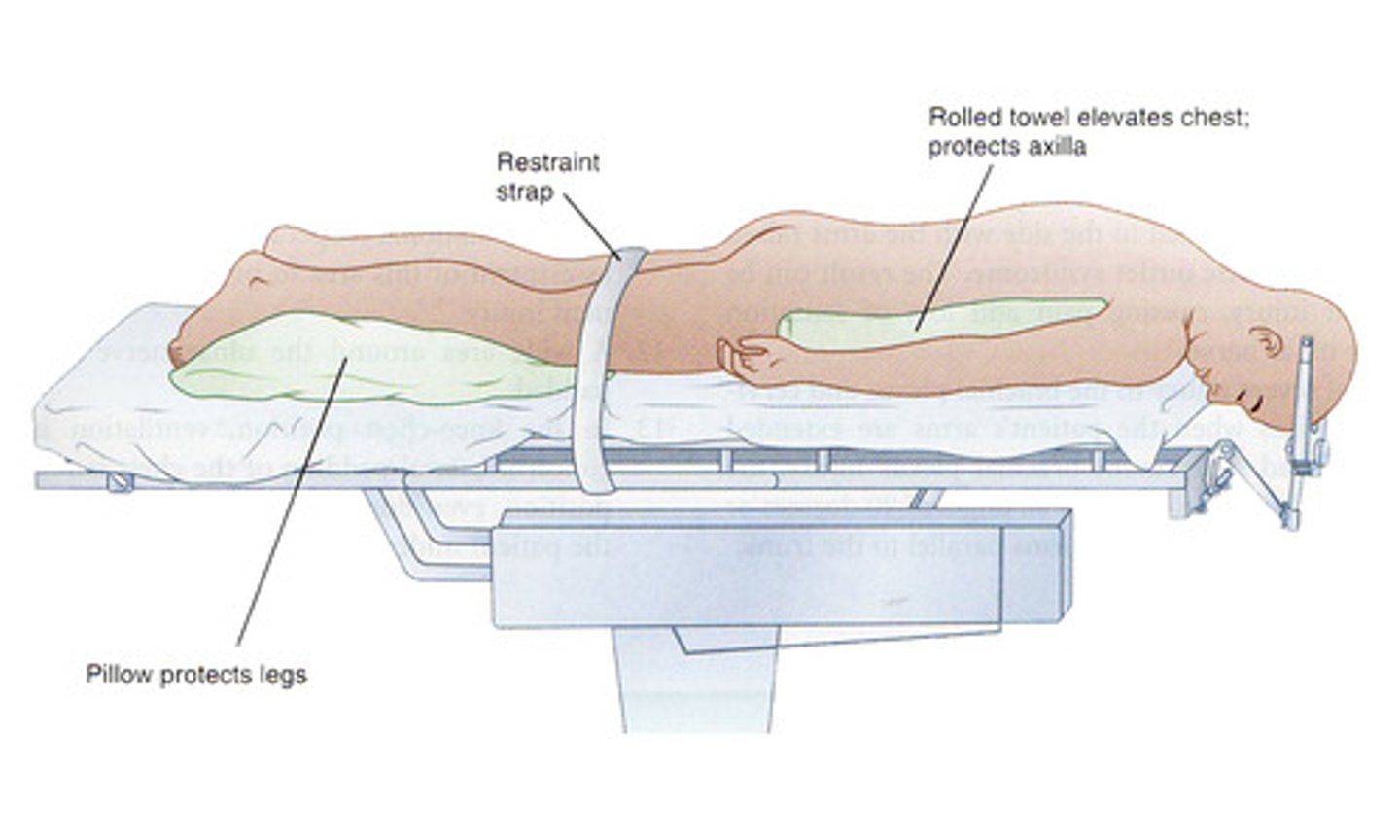
Prone
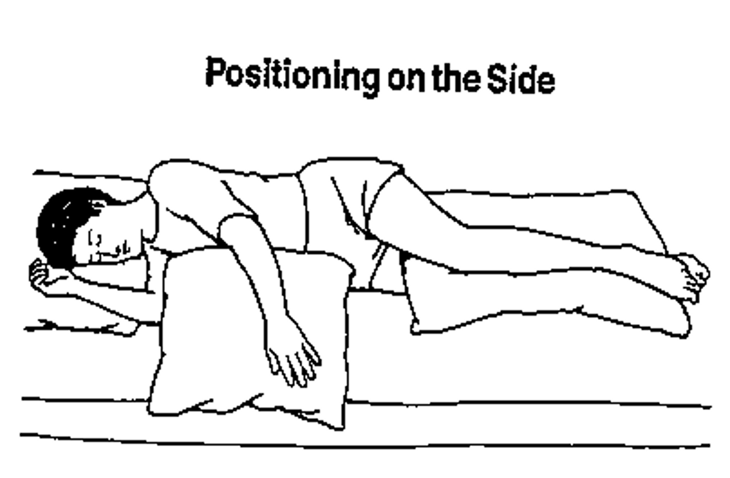
Lateral
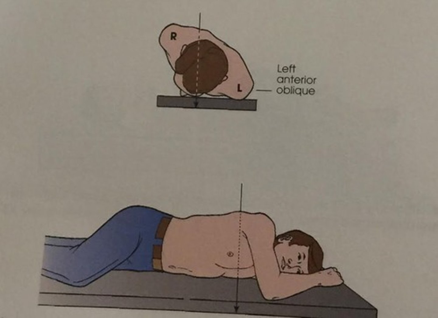
Oblique
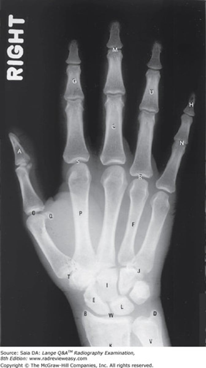
Radiograph
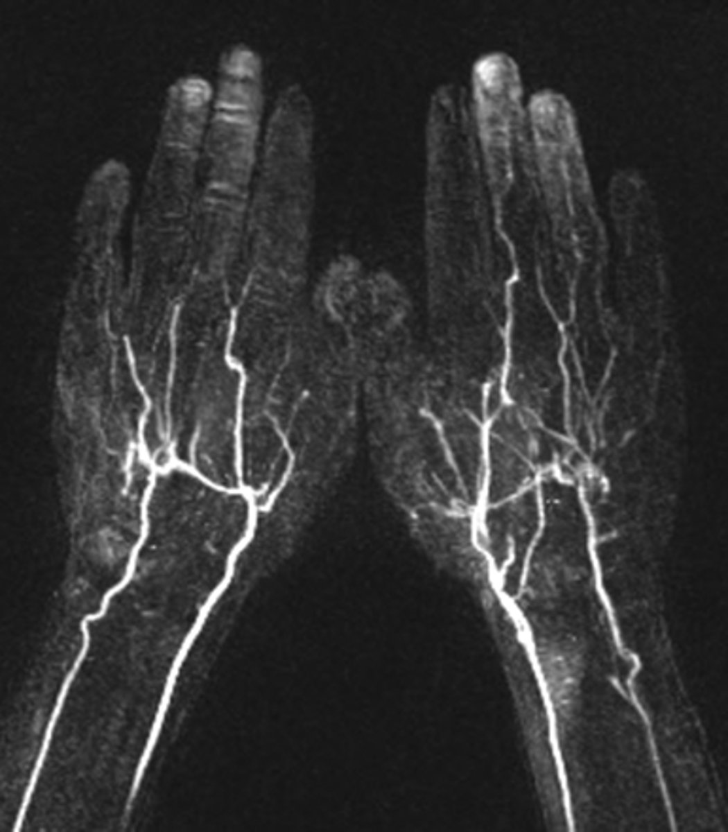
Angiograph
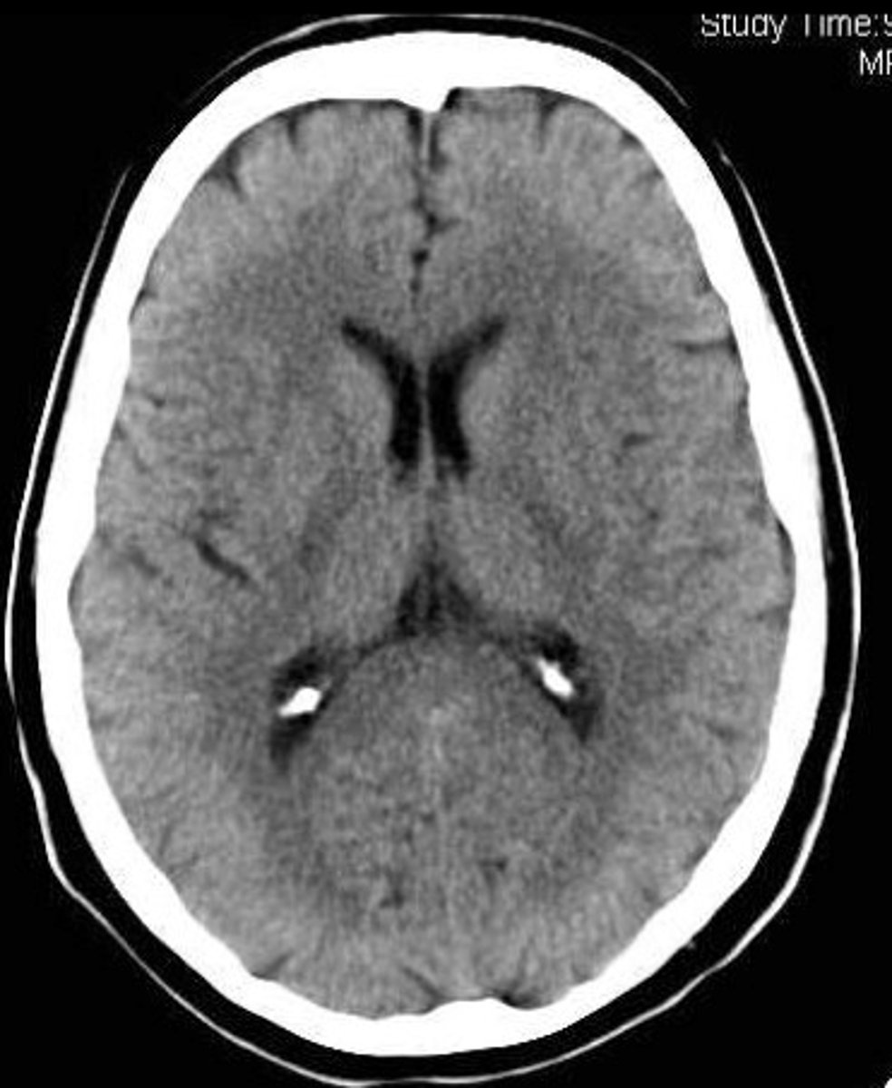
CT
MRI