Anatomy - Module 14 2026 Ratio
1/115
There's no tags or description
Looks like no tags are added yet.
Name | Mastery | Learn | Test | Matching | Spaced |
|---|
No study sessions yet.
116 Terms
A. Foramen magnum
This foramen within the skull base transmits the vertebral artery, anterior spinal artery, posterior spinal artery, and spinal cord:
A. Foramen magnum
B. Carotid canal
C. Jugular foramen
D. Foramen ovale
D. Superior orbital fissure
This foramen within the middle cranial fossa transmits cranial nerves III, IV, V1, VI, ophthalmic veins, and sympathetic fibers:
A. Foramen spinosum
B. Foramen rotundum
C. Foramen ovale
D. Superior orbital fissure
C. Sagittal suture
A non-movable, fibrous joint joining the two parietal bones in the skull:
A. Coronal suture
B. Metopic suture
C. Sagittal suture
D. Lambdoid suture
D. Coronal suture
A non-movable, fibrous joint joining the frontal and parietal bones
A. Metopic suture
B. Sagittal suture
C. Lambdoid suture
D. Coronal suture
D. Metopic suture
A non-movable, fibrous joint which represents a persistent frontal suture
A. Coronal suture
B. Sagittal suture
C. Lambdoid suture
D. Metopic suture
C. Posterior fontanelle
This structure joins the sagittal and lambdoid sutures, and closes within 2-3 months of age.
A. Mastoid fontanelle
B. Anterior fontanelle
C. Posterior fontanelle
D. Sphenoidal fontanelle
D. Premature closure leads to craniosynostosis.
TRUE of the fontanelles of the skull:
A. They allow for the overlapping of the occipital and frontal bones as the newborn passes through the birth canal.
B. All fontanelles are paired.
C. The anterior fontanelle is triangle-shaped, and closes between 1-2 years of age.
D. Premature closure leads to craniosynostosis.
D. Lacrimal bone
This structure lies anteriorly in the medial wall of the orbits, and is considered the smallest of the cranial bones.
A. Sphenoid bone
B. Ethmoid bone
C. Palatine bone
D. Lacrimal bone
B. Maxillary bone
This bone contributes to the roof of the mouth, floor of the orbit, and floor and lateral walls of the nasal cavity.
A. Sphenoid bone
B. Maxillary bone
C. Orbital bone
D. Ethmoid bone
C. Nasal bone
A 23 year old male comes to the ER after getting hit in the face with an opponent’s elbow while playing basketball. You want to rule out a facial fracture in this case. Which of the facial bones is the one that is most commonly fractured?
A. Orbital bones
B. Maxillary bone
C. Nasal bone
D. Frontal bone
C. Transmits the optic nerve and ophthalmic artery
TRUE of the optic canal:
A. Transmits cranial nerves III, IV, and VI
B. Transmits the optic nerve and maxillary artery
C. Transmits the optic nerve and ophthalmic artery
D. Transmits the maxillary nerve only
A. Periosteum
Among the 5 layers of the scalp, which layer is firmly adherent to the calvaria?
A. Periosteum
B. Connective tissue
C. Aponeurosis
D. Loose areolar tissue
A. Galea aponeurotica
This structure is thin and tendinous, and serves as the insertion site for the occipitofrontalis muscle.
A. Galea aponeurotica
B. Fibroadipose tissue
C. Platysma
D. Loose connective tissue layer
D. Saddle nose deformity
A 35 year old male who figured in a motor vehicular accident was rushed to the ER due to epistaxis. Upon physical examination, you noted the presence of a nasal septal hematoma. If this hematoma is not properly drained immediately, what consequence would you expect?
A. Worsening of an existing nasal bone fracture
B. Development of chronic rhinosinusitis
C. Increased risk for the development of allergic rhinitis
D. Saddle nose deformity
C. Relaxed skin tension lines
A 22 year old female comes to the OPD due to a small, palpable mass on her forehead. You suspect an epidermal inclusion cyst, and suggest excision of the mass. What guide would you follow in planning the incision, to make sure that the resulting scar is aesthetically acceptable?
A. SMAS
B. Ohngren’s line
C. Relaxed skin tension lines
D. Dynamic lines
C. Cranial nerve V
Sensory innervation of the face is mainly supplied by this cranial nerve:
A. Cranial nerve IV
B. Cranial nerve VI
C. Cranial nerve V
D. Cranial nerve VII
D. Nasalis muscle
This facial muscle originates from the maxilla, and acts to flare the nostrils:
A. Risorius muscle
B. Procerus muscle
C. Depressor septi nasi
D. Nasalis muscle
C. Inferior orbital margin
The levator labii superioris, which elevates the upper lip, originates from which structure?
A. Mandible
B. Zygomatic bone
C. Inferior orbital margin
D. Incisive fossa of the maxilla
C. Temporal branch
Of the five branches of the facial nerve, this branch is responsible for innervating the corrugator supercilii muscle:
A. Mandibular branch
B. Zygomatic branch
C. Temporal branch
D. Buccal branch
B. Lateral pterygoid muscle
Which muscle of mastication allows for the protrusion and lateral movements of the mandible?
A. Masseter muscle
B. Lateral pterygoid muscle
C. Temporalis muscle
D. Medial pterygoid muscle
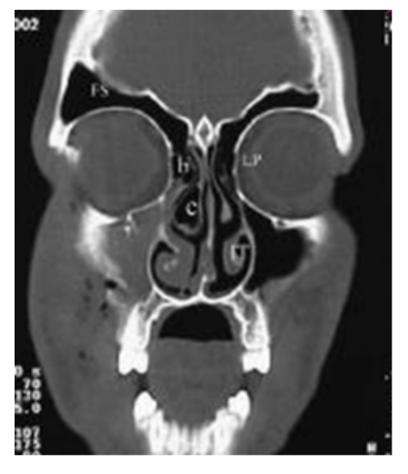
A. Lamina Papyracea
An ENT surgeon experienced extensive bleeding while performing complicated endoscopic intranasal surgery. 1 day after surgery, he noted a hematoma at the medial canthal area of his patient’s left eye that limits the patient’s range of orbital motion. What structure is most likely to have been damaged or penetrated.
A. Lamina Papyracea
B. Skull base
C. The nasal septum
D. Cribriform plate of the ethmoid
C. The Trigeminal Nerve is intact
A patient working at a chemical plant is claiming compensation for work related anosmia. You were tasked to determine the authenticity of his claim. He declares that he cannot smell coffee nor tobacco. But, as you introduce the spirit of ammonia to his nose, he withdraws vigorously due to disturbing intranasal pain. These findings prove that...
A. The patient is lying
B. The olfactory nerve is intact
C. The Trigeminal Nerve is intact
D. it is false anosmia
D. sphenoid sinus
This sinus if penetrated during intranasal sinus surgery may produce blindness and fatal bleeding due to it being intimately related to the optic nerve and internal carotid artery.
A. maxillary sinus
B. ethmoidal recess
C. frontal sinus
D. sphenoid sinus
B. middle meatus
Your patient consulted you for fever, congestion and purulent nasal discharge with facial pain and a CT scan of the sinuses showed pansinusitis involving the frontal, maxillary and anterior ethmoid sinuses. The most likely compromised structure anatomically obstructed is the...
A. Sphenoid ostium
B. middle meatus
C. facial vein
D. frontal recess
B. Possibility of cavernous sinus thrombophlebitis.
Your patient visits your clinic due to swelling and cellulitis between his orbits. CT of the sinuses showed an abscess formation of the nasal septum encroaching onto the upper lips. You advised emergency admission for aggressive medical/surgical management and the patient asked you the indication for admission. Your reply is...
A. Patient convenience
B. Possibility of cavernous sinus thrombophlebitis.
C. Patient treatment compliance
D. Cosmetic deformity
D. Facial vein
What is the most likely PATHWAY OF INFECTION TO THE CAVERNOUS SINUS in a patient with facial cellulitis?
A. internal jugular vein
B. Superficial temporal artery
C. common carotid artery
D. Facial vein
C. CT Scan
An intoxicated motorcycle rider hit his face on the curb while texting while riding his vehicle and suffered facial fractures. What is The DIAGNOSTIC IMAGING OF CHOICE for trauma OF THE PNS/facial bones.
A. plain radiographs
B. MRI
C. CT Scan
D. ultrasound
B. Maxillary sinusitis
A patient with foul rhinorrhea, maxillary pain on the right with fever consults you. He was seen by another MD and CT scan was done. What sinus abnormality is clearly demonstrated in this CT scan of the PNS?
A. Concha bullosa
B. Maxillary sinusitis
C. normal PNS CT scan
D. none of the above
A. The little’s area
A patient consults you with anterior epistaxis. You were taught in 1st aid class to pinch the nose and apply digital pressure onto the nasal ala. What area are you applying to help control bleeding?
A. The little’s area
B. jacobson’s area
C. perpendicular ethmoidal plate area
D. Pyriform aperture
C. Anterior ethmoidal artery, posterior ethmoidal artery, sphenopalatine artery
The blood supply of the nasal septum has contributions from the external and internal carotid artery systems. What arteries mentioned are branches that specifically supply the nasal septum.
A. Superior labial artery, ascending pharyngeal artery, tonsillar artery
B. Maxillary artery, lingual artery, temporal artery
C. Anterior ethmoidal artery, posterior ethmoidal artery, sphenopalatine artery
D. Middle meningeal artery, circle of willy’s, basilar artery
B. Medial canthus to angle of the mandible
The Ohngren’s line is a landmark based line USED TO PROGNOSTICATE MAXILLARY SINUS CANCER. It is determined by land marks from the....
A. Mastoid tip to the angle of the mandible
B. Medial canthus to angle of the mandible
C. Superior nasal spine to the glabella
D. Nasal tip to the mentum
B. Fovea ethmoidalis
A novice ENT surgeon encountered difficulty in orienting himself during sinus surgery. Upon superior dissection of the ethmoidal sinus area. Clear to straw colored fluid began to ooze out of his surgical field. It was determined that the fluid is CSF. What structure was compromised during the surgery that SEPARATES THE ETHMOID SINUSES FROM THE ANTERIOR CRANIAL FOSSA?
A. Spheno ethmoidal recess
B. Fovea ethmoidalis
C. Frontal sinus
D. Sphenoid sinus
B. Pseudo stratified columnar epithelium with goblet cells
Mucus production and also muco-ciliary clearance is made possible by the epithelial lining of the paranasal sinuses. In particular the lining is...
A. simple cuboidal secretory epithelium
B. Pseudo stratified columnar epithelium with goblet cells
C. non-keratinizing squamous epithelium
D. simple squamous non ciliated epithelium
A. Purulent mucoid discharge at the middle meatus
A patient with maxillary and frontal and ethmoidal sinus pain, fever and foul muco purulent rhinorrhea consults you. What will be a likely finding during your PE intranasally?
A. Purulent mucoid discharge at the middle meatus
B. Hyperacusis
C. Epiphora
D. clear watery nasal discharge
B. Natural ostia of the pns
Muco-ciliary flow and clearance of the para nasal sinuses naturally drains via..
A. random directional flow
B. Natural ostia of the pns
C. gravity dependent flow
D. static flow
D. Via pterygoid plexus
A patient with a dental infection consults you for headache, fever, nausea and vomiting. He is toxic looking , weak and has signs of a central nervous infection. What is the possible posterior route of ascending infection to the CNS/cavernous sinus.
A. via facial vein
B. via jugular vein
C. via middle thyroid vein
D. Via pterygoid plexus
D. all of the above
Paranasal sinuses PARTLY SUPPLIED BY THE INTERNAL CAROTID ARTERY SYSTEM
A. frontal sinus
B. ethmoid sinus
C. sphenoid sinus
D. all of the above
D. Trigeminal nerve
A medical student disinfected his hands with 70% isoprpropyl alcohol. He suddenly covered his nose to smell his hands. You noted that he withdraws and frowns and coughs. What cranial nerve is stimulated by this action?
A. Facial nerve
B. Olfactory nerve
C. Vagus nerve
D. Trigeminal nerve
D. Osteomeatal unit
What is the final Common Pathway of Paranasal sinus Drainage particularly the Frontal Sinus, Anterior Ethmoids and Maxillary Sinus.
A. Spheno-ethmoidal recess
B. Frontal recess
C. Superior meatus
D. Osteomeatal unit
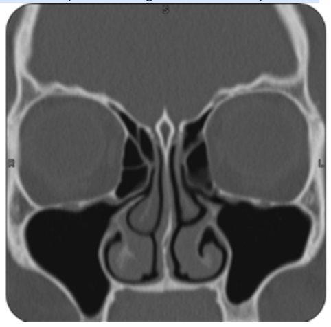
C. Normal pns ct scan
What is the specific findings in the CT scan pictured?
A. Sinusitis
B. Tumor
C. Normal pns ct scan
D. Post op surgical defect
C. Cranial nerve V
Which of the following cranial nerves supply the general sensation of the anterior 2/3 of the tongue?
A. Cranial nerve X
B. Cranial nerve IX
C. Cranial nerve V
D. Cranial nerve VII
C. Stensen’s duct
The parotid gland is in the infra-auricular area and secretes saliva via which of the following ducts?
A. Duct of Rouviere
B. Duct of Rivinus
C. Stensen’s duct
D. Wharton’s duct
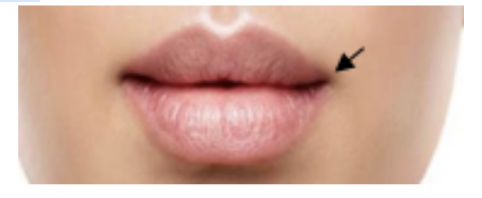
C. Oral commissure
This is the structure where the upper and lower lips meet?
A. Cupid’s bow
B. Vermillion border
C. Oral commissure
D. Philtral ridge
A. Marginal mandibular
A patient with parotid mass underwent surgery. You noticed that the patient cannot depress the ipsilateral lip. Which branch of the facial nerve is the most likely involved?
A. Marginal mandibular
B. Temporal
C. Zygomatic
D. Buccal
C. Orbicularis oris
Which of the following muscles of the lip is innervated by the buccal branch of the facial nerve?
A. Platysma
B. Depressor labii inferioris
C. Orbicularis oris
D. Mentalis
D. Retromolar trigone
The slit-like space bounded by the lips and buccal mucosa to the alveolar ridge and teeth communicates with the oral cavity proper behind the 3rd molar in which area?
A. Oral vestibule
B. Buccinator space
C. Vallecula
D. Retromolar trigone
A. Main trunk of inferior alveolar nerve
A patient complains toothache on the left mandibular 3rd molar and needs to undergo odontectomy. Which nerve needs to be blocked with anesthesia?
A. Main trunk of inferior alveolar nerve
B. Middle superior alveolar nerve
C. Anterior superior alveolar nerve
D. Incisive branch of the inferior alveolar nerve
C. Nasopalatine nerve
Which of the following supplies the palatal side of the upper mucosal canines and incisors?
A. Middle superior alveolar nerve
B. Greater palatine nerve
C. Nasopalatine nerve
D. Anterior superior alveolar nerve
C. Floor of mouth
This oral cavity subsite is a crescent-shaped region of mucosa overlying the mylohyoid and hyoglossus.
A. Buccal mucosa
B. Roof
C. Floor of mouth
D. Retromolar trigone
D. 2nd upper molar
The Stensen’s duct drain saliva in which area opposite to the buccal mucosa?
A. 3rd upper molar
B. 3rd lower molar
C. 2nd lower molar
D. 2nd upper molar
D. Frenulum
The ventral portion of the tongue is attached to the floor of the mouth by which anatomic structure?
A. Sulcus terminalis
B. Plica fimbriata
C. Circumvallate papillae
D. Frenulum
A. Genioglossus
Which of the following extrinsic muscles originates from the superior genial spine of the mandible?
A. Genioglossus
B. Styloglossus
C. Hyoglossus
D. Palatoglossus
D. Killian’s dehiscence
A patient complains of difficulty swallowing and was diagnosed with Zenker’s diverticulum. Which anatomic structure is most likely involved in this condition?
A. Cricopharyngeus
B. Superior pharyngeal constrictor
C. Middle pharyngeal constrictor
D. Killian’s dehiscence
A. Cricopharyngeus
This pharyngeal muscle originates in the lowest fibers of inferior constrictor muscle and serves as sphincter at the lower end of pharynx
A. Cricopharyngeus
B. Salpingopharyngeus
C. Stylopharyngeus
D. Palatopharyngeus
B. Nasopharynx
Which anatomical area has its boundary beginning at the level of skull base until the tip of uvula inferiorly?
A. Oropharynx
B. Nasopharynx
C. Laryngopharynx
D. Hypopharynx
C. V
Which cranial nerve supplies the Tensor veli palatini muscle?
A. X
B. VII
C. V
D. IX
D. Lingual
Which of the following branches of the external carotid artery supplies the palatine tonsils?
A. Occipital
B. Superficial temporal
C. Superior thyroid
D. Lingual
D. Pharyngeal
What phase of swallowing primarily propels bolus to the esophagus via anterograde peristaltic control?
A. Esophageal
B. Oral transit
C. Oral preparatory
D. Pharyngeal
C. IX
Which cranial nerve is the origin of the Jacobson’s Nerve responsible for referred otalgia or ear pain in patients with tonsillitis?
A. V
B. X
C. IX
D. VII
D. Mandible
The neck is bounded superiorly by the:
A. SCM
B. Maxilla
C. Hyoid bone
D. Mandible
A. Investing Fascia
Which among the following belongs to the most superficial of the deep cervical fascia?
A. Investing Fascia
B. Vertebral Fascia
C. Visceral Fascia
D. Pretracheal Fascia
C. Platysma
Which of the following is invested in the superficial cervical fascia?
A. Omohyoid
B. SCM
C. Platysma
D. Digastric
D. All of the above
What structures does the visceral fascia surround?
A. Trachea
B. Esophagus
C. Thyroid
D. All of the above
D. CN XI
What is the innervation of the SCM?
A. CN XII
B. CN X
C. CN IX
D. CN XI
C. SCM
What is the structure in the neck that divides it into the anterior and posterior triangle?
A. Trapezius
B. Digastric
C. SCM
D. Omohyoid
D. All of the above
The carotid sheath is formed by which fascia:
A. Superficial Deep Cervical Fascia
B. Middle Deep Cervical Fascia
C. Deep Layer of the Deep Cervical Fascia
D. All of the above
C. External Jugular vein
Which of the following is not contained inside the carotid space?
A. Vagus nerve
B. Internal Jugular vein
C. External Jugular vein
D. Carotid artery
D. Occipital
What is the fifth branch of the External Carotid artery?
A. Lingual
B. Inferior Thyroid
C. Posterior Auricular
D. Occipital
A. Retropharyngeal Space
What neck space is involved if an abscess is formed between the buccopharyngeal fascia and alar fascia?
A. Retropharyngeal Space
B. Parapharyngeal Space
C. Danger Space
D. Prevertebral Space
D. Coccyx
Abscess formation in the prevertebral space can potentially spread to what area?
A. Superior Mediastinum
B. Inferior Mediastinum
C. Diaphragm
D. Coccyx
B. It begins at the level of the Hyoid bone
Which of the following is true about the External Carotid artery:
A. It is the main blood supply of the brain
B. It begins at the level of the Hyoid bone
C. It crosses deep to the styloglossus muscle
D. It has 2 layers
C. the internal branch of the superior laryngeal nerve pierces thru the thyrohyoid membrane
Which of the following is true about the Vagus nerve:
A. it is a purely sensory nerve
B. the external branch of the superior laryngeal nerve supplies the sternothyroid
C. the internal branch of the superior laryngeal nerve pierces thru the thyrohyoid membrane
D. it has an anterior and posterior branch as it courses thru the neck
C. Acromial head of the clavicle
Which among the following is not an attachment of the SCM?
A. Sternal head of the clavicle
B. Sternal Head
C. Acromial head of the clavicle
D. Manubrium
C. Anterior border of the clavicle
Which among the following is not an attachment of the Trapezius?
A. Acromion process
B. Scapula
C. Anterior border of the clavicle
D. All of the above
D. Greater auricular
A 20 year old man was brought into the emergency room with a stab wound in the upper part of the neck. Although there was no major damage done, he lost sensation from the skin over the angle of the jaw. Which nerve has been cut?
A. Greater occipital
B. Suprascapular
C. Transverse cervical
D. Greater auricular
C. Recurrent Laryngeal
In repairing a damaged right subclavian artery, the surgeon notices and protects a large nerve passing around to the posterior surface of the artery. This nerve, which does not encircle the subclavian on the left side, is the:
A. Ansa cervicalis
B. Vagus
C. Recurrent Laryngeal
D. Phrenic
D. CN XI
An abscess was surgically removed from the middle of the posterior triangle on the right side. During recovery the patient noticed that her shoulder drooped and she could no longer raise her right hand above her head to brush her hair. Which nerve has been cut?
A. CN X
B. CN XII
C. Ansa cervicalis
D. CN XI
D. Superior Thyroid Artery
What is the arterial supply of the upper lobe of the thyroid?
A. Inferior Thyroid Artery
B. Thyroid Ima Artery
C. Middle Thyroid Artery
D. Superior Thyroid Artery
B. External Carotid Artery
The superior thyroid artery is a branch of what artery?
A. Brachiocephalic Artery
B. External Carotid Artery
C. Internal Carotid Artery
D. Subclavian Artery
D. Subclavian Artery
The inferior thyroid artery is a branch of what artery?
A. Internal Carotid Artery
B. External Carotid Artery
C. Brachiocephalic Artery
D. Subclavian Artery
C. Inferior Thyroid Artery
What is the primary blood supply of the parathyroid?
A. Subclavian Artery
B. Parathyroid Artery
C. Inferior Thyroid Artery
D. Superior Thyroid Artery
B. 4
How many parathyroids do most people have:
A. 3
B. 4
C. 1
D. 2
D. C6
At what Vertebral level does the trachea starts?
A. C7
B. C4
C. C5
D. C6
C. Recurrent Laryngeal Nerve
The trachea receives sensory innervation from what nerve?
A. Phrenic Nerve
B. Superior Laryngeal Nerve
C. Recurrent Laryngeal Nerve
D. Glossopharyngeal Nerve
A. C6
At what vertebral level does the cervical esophagus start?
A. C6
B. C5
C. C4
D. C7
A. T10
At what vertebral level does the thoracic esophagus end?
A. T10
B. T9
C. T8
D. T7
C. Thyroid Isthmus
During tracheostomy procedure, what structure superficial to the trachea can be potentially injured that can result to bleeding?
A. Carotid artery
B. Esophagus
C. Thyroid Isthmus
D. Pyramidal lobe
B. Upper third of the esophagus
Which anatomical segment of the esophagus is completely made up of striated muscle?
A. Thoracic esophagus
B. Upper third of the esophagus
C. Abdominal esophagus
D. Middle third of the esophagus
D. Upper Esophageal Sphincter
Esophageal constriction located 15 cm from the incisor is due to what structure?
A. Lower Esophageal Sphincter
B. Left Main stem bronchus
C. Arch of the Aorta
D. Upper Esophageal Sphincter
C. Left Main stem bronchus
Esophageal constriction located 27.5 cm from the incisor is due to what structure?
A. Lower Esophageal Sphincter
B. Upper Esophageal Sphincter
C. Left Main stem bronchus
D. Arch of the Aorta
B. Recurrent Laryngeal nerve
Exploration of the tracheoesophageal groove at the level of the thyroid gland would reveal what important structure bilaterally:
A. Phrenic nerve
B. Recurrent Laryngeal nerve
C. Inferior thyroid artery
D. Brachial plexus
B. Superior Laryngeal Nerve
Ligating the superior thyroid artery near the level of the hyoid can potentially injure what structure?
A. Phrenic Nerve
B. Superior Laryngeal Nerve
C. Vagus nerve
D. Recurrent Laryngeal Nerve
D. All of the above
Which of the following is true about the recurrent laryngeal nerve:
A. The right loops around the subclavian artery
B. The left loops around the aortic arch
C. The nerve travels along the tracheoesophageal groove as it ascends back to the larynx
D. All of the above
B. Recurrent Laryngeal nerve
A 35 year old woman was diagnosed with an adenoma of the thyroid gland. This required excision of the lower pole (left lobe) of the gland and ligation of the artery supplying that region. Which of the following nerves accompanying the artery is most likely to be damaged if the surgeon is not careful?
A. Internal branch of the Superior Laryngeal nerve
B. Recurrent Laryngeal nerve
C. Vagus proper
D. External branch of the Superior Laryngeal nerve
A. External branch of the Superior Laryngeal
Following surgery on the upper pole of the right lobe of the thyroid gland, a patient complains of hoarseness and weakness of voice. What nerve may have been injured?
A. External branch of the Superior Laryngeal
B. Recurrent Laryngeal
C. Ansa cervicalis
D. Internal branch of the Superior Laryngeal
C. just below the thyroid cartilage
A patient is brought into the Emergency Room in respiratory distress. It is quickly decided to create an emergency airway to restore respiration. At what level could you rapidly create an airway below the vocal cords with a minimum danger of hemorrhage?
A. just below the cricoid cartilage
B. just above the thyroid cartilage
C. just below the thyroid cartilage
D. just above the jugular notch
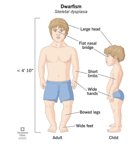
B. Neural crest cells
CASE: All MESODERM-derived bones (i.e., limbs, spinal column, and thoracic cage) are affected and shorter-than-normal amongst Achondroplastic/Skeletal Dysplastic patients (a type of dwarfism) but their faces are normal and almost adult-looking - which seem unaffected. This is because the “skeleton-of-the-face” called Viscerocranium and “forehead” called frontal bone of the neurocranium, which are not affected in most types of dwarfism, are derivatives of: (Kindly check illustration courtesy of Cleveland Clinic)
A. Neuroectodermal cells
B. Neural crest cells
C. Placode cells
D. Surface ectoderm cells
D. Endoderm cells
The “parathyroid glands”, “thymus gland”, and “thyroid gland” are derivatives of these germ cells:
A. Primordial Germ cells
B. Mesoderm cells
C. Ectoderm cells
D. Endoderm cells
C. PA 5
The only “Pharyngeal Arch” (PA) that will NOT provide any adult derivative due to early obliteration as early as 5th Week after Fertilization:
A. PA 4
B. PA 1
C. PA 5
D. PA 3
E. PA 2
B. PC 1
This “Pharyngeal Cleft” (PC) used to play direct and critical role in the formation of “external auditory meatus” or “external ear canal” according to former embryologists:
A. PC 2
B. PC 1
C. PC 4
D. PC 3