Section 3.2 Signaling and Cell Cycle Control

0.0(0)
Studied by 0 people
call kaiCall Kai
Locked
learnLearn
examPractice Test
spaced repetitionSpaced Repetition
heart puzzleMatch
flashcardsFlashcards
GameKnowt Play
Card Sorting

1/15

encourage image

There's no tags or description

Looks like no tags are added yet.

Last updated 10:00 PM on 8/25/26
Name
Mastery
Learn
Test
Matching
Spaced
Call with Kai
Chat

No analytics yet

Send a link to your students to track their progress

16 Terms

1
New cards

The Eukaryotic Cell Cycle

Cell division occurs in nearly every tissue during development: Specific events required to ensure every cell had everything it needs to live

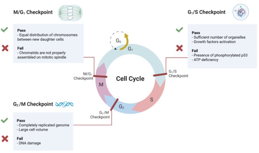
After passing M phase and into G1, a cell can either continue through another division or enters G0; The cell cycle has major checkpoints where the cell cycle stops to make sure everything is good before continuing

Differentiated cells stop dividing and enter the G0 phase = quiescent

Quiescent: Temporary G0 state, the cell can enter G1 again if it is signaled to

Senescence: The cell is in a permanent G0 state; it can no longer enter G1

Unregulated cellular division leads to cancer development
<p>Cell division occurs in nearly every tissue during development: Specific events required to ensure every cell had everything it needs to live</p><p>After passing M phase and into <strong>G1</strong>, a cell can either continue through another division or enters <strong>G0</strong>; The cell cycle has major checkpoints where the cell cycle stops to make sure everything is good before continuing</p><p>Differentiated cells stop dividing and enter the<strong> G0 phase = quiescent</strong></p><p>Quiescent: Temporary G0 state, the cell can enter G1 again if it is signaled to</p><p>Senescence: The cell is in a permanent G0 state; it can no longer enter G1</p><p>Unregulated cellular division leads to cancer development</p>
2
New cards

Mammary Gland Development (Not on exam; an example of G0 → replication)

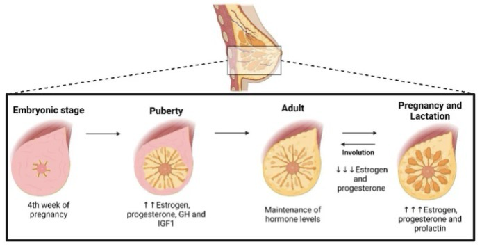
Mammary gland development is primarily post-natal:

  1. The ductal tree develops as an invagnication of ectoderm

  2. At birth, a rudimentary “ductal tree” structure is already present

  3. Very limited development occurs until the onset of puberty

Majority of development occurs in utero; this is an example of development outside utero/during puberty
<p>Mammary gland development is primarily post-natal:</p><ol><li><p>The ductal tree develops as an invagnication of ectoderm</p></li><li><p>At birth, a rudimentary “ductal tree” structure is already present</p></li><li><p>Very limited development occurs until the onset of puberty</p></li></ol><p>Majority of development occurs in utero; this is an example of development outside utero/during puberty</p>
3
New cards

Signals and Receptors

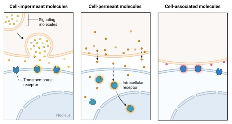
Protein signaling molecules require a cell membrane receptor (Causes secondary signaling to occur)

Hydrophobic signaling molecules, such as steroid hormones, use nuclear receptors (Binds to ligand and goes through the plasma membrane/No secondary signaling molecule required)

A cell that is growing to divide experiences many changes:

  1. Altered gene expression

  2. Increased translation

  3. Increased nutrient uptake and metabolic rate

  4. Altered morphology


<p>Protein <strong>signaling</strong> <strong>molecules</strong> require a cell membrane receptor (Causes secondary signaling to occur)</p><p>Hydrophobic signaling molecules, such as <strong>steroid hormones</strong>, use<strong> nuclear receptors</strong> (Binds to ligand and goes through the plasma membrane/No secondary signaling molecule required)</p><p>A cell that is growing to divide experiences many changes:</p><ol><li><p>Altered <strong>gene expression</strong></p></li><li><p>Increased <strong>translation</strong></p></li><li><p>Increased nutrient uptake and metabolic rate</p></li><li><p>Altered morphology</p></li></ol><p></p>
4
New cards

Nuclear Hormone Receptor: ER (Mammary gland development); What drug treats high estrogen in breast cancer cells

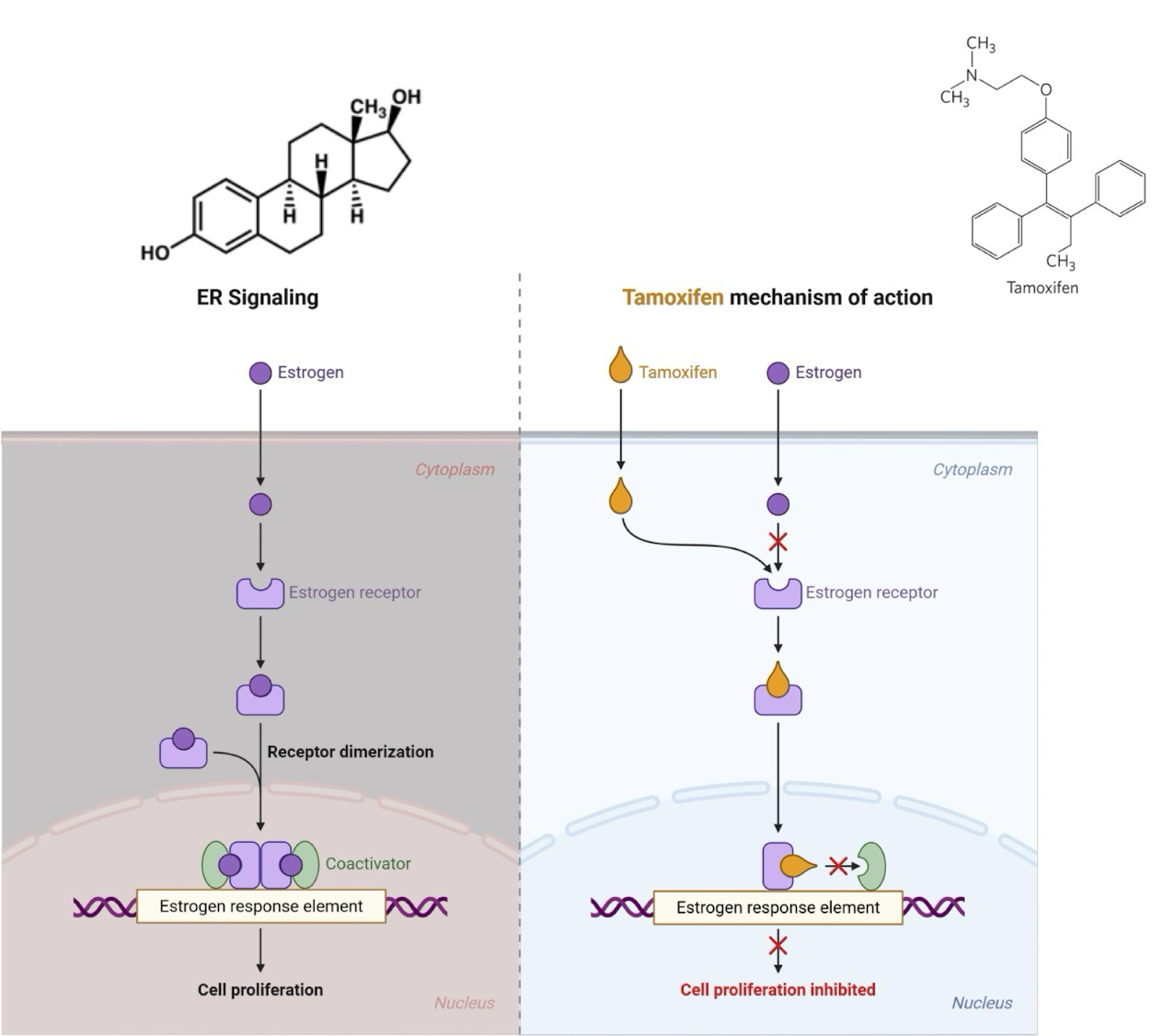
During puberty cells with estrogen binding receptors respond to increase in estrogen

Ex. Mammary epithelial cells on ductal tree lining

Estrogen passively diffuses through plasma membrane and binds to estrogen receptor in cytoplasm

Estrogen binding induces estrogen receptor (ER) dimerization

  • Receptor is then translocated to the nucleus

  • Binds to genes containing an estrogen response element (ERE)

  • ER interacts with coactivator proteins for the recruitment of transcription machinery

ER effects in combination with other transcription factors determines the levels expression for each gene in the cell

Tamoxifen: Estrogen antagonist competes with estrogen for binding to ER, stops dimerization from occurring
<p>During puberty cells with estrogen binding receptors respond to increase in estrogen</p><p>Ex. Mammary epithelial cells on ductal tree lining</p><p>Estrogen passively diffuses through plasma membrane and binds to estrogen receptor in cytoplasm</p><p><strong>Estrogen</strong> binding induces <strong>estrogen receptor (ER) </strong>dimerization</p><ul><li><p>Receptor is then translocated to the nucleus</p></li><li><p>Binds to genes containing an<strong> estrogen response element (ERE)</strong></p></li><li><p>ER interacts with <strong>coactivator</strong> proteins for the recruitment of transcription machinery</p></li></ul><p>ER effects in combination with other <strong>transcription</strong> <strong>factors</strong> determines the levels expression for each gene in the cell</p><p><strong>Tamoxifen</strong>: Estrogen antagonist competes with estrogen for binding to ER, stops dimerization from occurring</p>
5
New cards

RAS-Dependent Pathway Activation/Deactivation

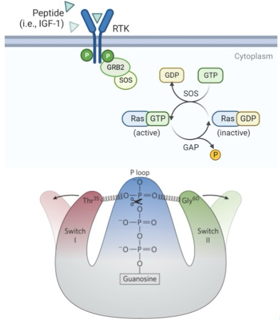
Peptide binding to RTKs (receptor tyrosine kinases) stimulates autophosphorylation; GRB2 bind phosphotyrosines; SOS is a guanine nucleotide exchange factor (GEF)

SOS activity results in GDP/GTP exchange by RAS; in a non-dividing cell RAS is an GDP-bound state; “active” RAS activates a signaling cascade for cell growth

RAS signaling is turned off by GTPase activating protein (GAP)

GTP → GDP + Pi

3rd phosphate in GTP binds to structure and allows function; when this P loop is hydrolyzed, the protein is no longer held up and is deactivated
<p>Peptide binding to <strong>RTKs (receptor tyrosine kinases)</strong> stimulates autophosphorylation; GRB2 bind phosphotyrosines; <strong>SOS</strong> is a <strong>guanine nucleotide exchange factor (GEF)</strong></p><p>SOS activity results in GDP/GTP exchange by <strong>RAS</strong>; in a non-dividing cell RAS is an GDP-bound state; “active” RAS activates a signaling cascade for cell growth</p><p>RAS signaling is turned off by <strong>GTPase activating protein (GAP)</strong></p><p>GTP → GDP + P<sub>i</sub></p><p>3rd phosphate in GTP binds to structure and allows function; when this P loop is hydrolyzed, the protein is no longer held up and is deactivated</p>
6
New cards

RAS-Dependent Pathway

Protein

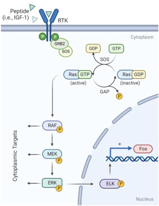
Pathway is activated by RTKs for many growth factors, such as IGF-1, EGF and PDGF

RAS-GTP interacts with RAF activating its protein kinase activity:

  1. RAF phosphorylates MEK

  2. MEK phosphorylates ERK

  3. ERK phosphorylates transcription factors in the nucleus

  4. ELK-1 binds Serum Response Element (SRE) located in pro-growth genes to activate their transcription; FOS is one of these genes

Sequential activation of protein kinases results in signal amplification and diversification, also known as a Signaling cascade

Phosphorylation and activity of target proteins reversed by protein phosphatases (Dephosphorylation)
<p>Protein </p><p>Pathway is activated by RTKs for many growth factors, such as IGF-1, EGF and PDGF</p><p><strong>RAS</strong>-GTP interacts with <strong>RAF</strong> activating its protein kinase activity:</p><ol><li><p>RAF phosphorylates <strong>MEK</strong></p></li><li><p>MEK phosphorylates <strong>ERK</strong></p></li><li><p>ERK phosphorylates transcription factors in the nucleus</p></li><li><p><strong>ELK-1</strong> binds Serum Response Element (SRE) located in pro-growth genes to activate their transcription; <strong>FOS</strong> is one of these genes</p></li></ol><p>Sequential activation of protein kinases results in signal amplification and diversification, also known as a <strong>Signaling cascade</strong></p><p>Phosphorylation and activity of target proteins reversed by <strong>protein phosphatases</strong> (Dephosphorylation)</p>
7
New cards
<p><strong>Activating Protein-1</strong></p>

Activating Protein-1

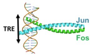
JUN and FOS = Activating protein-1 (AP-1)

This heterodimer is a very strong activator for the cell cycle

Transcriptional activator; binds TRE cis-acting element; regulates many genes needed for progression through cell cycle

One of the proteins resulting the AP-1 cascade is Cyclin D, an official signal that we are in G1

<p><strong>JUN</strong> and <strong>FOS</strong> = <strong>Activating protein-1 (AP-1)</strong></p><p>This heterodimer is a very strong activator for the cell cycle</p><p>Transcriptional <strong>activator</strong>; binds TRE<strong> cis-acting</strong> element; regulates many genes needed for progression through cell cycle</p><p>One of the proteins resulting the AP-1 cascade is Cyclin D, an official signal that we are in G1</p>
8
New cards

Myc/Max is Activator of Proliferation Genes

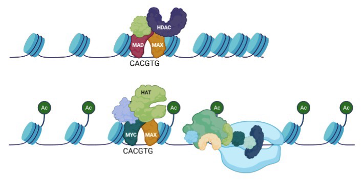
Mad/Max is a repressor of pro-growth genes (Recruits HDACs to close chromatin structure)

When AP1 is produced, it first results in MYC production; Myc competes with Mad for Max

Myc/Max complex is an activator of many pro-growth genes

  • i.e. cyclin D, cyclin E, E2F, PCNA, DNA pol ⍺ and stimulates RNA Pol II activity

The upregulation of cyclin D gene transcription by AP-1 and Myc “kick starts” the cell cycle
<p><strong>Mad/Max i</strong>s a repressor of pro-growth genes (Recruits HDACs to close chromatin structure)</p><p>When AP1 is produced, it first results in MYC production; Myc competes with Mad for Max</p><p><strong>Myc/Max </strong>complex is an activator of many pro-growth genes</p><ul><li><p>i.e. cyclin D, cyclin E, E2F, PCNA, DNA pol ⍺ and stimulates RNA Pol II activity</p></li></ul><p>The upregulation of<strong> cyclin D</strong> gene transcription by AP-1 and Myc “kick starts” the cell cycle</p>
9
New cards

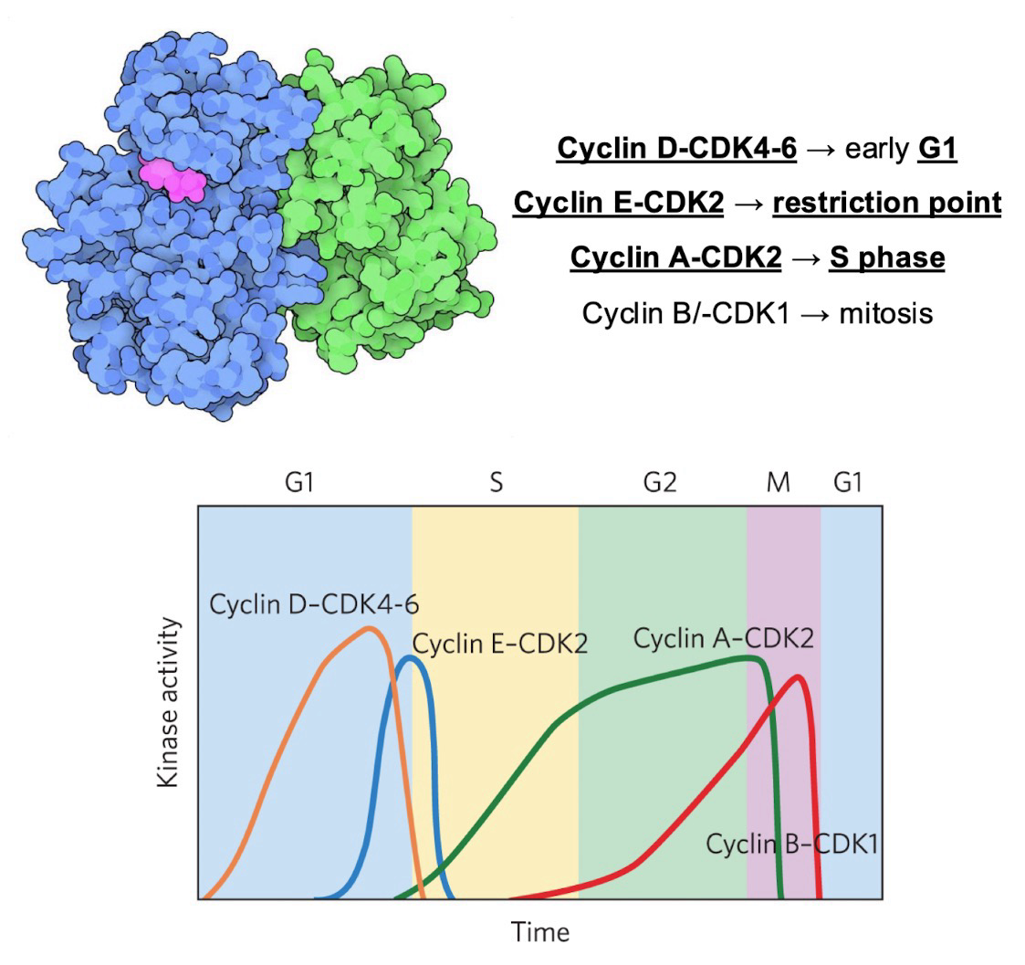
Cyclin-CDK Complex; Specific Cyclins and Their Respective Checkpoints; How are they regulated?

A family of kinases that controls cell cycle timing

  • Each active kinase has 2 subunits:

    • One catalytic subunit with a cyclin-dependent kinase (CDK)

      • CDK4, CDK6, CDK2, and CDK1

    • One regulatory subunit that requires cyclin

      • cyclin D, cyclin E, cyclin A, cyclin B

Specific Cyclins and their respective checkpoints:

  • Cyclin D-CDK4-6 → G1

  • Cyclin E-CDK2 → Restriction point (to enter S)

  • Cyclin A-CDK2 → S

  • Cyclin B/-CDK1 → M/Mitosis

Oscillation of Cyclin-CDK activity are the result of:

  • CDK phosphorylation or dephosphorylation

  • Controlled degradation of the cyclin subunit via ubiquitin (Ubiquitination)

  • Periodic synthesis of CDKs and cyclins

  • The action of specific CDK-inhibiting proteins (CIP)

<p>A family of <strong>kinases</strong> that controls cell cycle timing</p><ul><li><p>Each active kinase has 2 subunits:</p><ul><li><p>One catalytic subunit with a <strong>cyclin-dependent kinase (CDK)</strong></p><ul><li><p>CDK4, CDK6, CDK2, and CDK1</p></li></ul></li><li><p>One regulatory subunit that requires <strong>cyclin</strong></p><ul><li><p>cyclin D, cyclin E, cyclin A, cyclin B</p></li></ul></li></ul></li></ul><p>Specific Cyclins and their respective checkpoints:</p><ul><li><p><strong>Cyclin D-CDK4-6 → G1</strong></p></li><li><p><strong>Cyclin E-CDK2 → Restriction point</strong> (to enter S)</p></li><li><p><strong>Cyclin A-CDK2 → S</strong></p></li><li><p>Cyclin B/-CDK1 → M/Mitosis</p></li></ul><p>Oscillation of Cyclin-CDK activity are the result of:</p><ul><li><p>CDK phosphorylation or dephosphorylation</p></li><li><p>Controlled degradation of the cyclin subunit via <strong>ubiquitin </strong>(Ubiquitination)</p></li><li><p>Periodic synthesis of CDKs and cyclins</p></li><li><p>The action of specific <strong>CDK-inhibiting proteins (CIP)</strong></p></li></ul><p></p>
10
New cards

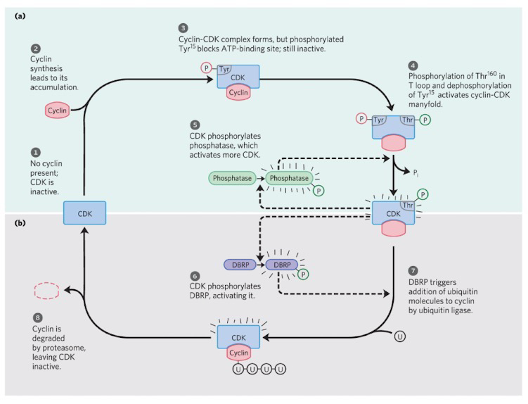
CDK Regulation by Phosphorylation and Proteolysis

Ubiquitin = Regulatory protein that targets proteins for destruction

Proteasomes = Proteolytic enzyme complexes

DBRP = Destruction box recognizing protein
<p><strong>Ubiquitin</strong> = Regulatory protein that targets proteins for destruction</p><p><strong>Proteasomes</strong> = Proteolytic enzyme complexes</p><p><strong>DBRP</strong> = Destruction box recognizing protein</p>
11
New cards

Cell Cycle Checkpoints

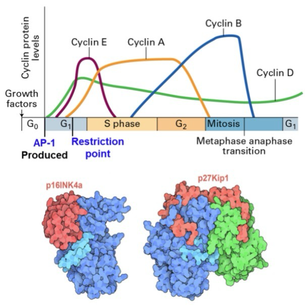
Restriction point: Checkpoint before DNA replication (Between G1 and S); If a cell passes this point, it is committed to S phase and cellular division

At this point cyclin-dependent kinase activity inhibitors are expressed in G1 termed CDK inhibitors (CKI)

  • Inhibitor of cdk4 (INK4) proteins bind to CDK4/6 in 1:1 stoichiometry, blocking cyclin D binding; (Ex, p16INK4a and p14ARF)

  • CDK interacting protein (CIP) binds to and inhibits the activities of intact (Active) CDK-cyclin complexes; (Ex, p21 and p27)

Overall works to inhibit S phase/cellular division
<p><strong>Restriction point:</strong> Checkpoint before <strong>DNA replication</strong> (Between G1 and S); If a cell passes this point, it is committed to S phase and cellular division</p><p>At this point cyclin-dependent kinase activity inhibitors are expressed in <strong>G1</strong> termed <strong>CDK inhibitors (CKI)</strong></p><ul><li><p><strong>Inhibitor of cdk4 (INK4)</strong> proteins bind to CDK4/6 in 1:1 stoichiometry, blocking cyclin D binding; (Ex, p16INK4a and p14ARF)</p></li><li><p><strong>CDK interacting protein (CIP) </strong>binds to and inhibits the activities of intact (Active) CDK-cyclin complexes; (Ex, <strong>p21</strong> and p27)</p></li></ul><p>Overall works to inhibit S phase/cellular division</p>
12
New cards

Restriction Point

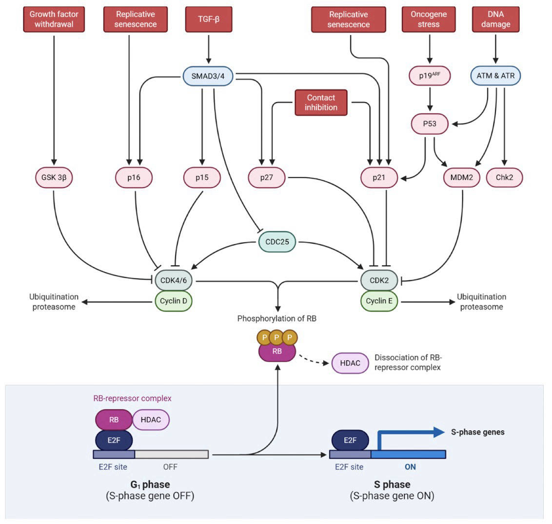
Retinoblastoma protein (pRb): Corepressor that binds the activator E2F to arrest/stop cell division; Also recruits HDACs

Cyclin D-CDK4/6 and cyclin E-CDK2 both hyperphosphorylates Rb, releasing Rb from E2F

E2F is an activator of genes needed in S phase like:

  • Cell cycle machinery genes (cyclins, cyclin-dependent kinases)

  • pro-growth transcription factors (Myc, E2F family)

  • component of the DNA replication machinery (replisome) and chromatin proteins (histones)

<p><strong>Retinoblastoma protein (pRb)</strong>: <strong>Corepressor</strong> that binds the <strong>activator E2F</strong> to arrest/stop cell division; Also recruits HDACs</p><p><strong>Cyclin D-CDK4/6 </strong>and<strong> cyclin E-CDK2</strong> both hyperphosphorylates Rb, releasing Rb from <strong>E2F</strong></p><p><strong>E2F</strong> is an activator of genes needed in <strong>S phase</strong> like:</p><ul><li><p>Cell cycle machinery genes (<strong>cyclins</strong>, <strong>cyclin-dependent kinases</strong>)</p></li><li><p>pro-growth transcription factors (<strong>Myc</strong>,<strong> E2F family</strong>)</p></li><li><p>component of the DNA replication machinery (<strong>replisome</strong>) and chromatin proteins (<strong>histones</strong>)</p></li></ul><p></p>
13
New cards

Restriction Point Specific in Blocking Progress When DNA Damage is Detected

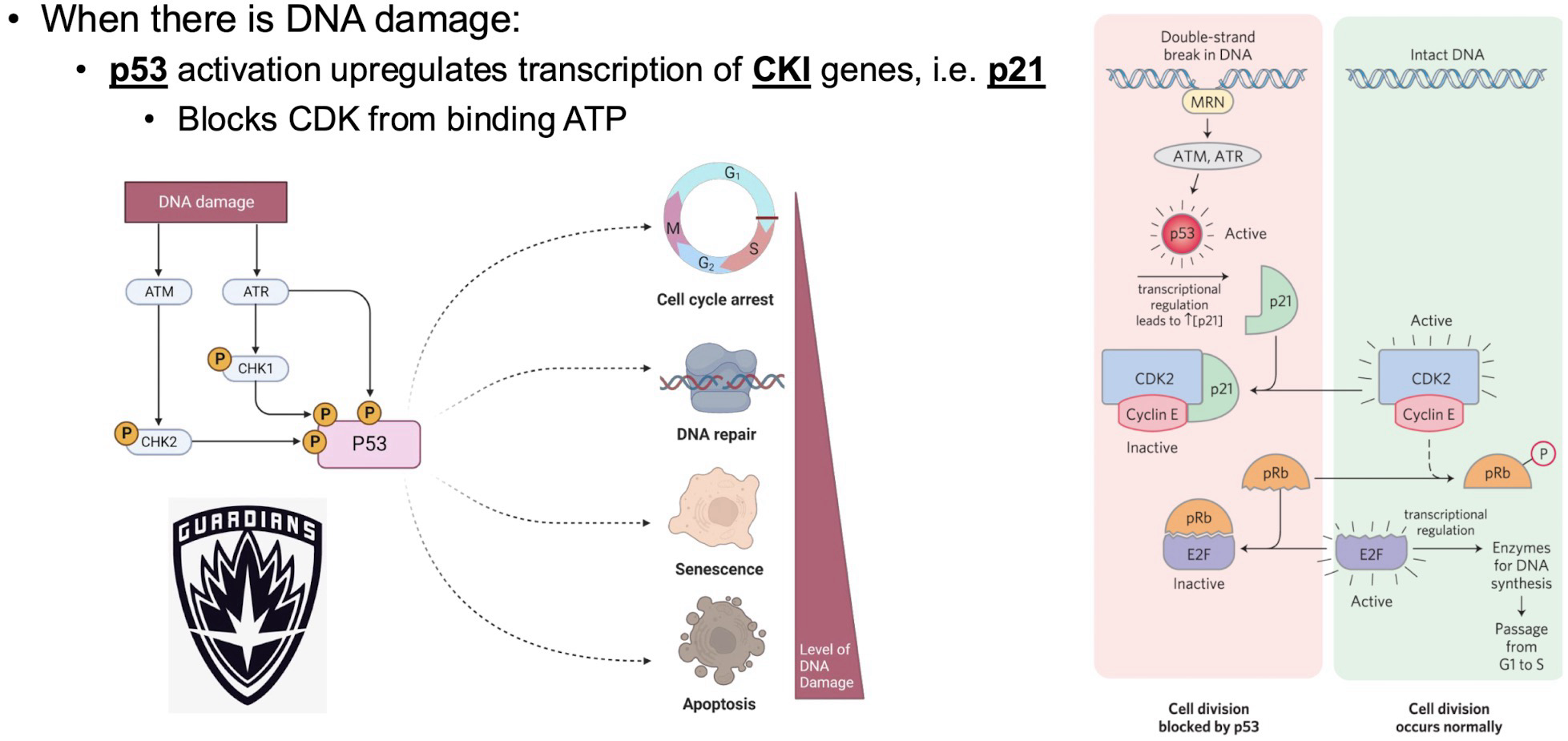
When DNA is damaged, p53 is activated and upregulates transcription of CKI genes (i.e. p21), which blocks CDK from binding ATP

p53 also upregulates cell cycle arrest proteins and DNA repair proteins

If damage cannot be repaired, p53 activates senescence

If the cell is unviable p53 activates apoptosis pathway
<p>When DNA is damaged, <strong>p53</strong> is activated and upregulates transcription of CKI genes (i.e. p21), which blocks CDK from binding ATP </p><p>p53 also upregulates cell cycle arrest proteins and DNA repair proteins</p><p>If damage cannot be repaired, p53 activates senescence</p><p>If the cell is unviable p53 activates apoptosis pathway</p>
14
New cards

Retinoblastoma

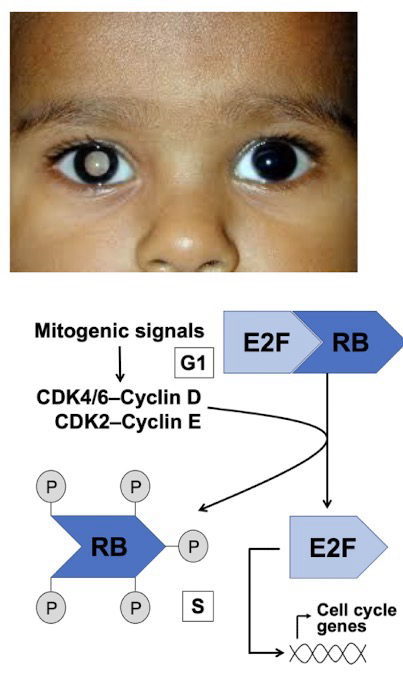
Mutation in RB1 gene inactivates retinoblastoma protein (pRb) function/production, a key regulator of G1/S checkpoint

pRb typically inhibits E2F transcription, preventing early entry to the S phase

This loss of cell cycle control results in excessive proliferation and tumor formation (Because cells no longer stops to fix DNA damage)

Two-hit hypothesis: A defective allele is inherited and the second is lost through somatic mutation
<p>Mutation in RB1 gene inactivates <strong>retinoblastoma protein (pRb)</strong> function/production, a key regulator of <strong>G1/S checkpoint</strong></p><p>pRb typically inhibits <strong>E2F</strong> transcription, preventing early entry to the <strong>S phase</strong></p><p>This loss of cell cycle control results in excessive proliferation and tumor formation (Because cells no longer stops to fix DNA damage)</p><p><strong>Two-hit hypothesis</strong>: A defective allele is inherited and the second is lost through somatic mutation</p>
15
New cards

G1 → S Phase Transition

Synthesis of cyclin A is required to transition to the S phase; E2F is a transcriptional activator of cyclin A

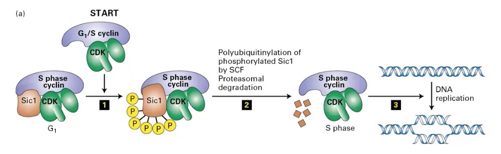
Requires enough active cyclin E-CDK2 to activate cyclin A-CDK2

This is because cyclin E-CDK2 is required for the cyclin E-CDK2-dependent destruction of cyclinA/cdk2 inhibitor Sic1

E-CDK2 phosphorylates Sic1, making it susceptible to ubiquitin ligase → ubiquitin-mediated degradation
<p>Synthesis of <strong>cyclin A</strong> is required to transition to the<strong> S phase</strong>;<strong> E2F</strong> is a transcriptional activator of cyclin A</p><p>Requires enough <strong>active cyclin E-CDK2</strong> to activate<strong> cyclin A-CDK2</strong></p><p>This is because cyclin E-CDK2 is required for the cyclin E-CDK2-dependent destruction of cyclinA/cdk2 inhibitor <strong>Sic1</strong></p><p>E-CDK2 phosphorylates Sic1, making it susceptible to<strong> ubiquitin ligase</strong> → ubiquitin-mediated degradation</p>
16
New cards

How Does Cyclin A-CDK2 Relate to Licensing Coordination in DNA replication

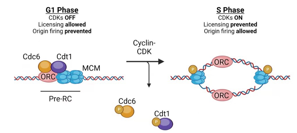
Recall that the ORC binds tightly to DNA in G1

Regulating complexes Cdc6 and Cdt1 stops replication from occurring

In S phase, Cyclin A-CDK2 phosphorylates these complexes (and MCM) to begin replication
<p>Recall that the ORC binds tightly to DNA in G1</p><p>Regulating complexes Cdc6 and Cdt1 stops replication from occurring</p><p>In S phase, <strong>Cyclin A-CDK2</strong> phosphorylates these complexes (and MCM) to begin replication</p>