🪢 Lecture 3: Neurophysiology II

0.0(0)
Studied by 0 people
call kaiCall Kai
Locked
learnLearn
examPractice Test
spaced repetitionSpaced Repetition
heart puzzleMatch
flashcardsFlashcards
GameKnowt Play
Card Sorting

1/28

encourage image

There's no tags or description

Looks like no tags are added yet.

Last updated 11:05 PM on 9/18/26
Name
Mastery
Learn
Test
Matching
Spaced
Call with Kai
Chat

No analytics yet

Send a link to your students to track their progress

29 Terms

1
New cards

Organization of the CNS

Central Nervous System (CNS)
  • Brain
  • Spinal cord

Brain
  • Cerebrum (Forebrain)
  • Diencephalon
    • Thalamus
    • Hypothalamus
  • Cerebellum
  • Brain stem
    • Mesencephalon
    • Pons
    • Medulla oblongata

<p><strong>Central Nervous System (CNS)</strong><br>  • <strong>Brain</strong><br>  • <strong>Spinal cord</strong></p><p><strong>Brain</strong><br>  • <strong>Cerebrum (Forebrain)</strong><br>  • <strong>Diencephalon</strong><br>    • <strong>Thalamus</strong><br>    • <strong>Hypothalamus</strong><br>  • <strong>Cerebellum</strong><br>  • <strong>Brain stem</strong><br>    • <strong>Mesencephalon</strong><br>    • <strong>Pons</strong><br>    • <strong>Medulla oblongata</strong></p>
2
New cards

Tasks of the CNS

Receives Information
  • Receives a continuous flow of information
  • Information comes from outside and inside the body

Processes Information
  • Analyzes and interprets the information
  • Makes sense of internal and external information

Sends Commands
  • Sends commands to the body to:
    • Maintain function of vital systems and organs
    • Respond to the external environment

Complex Organization
  • Example → sequence of events required for a horse to jump a fence
  • Although very complex, the brain can be organized by:
    • Structural (anatomical) features
    • Function

<p><strong>Receives Information</strong><br>  • Receives a continuous flow of <strong>information</strong><br>  • Information comes from <strong>outside and inside the body</strong></p><p><strong>Processes Information</strong><br>  • <strong>Analyzes and interprets</strong> the information<br>  • Makes sense of <strong>internal and external information</strong></p><p><strong>Sends Commands</strong><br>  • Sends commands to the body to:<br>    • Maintain function of <strong>vital systems and organs</strong><br>    • <strong>Respond to the external environment</strong></p><p><strong>Complex Organization</strong><br>  • Example → sequence of events required for a <strong>horse to jump a fence</strong><br>  • Although very complex, the brain can be organized by:<br>    • <strong>Structural (anatomical) features</strong><br>    • <strong>Function</strong></p>
3
New cards

Meninges and Cerebrospinal Fluid

Meninges
  • 3 layers surrounding the brain and spinal cord

Outer Meninx – Dura Mater
  • Thick and tough
  • Supports the brain
  • Inserted between the cerebral hemispheres
  • Forms the venous sinus
    • Collects most of the blood and cerebrospinal fluid (CSF) from the brain

Middle Meninx – Arachnoid
  • Middle layer of the meninges

Inner Meninx – Pia Mater
  • Tightly attached to the neural tissue

Subarachnoid Space
  • Located between the arachnoid and pia mater
  • Filled with cerebrospinal fluid (CSF)

<p><strong>Meninges</strong><br>  • <strong>3 layers</strong> surrounding the <strong>brain and spinal cord</strong></p><p><strong>Outer Meninx – Dura Mater</strong><br>  • <strong>Thick and tough</strong><br>  • <strong>Supports the brain</strong><br>  • Inserted between the <strong>cerebral hemispheres</strong><br>  • Forms the <strong>venous sinus</strong><br>    • Collects most of the <strong>blood and cerebrospinal fluid (CSF)</strong> from the brain</p><p><strong>Middle Meninx – Arachnoid</strong><br>  • Middle layer of the meninges</p><p><strong>Inner Meninx – Pia Mater</strong><br>  • Tightly attached to the <strong>neural tissue</strong></p><p><strong>Subarachnoid Space</strong><br>  • Located between the <strong>arachnoid and pia mater</strong><br>  • Filled with <strong>cerebrospinal fluid (CSF)</strong></p>
4
New cards

Meninges and Venous Sinus

Meninges
  • Dura mater → outer layer
  • Arachnoid → middle layer
  • Pia mater → inner layer, tightly attached to brain tissue

Structures
  • Venous sinus → located within the dura mater
  • Falx cerebri → fold of dura mater between the cerebral hemispheres
  • Arachnoid villus → projects into the venous sinus
  • Subarachnoid space → between arachnoid and pia mater
    • Contains cerebrospinal fluid (CSF)
  • Brain → covered by the pia mater
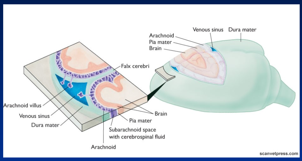
<p><strong>Meninges</strong><br>  • <strong>Dura mater</strong> → outer layer<br>  • <strong>Arachnoid</strong> → middle layer<br>  • <strong>Pia mater</strong> → inner layer, tightly attached to brain tissue</p><p><strong>Structures</strong><br>  • <strong>Venous sinus</strong> → located within the dura mater<br>  • <strong>Falx cerebri</strong> → fold of dura mater between the cerebral hemispheres<br>  • <strong>Arachnoid villus</strong> → projects into the venous sinus<br>  • <strong>Subarachnoid space</strong> → between arachnoid and pia mater<br>    • Contains <strong>cerebrospinal fluid (CSF)</strong><br>  • <strong>Brain</strong> → covered by the pia mater</p>
5
New cards

Brain Ventricles and Cerebrospinal Fluid

Brain Structures
  • Right cerebral hemisphere
  • Cerebellum
  • Venous sinus
  • Arachnoid villus
  • Subarachnoid space → contains cerebrospinal fluid (CSF)

Ventricles and CSF Pathway
  • 3rd ventricle
  • Aqueduct → connects the 3rd and 4th ventricles
  • 4th ventricle
  • Central canal → continues from the 4th ventricle into the spinal cord

<p><strong>Brain Structures</strong><br>  • <strong>Right cerebral hemisphere</strong><br>  • <strong>Cerebellum</strong><br>  • <strong>Venous sinus</strong><br>  • <strong>Arachnoid villus</strong><br>  • <strong>Subarachnoid space</strong> → contains cerebrospinal fluid (CSF)</p><p><strong>Ventricles and CSF Pathway</strong><br>  • <strong>3rd ventricle</strong><br>  • <strong>Aqueduct</strong> → connects the 3rd and 4th ventricles<br>  • <strong>4th ventricle</strong><br>  • <strong>Central canal</strong> → continues from the 4th ventricle into the spinal cord</p>
6
New cards

Cerebrospinal Fluid (CSF)

Formation
  • Formed by capillaries of specialized areas of the pia mater called the choroid plexus

Circulation
  • Circulates from the brain’s ventricles down to the central canal of the spinal cord
  • Diffuses from the 4th ventricle into the subarachnoid space
  • Circulates around the brain and spinal cord
  • Drained into the venous sinus before entering the blood circulation

Roles of CSF
  • Provides nutrients to the CNS
  • Protection → acts as a shock absorber
  • Maintains an ionic concentration ideal for optimal neuronal function

Blood–CSF Barrier
  • Located at the choroid plexus
  • Controls the uptake of nutrients and drugs into the CSF

<p><strong>Formation</strong><br>  • Formed by <strong>capillaries</strong> of specialized areas of the <strong>pia mater</strong> called the <strong>choroid plexus</strong></p><p><strong>Circulation</strong><br>  • Circulates from the brain’s <strong>ventricles</strong> down to the <strong>central canal</strong> of the spinal cord<br>  • Diffuses from the <strong>4th ventricle</strong> into the <strong>subarachnoid space</strong><br>  • Circulates around the <strong>brain and spinal cord</strong><br>  • Drained into the <strong>venous sinus</strong> before entering the <strong>blood circulation</strong></p><p><strong>Roles of CSF</strong><br>  • Provides <strong>nutrients</strong> to the CNS<br>  • <strong>Protection</strong> → acts as a shock absorber<br>  • Maintains an <strong>ionic concentration</strong> ideal for optimal <strong>neuronal function</strong></p><p><strong>Blood–CSF Barrier</strong><br>  • Located at the <strong>choroid plexus</strong><br>  • Controls the <strong>uptake of nutrients and drugs</strong> into the CSF</p>
7
New cards

Blood–Brain Barrier

What Can Pass Through
  • No cells or proteins can pass through
  • Low amino acid levels
  • Only liposoluble substances can diffuse
  • Example → gases

<p><strong>What Can Pass Through</strong><br>  • <strong>No cells or proteins</strong> can pass through<br>  • <strong>Low amino acid</strong> levels<br>  • Only <strong>liposoluble substances</strong> can diffuse<br>  • Example → <strong>gases</strong></p>
8
New cards

Cerebrum

Cerebral Cortex – Grey Matter
  • Most superficial layer of the cerebrum
  • Acquired late in evolution
  • Responsible for the conscious experience of sensory input
  • Site of high nervous association → integrates and connects information
  • Marked by a high degree of educability → ability to learn
  • Primary cortical areas have been mapped, mainly in humans
<p><strong>Cerebral Cortex – Grey Matter</strong><br>  • <strong>Most superficial layer</strong> of the cerebrum<br>  • Acquired <strong>late in evolution</strong><br>  • Responsible for the <strong>conscious experience of sensory input</strong><br>  • Site of <strong>high nervous association</strong> → integrates and connects information<br>  • Marked by a high degree of <strong>educability</strong> → ability to learn<br>  • <strong>Primary cortical areas</strong> have been mapped, mainly in <strong>humans</strong></p>
9
New cards

Cerebral Cortex – Functional Areas

Based on Anatomical Structure
  • Cerebral cortex can be organized based on anatomical structure

Based on Response to Stimulation
  • Can also be organized based on response to stimulation
  • Somesthetic → related to body sensations

Association Cortex
  • Areas without defined reactions to stimulation
  • Called the association cortex
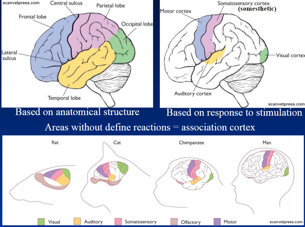
<p><strong>Based on Anatomical Structure</strong><br>  • Cerebral cortex can be organized based on <strong>anatomical structure</strong></p><p><strong>Based on Response to Stimulation</strong><br>  • Can also be organized based on <strong>response to stimulation</strong><br>  • <strong>Somesthetic</strong> → related to body sensations</p><p><strong>Association Cortex</strong><br>  • Areas without <strong>defined reactions</strong> to stimulation<br>  • Called the <strong>association cortex</strong></p>
10
New cards

Cerebrum – Brain Regions

Neocortex
  • Most complex part of the cerebrum

Rhinencephalon
  • Olfactory brain → involved in processing smell

Mesencephalon
  • Part of the midbrain
  • Processes visual information in birds
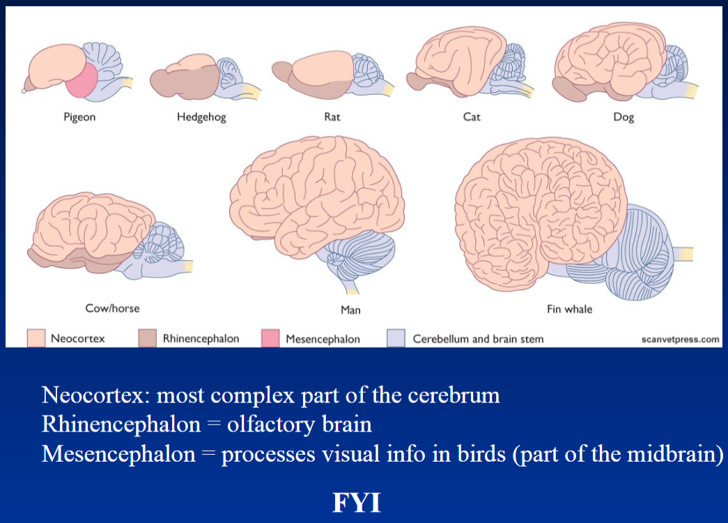
<p><strong>Neocortex</strong><br>  • <strong>Most complex part</strong> of the cerebrum</p><p><strong>Rhinencephalon</strong><br>  • <strong>Olfactory brain</strong> → involved in processing smell</p><p><strong>Mesencephalon</strong><br>  • Part of the <strong>midbrain</strong><br>  • Processes <strong>visual information in birds</strong></p>
11
New cards

Cerebral Cortex – Body Representation

Somatosensory Cortex
  • Represents different body parts in the somatosensory cortex
  • Different areas of the body have different representations

Motor Cortex
  • Represents different body parts in the motor cortex
  • Different areas of the body have different representations

Rabbit and Cat
  • Equivalent body-part representations are found in the somatosensory cortex of both the rabbit and cat

<p><strong>Somatosensory Cortex</strong><br>  • Represents different <strong>body parts</strong> in the <strong>somatosensory cortex</strong><br>  • Different areas of the body have different representations</p><p><strong>Motor Cortex</strong><br>  • Represents different <strong>body parts</strong> in the <strong>motor cortex</strong><br>  • Different areas of the body have different representations</p><p><strong>Rabbit and Cat</strong><br>  • <strong>Equivalent body-part representations</strong> are found in the <strong>somatosensory cortex</strong> of both the <strong>rabbit and cat</strong></p>
12
New cards

Cerebrum – White Matter and Basal Nuclei

White Matter
  • Located beneath the cortex
  • Composed of myelinated nerve fibers → nerve fibers covered by a protective myelin sheath
  • Includes:
    • Association fibers → connect different parts of the same cortex
    • Commissural fibers → connect the two cerebral hemispheres
    • Projection fibers → connect the cortex to other brain structures and spinal cord

Basal Nuclei
  • Located deep within the cerebrum, close to the thalamus
  • Paired nuclei → groups of neurons found on both sides of the brain
  • Control complex semi-voluntary movements → movements that are partly automatic, such as walking and running
  • Work together with the cortex and cerebellum
  • In birds, also control voluntary movements

<p><strong>White Matter</strong><br>  • Located <strong>beneath the cortex</strong><br>  • Composed of <strong>myelinated nerve fibers</strong> → nerve fibers covered by a protective myelin sheath<br>  • Includes:<br>    • <strong>Association fibers</strong> → connect different parts of the <strong>same cortex</strong><br>    • <strong>Commissural fibers</strong> → connect the <strong>two cerebral hemispheres</strong><br>    • <strong>Projection fibers</strong> → connect the <strong>cortex</strong> to other <strong>brain structures and spinal cord</strong></p><p><strong>Basal Nuclei</strong><br>  • Located <strong>deep within the cerebrum</strong>, close to the <strong>thalamus</strong><br>  • <strong>Paired nuclei</strong> → groups of neurons found on both sides of the brain<br>  • Control complex <strong>semi-voluntary movements</strong> → movements that are partly automatic, such as <strong>walking and running</strong><br>  • Work together with the <strong>cortex and cerebellum</strong><br>  • In <strong>birds</strong>, also control <strong>voluntary movements</strong></p>
13
New cards

Cerebellum

Main Role
  • Not involved in consciousness or sensation
  • Main role → prevents distortion of intended movement
  • Helps make movements smooth and accurate

Information Received
  • Receives information from the inner earequilibrium/balance
  • Receives information from proprioceptive receptors → receptors in muscles, tendons, and joints that provide information about body position
  • Receives information from the cortexvisual and motor information

Movement Adjustment
  • Receives information about the status of the body and the intended movement
  • Makes adjustments for a smooth completion of movement
  • Corrective signals are sent through the thalamus to the cortex and spinal nuclei that command the muscles

Development
  • Well developed in animals that require precise adjustment for locomotion
  • Important for locomotion through air, ground, and water
<p><strong>Main Role</strong><br>  • Not involved in <strong>consciousness or sensation</strong><br>  • Main role → prevents <strong>distortion of intended movement</strong><br>  • Helps make movements <strong>smooth and accurate</strong></p><p><strong>Information Received</strong><br>  • Receives information from the <strong>inner ear</strong> → <strong>equilibrium/balance</strong><br>  • Receives information from <strong>proprioceptive receptors</strong> → receptors in <strong>muscles, tendons, and joints</strong> that provide information about body position<br>  • Receives information from the <strong>cortex</strong> → <strong>visual and motor</strong> information</p><p><strong>Movement Adjustment</strong><br>  • Receives information about the <strong>status of the body</strong> and the <strong>intended movement</strong><br>  • Makes adjustments for a <strong>smooth completion of movement</strong><br>  • <strong>Corrective signals</strong> are sent through the <strong>thalamus</strong> to the <strong>cortex and spinal nuclei</strong> that command the muscles</p><p><strong>Development</strong><br>  • Well developed in animals that require <strong>precise adjustment for locomotion</strong><br>  • Important for locomotion through <strong>air, ground, and water</strong></p>
14
New cards

Diencephalon

Diencephalon – “Interbrain”
  • Also called the interbrain
  • Includes the hypothalamus, thalamus, and epithalamus

Thalamus
  • Acts as a relay station for:
    • Sensory information
    • Information from the cerebellum and basal ganglia
    • Information going to and from the cerebral cortex

Epithalamus
  • Contains the olfactory correlation center
  • Contains the pineal gland

Hypothalamus
  • Produces neuropeptides that control the anterior pituitary
  • Produces neurohormones released by the posterior pituitary
  • Principal regulator of the autonomic nervous system
  • Along with the brain stem, it is the most important region for controlling homeostasis → maintaining stable internal conditions

<p><strong>Diencephalon – “Interbrain”</strong><br>  • Also called the <strong>interbrain</strong><br>  • Includes the <strong>hypothalamus, thalamus, and epithalamus</strong></p><p><strong>Thalamus</strong><br>  • Acts as a <strong>relay station</strong> for:<br>    • <strong>Sensory information</strong><br>    • Information from the <strong>cerebellum and basal ganglia</strong><br>    • Information going to and from the <strong>cerebral cortex</strong></p><p><strong>Epithalamus</strong><br>  • Contains the <strong>olfactory correlation center</strong><br>  • Contains the <strong>pineal gland</strong></p><p><strong>Hypothalamus</strong><br>  • Produces <strong>neuropeptides</strong> that control the <strong>anterior pituitary</strong><br>  • Produces <strong>neurohormones</strong> released by the <strong>posterior pituitary</strong><br>  • Principal regulator of the <strong>autonomic nervous system</strong><br>  • Along with the <strong>brain stem</strong>, it is the most important region for controlling <strong>homeostasis</strong> → maintaining stable internal conditions</p>
15
New cards

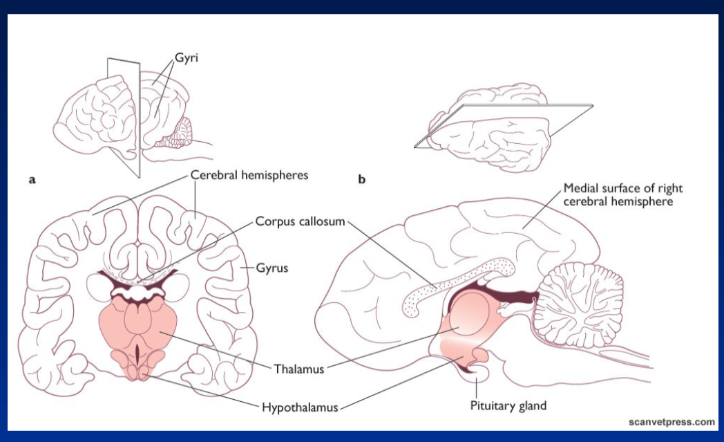
Cerebrum – Medial Surface

Cerebral Hemispheres
  • The brain is divided into cerebral hemispheres
  • Right cerebral hemisphere shown from the medial surface

Gyri and Gyrus
  • Gyri → folds or raised areas of the cerebral cortex
  • Gyrus → singular form of gyri

Corpus Callosum
  • Structure connecting the two cerebral hemispheres

Diencephalon Structures
  • Thalamus
  • Hypothalamus
  • Pituitary gland
<p><strong>Cerebral Hemispheres</strong><br>  • The brain is divided into <strong>cerebral hemispheres</strong><br>  • <strong>Right cerebral hemisphere</strong> shown from the <strong>medial surface</strong></p><p><strong>Gyri and Gyrus</strong><br>  • <strong>Gyri</strong> → folds or raised areas of the cerebral cortex<br>  • <strong>Gyrus</strong> → singular form of gyri</p><p><strong>Corpus Callosum</strong><br>  • Structure connecting the <strong>two cerebral hemispheres</strong></p><p><strong>Diencephalon Structures</strong><br>  • <strong>Thalamus</strong><br>  • <strong>Hypothalamus</strong><br>  • <strong>Pituitary gland</strong></p>
16
New cards

Brain Stem

Brain Stem
  • Includes nuclei that govern body functions through reflexes
  • Contains a large amount of white matter → acts as conveyers, carrying signals between brain regions and the body

Main Parts
  • Mesencephalon → midbrain
  • Pons
  • Medulla oblongata

<p><strong>Brain Stem</strong><br>  • Includes <strong>nuclei</strong> that govern body functions through <strong>reflexes</strong><br>  • Contains a large amount of <strong>white matter</strong> → acts as <strong>conveyers</strong>, carrying signals between brain regions and the body</p><p><strong>Main Parts</strong><br>  • <strong>Mesencephalon</strong> → midbrain<br>  • <strong>Pons</strong><br>  • <strong>Medulla oblongata</strong></p>
17
New cards

Mesencephalon – Midbrain

Visual Reflex Center
  • Receives visual information
  • Controls eye movements for focus

Auditory Reflex Center
  • Responds to sounds
  • Helps control movements such as turning the head toward a noise

Birds
  • Well developed in birds
  • Important for processing visual information

Mammals
  • Most mammals have a very small midbrain
  • It is covered by the cortex

<p><strong>Visual Reflex Center</strong><br>  • Receives <strong>visual information</strong><br>  • Controls <strong>eye movements</strong> for <strong>focus</strong></p><p><strong>Auditory Reflex Center</strong><br>  • Responds to <strong>sounds</strong><br>  • Helps control movements such as <strong>turning the head toward a noise</strong></p><p><strong>Birds</strong><br>  • <strong>Well developed</strong> in birds<br>  • Important for processing <strong>visual information</strong></p><p><strong>Mammals</strong><br>  • Most mammals have a <strong>very small midbrain</strong><br>  • It is <strong>covered by the cortex</strong></p>
18
New cards

Pons and Medulla Oblongata

Postural Reflexes
  • Contain centers for postural reflexes
    • Hopping
    • Righting
    • Placing

Respiration
  • Control respiratory movements → breathing movements

Medulla Oblongata
  • Crucial for control of:
    • Heart activity
    • Blood pressure
    • Relative distribution of blood to organs
<p><strong>Postural Reflexes</strong><br>  • Contain centers for <strong>postural reflexes</strong><br>    • <strong>Hopping</strong><br>    • <strong>Righting</strong><br>    • <strong>Placing</strong></p><p><strong>Respiration</strong><br>  • Control <strong>respiratory movements</strong> → breathing movements</p><p><strong>Medulla Oblongata</strong><br>  • Crucial for control of:<br>    • <strong>Heart activity</strong><br>    • <strong>Blood pressure</strong><br>    • <strong>Relative distribution of blood to organs</strong></p>
19
New cards

Cranial Nerves

General
  • 12 pairs of cranial nerves

Cranial Nerves I and II
  • I – Olfactory → purely sensory
  • II – Optic → purely sensory
  • Both remain in the brain

Cranial Nerves III–XII
  • Exit from the brain stem
  • Innervate structures of the head and neck
  • Most are mixed nerves → both sensory and motor

Exception – X: Vagus Nerve
  • Most widely distributed nerve throughout the body
  • Extends from the neck to the abdomen
  • Carries parasympathetic fibers to visceral structures in the thorax and abdomen
<p><strong>General</strong><br>  • <strong>12 pairs</strong> of cranial nerves</p><p><strong>Cranial Nerves I and II</strong><br>  • <strong>I – Olfactory</strong> → purely <strong>sensory</strong><br>  • <strong>II – Optic</strong> → purely <strong>sensory</strong><br>  • Both remain in the <strong>brain</strong></p><p><strong>Cranial Nerves III–XII</strong><br>  • Exit from the <strong>brain stem</strong><br>  • Innervate structures of the <strong>head and neck</strong><br>  • Most are <strong>mixed nerves</strong> → both <strong>sensory and motor</strong></p><p><strong>Exception – X: Vagus Nerve</strong><br>  • <strong>Most widely distributed nerve</strong> throughout the body<br>  • Extends from the <strong>neck to the abdomen</strong><br>  • Carries <strong>parasympathetic fibers</strong> to visceral structures in the <strong>thorax and abdomen</strong></p>
20
New cards

Cranial Nerves – I–XII

I – Olfactory Nerve
  • Smell

II – Optic Nerve
  • Vision

III – Oculomotor Nerve
  • Eye movement

IV – Trochlear Nerve
  • Eye movement

V – Trigeminal Nerve
  • Sensory and motor functions of the face

VI – Abducent Nerve
  • Eye movement

VII – Facial Nerve
  • Facial movement

VIII – Vestibulocochlear Nerve
  • Hearing and balance

IX – Glossopharyngeal Nerve
  • Sensory and motor functions involving the throat

X – Vagus Nerve
  • Parasympathetic control of visceral organs
  • Extends from the neck to the abdomen

XI – Accessory Nerve
  • Motor control of certain neck and shoulder muscles

XII – Hypoglossal Nerve
  • Motor control of the tongue

<p><strong>I – Olfactory Nerve</strong><br>  • <strong>Smell</strong></p><p><strong>II – Optic Nerve</strong><br>  • <strong>Vision</strong></p><p><strong>III – Oculomotor Nerve</strong><br>  • <strong>Eye movement</strong></p><p><strong>IV – Trochlear Nerve</strong><br>  • <strong>Eye movement</strong></p><p><strong>V – Trigeminal Nerve</strong><br>  • <strong>Sensory and motor</strong> functions of the face</p><p><strong>VI – Abducent Nerve</strong><br>  • <strong>Eye movement</strong></p><p><strong>VII – Facial Nerve</strong><br>  • <strong>Facial movement</strong></p><p><strong>VIII – Vestibulocochlear Nerve</strong><br>  • <strong>Hearing and balance</strong></p><p><strong>IX – Glossopharyngeal Nerve</strong><br>  • <strong>Sensory and motor</strong> functions involving the throat</p><p><strong>X – Vagus Nerve</strong><br>  • <strong>Parasympathetic</strong> control of visceral organs<br>  • Extends from the <strong>neck to the abdomen</strong></p><p><strong>XI – Accessory Nerve</strong><br>  • <strong>Motor</strong> control of certain neck and shoulder muscles</p><p><strong>XII – Hypoglossal Nerve</strong><br>  • <strong>Motor control of the tongue</strong></p>
21
New cards

Spinal Cord – Vertebral Column

Vertebral Column
  • Protects and surrounds the spinal cord
  • Divided into cervical, thoracic, lumbar, sacral, and coccygeal regions
  • Number of vertebrae differs between species

Main Structures
  • Spinous process → dorsal projection of the vertebra
  • Vertebral body → main, weight-bearing part of the vertebra
  • Transverse process → lateral projection of the vertebra
  • Intervertebral disc → located between vertebrae
  • Intervertebral foramen → opening between vertebrae where spinal nerves pass

Orientation
  • Cranial → toward the head
  • Caudal → toward the tail
  • Dorsal → toward the back

<p><strong>Vertebral Column</strong><br>  • Protects and surrounds the <strong>spinal cord</strong><br>  • Divided into <strong>cervical, thoracic, lumbar, sacral, and coccygeal regions</strong><br>  • Number of vertebrae <strong>differs between species</strong></p><p><strong>Main Structures</strong><br>  • <strong>Spinous process</strong> → dorsal projection of the vertebra<br>  • <strong>Vertebral body</strong> → main, weight-bearing part of the vertebra<br>  • <strong>Transverse process</strong> → lateral projection of the vertebra<br>  • <strong>Intervertebral disc</strong> → located between vertebrae<br>  • <strong>Intervertebral foramen</strong> → opening between vertebrae where spinal nerves pass</p><p><strong>Orientation</strong><br>  • <strong>Cranial</strong> → toward the head<br>  • <strong>Caudal</strong> → toward the tail<br>  • <strong>Dorsal</strong> → toward the back</p>
22
New cards

Vertebral Column – Number of Vertebrae by Species

Horse
  • Cervical (C): 7
  • Thoracic (T): 18
  • Lumbar (L): 6
  • Sacral (S): 5
  • Coccygeal (Co): 15–20

Ox
  • C: 7
  • T: 13
  • L: 6
  • S: 5
  • Co: 18–20

Sheep
  • C: 7
  • T: 13
  • L: 6–7
  • S: 4
  • Co: 16–18

Goat
  • C: 7
  • T: 13
  • L: 7
  • S: 4
  • Co: 12

Hog
  • C: 7
  • T: 14–15
  • L: 6–7
  • S: 4
  • Co: 20–23

Dog
  • C: 7
  • T: 13
  • L: 7
  • S: 3
  • Co: 20–23

Chicken
  • C: 14
  • T: 7
  • L: 14
  • S: 6

Human
  • C: 7
  • T: 12
  • L: 5
  • S: 5
  • Co: 4

<p><strong>Horse</strong><br>  • <strong>Cervical (C): 7</strong><br>  • <strong>Thoracic (T): 18</strong><br>  • <strong>Lumbar (L): 6</strong><br>  • <strong>Sacral (S): 5</strong><br>  • <strong>Coccygeal (Co): 15–20</strong></p><p><strong>Ox</strong><br>  • <strong>C: 7</strong><br>  • <strong>T: 13</strong><br>  • <strong>L: 6</strong><br>  • <strong>S: 5</strong><br>  • <strong>Co: 18–20</strong></p><p><strong>Sheep</strong><br>  • <strong>C: 7</strong><br>  • <strong>T: 13</strong><br>  • <strong>L: 6–7</strong><br>  • <strong>S: 4</strong><br>  • <strong>Co: 16–18</strong></p><p><strong>Goat</strong><br>  • <strong>C: 7</strong><br>  • <strong>T: 13</strong><br>  • <strong>L: 7</strong><br>  • <strong>S: 4</strong><br>  • <strong>Co: 12</strong></p><p><strong>Hog</strong><br>  • <strong>C: 7</strong><br>  • <strong>T: 14–15</strong><br>  • <strong>L: 6–7</strong><br>  • <strong>S: 4</strong><br>  • <strong>Co: 20–23</strong></p><p><strong>Dog</strong><br>  • <strong>C: 7</strong><br>  • <strong>T: 13</strong><br>  • <strong>L: 7</strong><br>  • <strong>S: 3</strong><br>  • <strong>Co: 20–23</strong></p><p><strong>Chicken</strong><br>  • <strong>C: 14</strong><br>  • <strong>T: 7</strong><br>  • <strong>L: 14</strong><br>  • <strong>S: 6</strong></p><p><strong>Human</strong><br>  • <strong>C: 7</strong><br>  • <strong>T: 12</strong><br>  • <strong>L: 5</strong><br>  • <strong>S: 5</strong><br>  • <strong>Co: 4</strong></p>
23
New cards

Vertebral Column – Regions and Vertebrae

Cervical (Neck)
  • 7 vertebrae

Thoracic (Chest)
  • 13 vertebrae

Lumbar (Low Back)
  • 7 vertebrae

Sacrum (Pelvic)
  • 3 vertebrae

Tail
  • Vertebrae extend into the tail

Intervertebral Disks
  • Vertebrae in this area have intervertebral disks
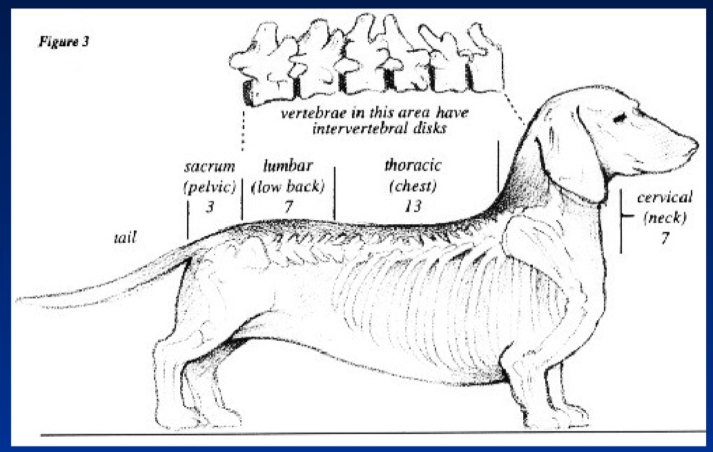
<p><strong>Cervical (Neck)</strong><br>  • <strong>7 vertebrae</strong></p><p><strong>Thoracic (Chest)</strong><br>  • <strong>13 vertebrae</strong></p><p><strong>Lumbar (Low Back)</strong><br>  • <strong>7 vertebrae</strong></p><p><strong>Sacrum (Pelvic)</strong><br>  • <strong>3 vertebrae</strong></p><p><strong>Tail</strong><br>  • Vertebrae extend into the <strong>tail</strong></p><p><strong>Intervertebral Disks</strong><br>  • Vertebrae in this area have <strong>intervertebral disks</strong></p>
24
New cards

Spinal Cord – Cross Section

Central Canal
  • Located in the center of the spinal cord
  • Contains cerebrospinal fluid (CSF)

Dorsal Surface
  • Back/top side of the spinal cord

Dorsal Horn
  • Located on the dorsal side of the spinal cord

Ventral Horn
  • Located on the ventral side of the spinal cord
  • Ventral = belly/front side

<p><strong>Central Canal</strong><br>  • Located in the <strong>center</strong> of the spinal cord<br>  • Contains <strong>cerebrospinal fluid (CSF)</strong></p><p><strong>Dorsal Surface</strong><br>  • <strong>Back/top side</strong> of the spinal cord</p><p><strong>Dorsal Horn</strong><br>  • Located on the <strong>dorsal side</strong> of the spinal cord</p><p><strong>Ventral Horn</strong><br>  • Located on the <strong>ventral side</strong> of the spinal cord<br>  • <strong>Ventral = belly/front side</strong></p>
25
New cards

Spinal Cord – White Matter and Grey Matter

White Matter
  • Contains myelinated nerve fibers → gives white appearance
  • Contains unmyelinated nerve fibers (axons)
  • Arranged in columns or “tracts” → bundles of nerve fibers

Grey Matter
  • Contains cell bodies and dendrites
  • Shaped like a butterfly

Dorsal Horn
  • Sensory → receives sensory information

Ventral Horn
  • Motor → controls skeletal muscles
  • Autonomic fibers → control automatic body functions

Middle Zone
  • Association → connects different areas of the spinal cord

<p class="PDq2pG_selectionAnchorContainer"><strong>White Matter</strong><br>  • Contains <strong>myelinated</strong> nerve fibers → gives white appearance<br>  • Contains <strong>unmyelinated nerve fibers (axons)</strong><br>  • Arranged in <strong>columns or “tracts”</strong> → bundles of nerve fibers</p><p><strong>Grey Matter</strong><br>  • Contains <strong>cell bodies and dendrites</strong><br>  • Shaped like a <strong>butterfly</strong></p><p><strong>Dorsal Horn</strong><br>  • <strong>Sensory</strong> → receives sensory information</p><p><strong>Ventral Horn</strong><br>  • <strong>Motor</strong> → controls skeletal muscles<br>  • <strong>Autonomic fibers</strong> → control automatic body functions</p><p><strong>Middle Zone</strong><br>  • <strong>Association</strong> → connects different areas of the spinal cord</p>
26
New cards

Spinal Cord – Main Structures

Grey Matter
  • Contains cell bodies and dendrites

Dorsal Horn
  • Sensory → receives sensory information

Ventral Horn
  • Motor → controls skeletal muscles
  • Contains autonomic fibers → control automatic body functions

White Matter
  • Contains myelinated and unmyelinated nerve fibers (axons)
  • Arranged in columns or “tracts”

Dorsal Root Ganglion
  • Contains sensory nerve cell bodies

Spinal Cord
  • Part of the central nervous system

Spinal Nerve
  • Contains both sensory and motor nerve fibers

Ventral Root
  • Carries motor signals away from the spinal cord

Vertebra
  • Bone that surrounds and protects the spinal cord

<p><strong>Grey Matter</strong><br>  • Contains <strong>cell bodies and dendrites</strong></p><p><strong>Dorsal Horn</strong><br>  • <strong>Sensory</strong> → receives sensory information</p><p><strong>Ventral Horn</strong><br>  • <strong>Motor</strong> → controls skeletal muscles<br>  • Contains <strong>autonomic fibers</strong> → control automatic body functions</p><p><strong>White Matter</strong><br>  • Contains <strong>myelinated and unmyelinated nerve fibers (axons)</strong><br>  • Arranged in <strong>columns or “tracts”</strong></p><p><strong>Dorsal Root Ganglion</strong><br>  • Contains <strong>sensory nerve cell bodies</strong></p><p><strong>Spinal Cord</strong><br>  • Part of the <strong>central nervous system</strong></p><p><strong>Spinal Nerve</strong><br>  • Contains both <strong>sensory and motor nerve fibers</strong></p><p><strong>Ventral Root</strong><br>  • Carries <strong>motor signals</strong> away from the spinal cord</p><p><strong>Vertebra</strong><br>  • <strong>Bone</strong> that surrounds and protects the spinal cord</p>
27
New cards

Spinal Cord – Nerve Pathways

White Matter
  • Contains nerve fibers (axons)

Grey Matter
  • Contains cell bodies and dendrites

Dorsal Horn
  • Sensory → receives sensory information

Ventral Horn
  • Motor → controls skeletal muscles

Lateral Horn
  • Contains preganglionic autonomic neurons

Spinal Ganglion
  • Contains sensory neuron cell bodies

Sensory Neuron
  • Carries sensory information toward the spinal cord

Somatic Motor Neuron
  • Carries motor signals to skeletal muscles

Preganglionic Autonomic Neuron
  • Carries signals from the CNS to an autonomic ganglion
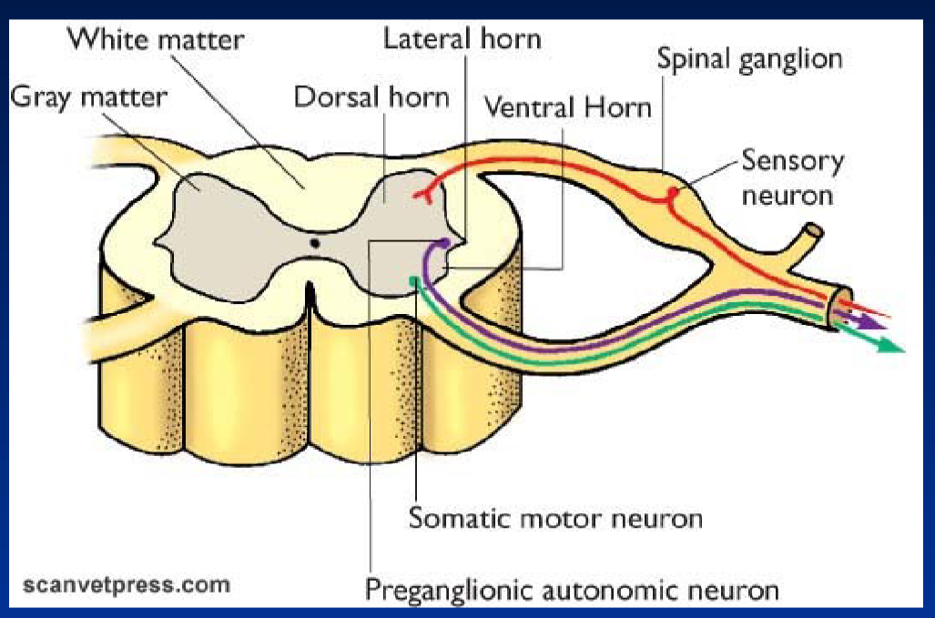
<p><strong>White Matter</strong><br>  • Contains <strong>nerve fibers (axons)</strong></p><p><strong>Grey Matter</strong><br>  • Contains <strong>cell bodies and dendrites</strong></p><p><strong>Dorsal Horn</strong><br>  • <strong>Sensory</strong> → receives sensory information</p><p><strong>Ventral Horn</strong><br>  • <strong>Motor</strong> → controls skeletal muscles</p><p><strong>Lateral Horn</strong><br>  • Contains <strong>preganglionic autonomic neurons</strong></p><p><strong>Spinal Ganglion</strong><br>  • Contains <strong>sensory neuron cell bodies</strong></p><p><strong>Sensory Neuron</strong><br>  • Carries <strong>sensory information</strong> toward the spinal cord</p><p><strong>Somatic Motor Neuron</strong><br>  • Carries <strong>motor signals</strong> to <strong>skeletal muscles</strong></p><p><strong>Preganglionic Autonomic Neuron</strong><br>  • Carries signals from the <strong>CNS</strong> to an <strong>autonomic ganglion</strong></p>
28
New cards

Spinal Nerves

Function
  • Entry and exit of neurons from the spinal cord

Formation
  • Formed when the dorsal and ventral roots join
  • Roots merge within the spinal canal

Number
  • Number corresponds to the vertebra they originate from

Exit
  • Emerge from the intervertebral foramina → openings between vertebrae

Spinal Segments
  • Each spinal segment receives sensory information from a section of the skin called a dermatome
  • Sends information to muscles called myotomes

Spinal Cord Injury
  • The organization of spinal nerves helps predict the effects of a spinal cord injury

<p><strong>Function</strong><br>  • <strong>Entry and exit of neurons</strong> from the spinal cord</p><p><strong>Formation</strong><br>  • Formed when the <strong>dorsal and ventral roots</strong> join<br>  • Roots merge within the <strong>spinal canal</strong></p><p><strong>Number</strong><br>  • Number corresponds to the <strong>vertebra they originate from</strong></p><p><strong>Exit</strong><br>  • Emerge from the <strong>intervertebral foramina</strong> → openings between vertebrae</p><p><strong>Spinal Segments</strong><br>  • Each spinal segment receives <strong>sensory information</strong> from a section of the skin called a <strong>dermatome</strong><br>  • Sends information to <strong>muscles</strong> called <strong>myotomes</strong></p><p><strong>Spinal Cord Injury</strong><br>  • The organization of spinal nerves helps <strong>predict the effects of a spinal cord injury</strong></p>
29
New cards

FYI

knowt flashcard image



