IB BIOLOGY- B2
1/58
Earn XP
Description and Tags
Name | Mastery | Learn | Test | Matching | Spaced |
|---|
No study sessions yet.
59 Terms
Organelles
Membrane bound structures in cells that are adapted to perform one or more vital functions.
Efficient → specialized for limited range of functions.

what structures are organelles?
Nuclei
Vesicles
Ribosomes
Plasma Membrane
what structures are NOT organelles?
Cell Wall
Cytoskeletons
Cytoplasm
Transcription
Uses DNA to make mRNA
Translation
Uses mRNA to make Proteins
Advantage of the separation of the nucleus
-Keeps DNA safe
-It facilitates efficient post-transcriptional modification of mRNA.

Advantage of compartmentalization in the cytoplasm of cells.
Helps prevent incompatible biochemical processes
Background Information for #2:
mRNA
carries protein information from the DNA in a cell's nucleus to the cell's cytoplasm.
What’s the range of cell size in humans?
Human cells range in size from 7.5µm to 150µm
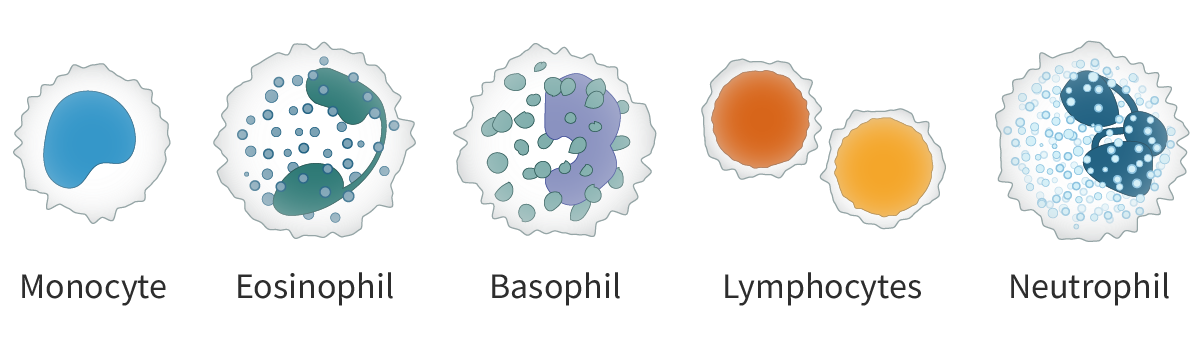
White blood cells (fight infection)
-Monocyte
-Eosinophil
-Basophil
-Lymphocytes
-Neutrophil
Sperm
-Size (diameter): 50µm
-Adaption to Function: Narrowness and small volume reduce resistance and make it easier to swim.
Egg
-Size (diameter): 110µm
-Adaption to Function: large quantities of food reserves to be stored in the cytoplasm
Red blood cells
-Size (diameter): 6µm to 8µm
-allow passage along narrow capillaries and gives a large surface area to volume ratio, so loading and unloading of oxygen is faster.
Why is a high surface area-to-volume ratio beneficial for cells?
It allows for more efficient nutrient intake and waste elimination.
Membrane structure
Membranes contain
Lipids (phospholipids, glycolipids and sterols)
Proteins
Carbohydrates (glycolipids & glycoproteins)
Lipid Bilayers
-Basis of cell membranes
-Contain 3 major classes of lipids - phospholipids, glycolipids and sterols like cholesterol.
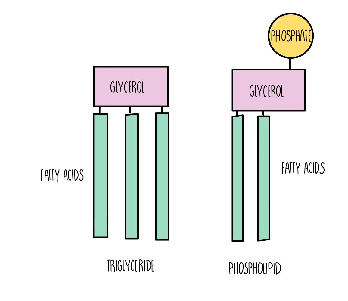
Phospholipids
Most abundant in plasma membrane
Glycerol
Phosphate
2 Fatty acids
Naturally form bilayers
Why do phospholipids form bilayers?
Because they are amphipathic
Lipid Bilyarers as barriers
Hydrophobic hydrocarbon chains that form the core of a membrane have low permeability to large molecules and hydrophilic particles. Including ions and polar molecules, so membrane’s function as effective barriers between aqueous solutions.
Integral protein
Embedded in the lipid bilayer
Amphipathic → interact with phospholipids in bilayer
Transmembrane = goes all the way across/through membrane
Peripheral protein
Found on outer surface of bilayer
Hydrophilic
What function do proteins serve in the plasma membrane?
1- Transport (channel, carrier)
2- Recognition
3-Receptors
4-Enzymes
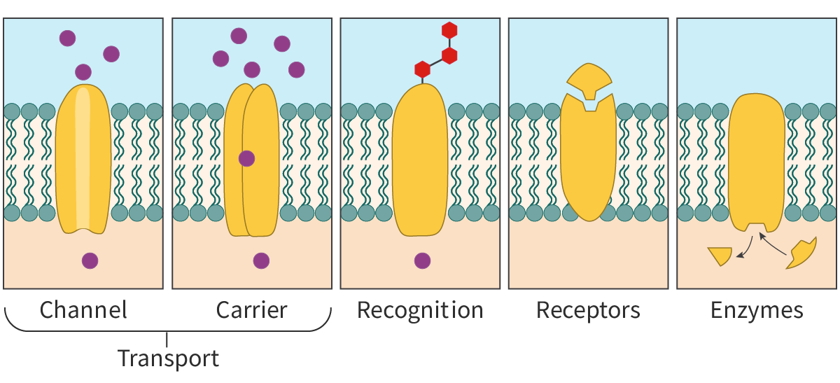
Transport proteins.
Membrane proteins facilitate the movement of molecules in and out of the cell. These include both channel proteins and carrier proteins.
Channel proteins
transmembrane proteins that form channels or pores for the passage of molecules
Carrier proteins
undergoes a conformational change to transfer the molecules from one side of the membrane to the other.
Recognition
Membrane proteins help in cell–cell recognition acting as ‘name tags’ for the cells. This is essential, especially in the functioning of the immune system, as it helps to distinguish between self and non-self cells.
Receptors
Membrane proteins act as receptors for chemical signals and are binding sites for molecules like hormones and neurotransmitters. Often, binding of these molecules triggers a chain of intracellular reactions.
Enzymes
Membrane proteins show enzymatic activity and catalyse reactions. For example, glucose-6-phosphatase is a membrane-bound enzyme found in the endoplasmic reticulum. They can help in cell adhesion to other cells or to the environment and play a role in cell motility.
Glycolipids
carbohydrate + lipid, Extends to extracellular space
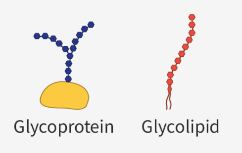
Glycoprotein
carbohydrate + protein, Extends to extracellular space.
Cell recognition
‘markers’ on the cell surface help cells of the body recognize each other; also, immune system.
Cell adhesion
attach and bind to other cells to form tissues.
Cell signalling
receptors for enzymes and other molecules helping in cell signaling, i.e. receiving and transmitting chemical signals.
Fluid mosaic model of membrane structure

Simple Diffusion
-Particles move from high to low concentration (down concentration gradient)
No energy needed for this to happen.
concentration gradient
occurs when the concentration of particles is higher in one area than another.
plasma membrane
selectively permeable and allows some molecules like O2 or CO2 to pass through via simple diffusion.

Osmosis
net movement of water in or out of a cell through the plasma membrane
Directly through membrane bilayer
With help of water selective pore called an aquaporin
Water moves toward higher concentration of solute.
Movement stops when solute concentration inside the cell equals solute concentration outside the cell.
Hypertonic solution
greater solute concentration
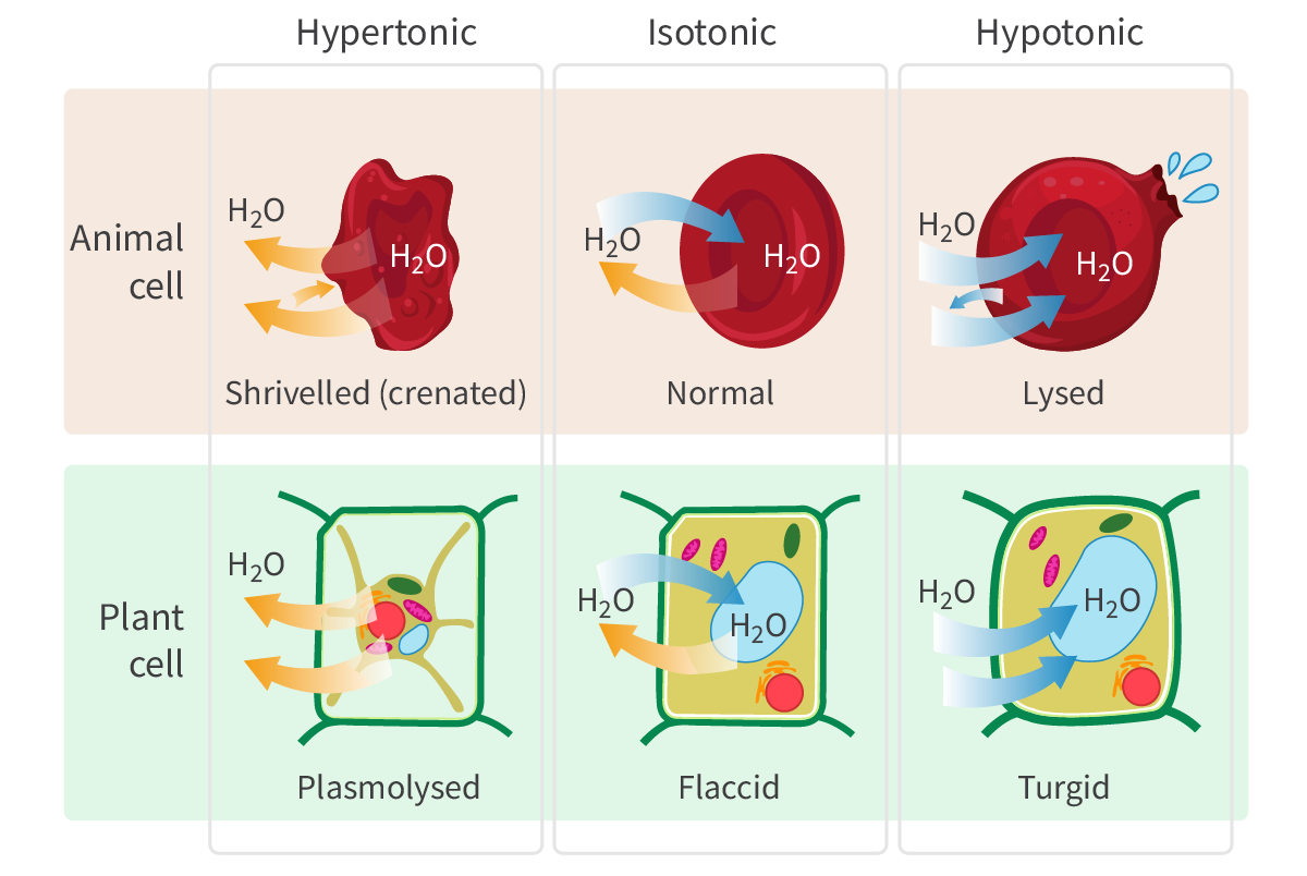
Isotonic solution
same solute concentration
Hypotonic solution
lower solute concentration than cell
Hypertonic, Isotonic, Hypotonic

Facilitated Diffusion
-Molecules move from high to low concentration (down the concentration gradient)
-facilitated by transport proteins.
Channel Protein
Highly selective
Size
Chemically specific binding sites
Open and close in response chemical signals
Ions can’t move through unless open
Carrier Proteins
binds to the solute molecules
↓
undergoes a conformational change
↓
transfers the molecules to the other side of the membrane
Channels are highly selective and only open/close in response to stimuli like
changes in voltage across the membrane or voltage-gated channels
binding of small molecules to the channel proteins or ligand-gated channels
mechanical forces like pressure.
Active Transport
molecules transported from low to high concentration (i.e. against their concentration gradient.)
Requires energy in the form of ATP
Utilizes protein (transport) pump
Simple diffusion
-Dependent on size and affinity for water (hydrophobic or hydrophilic)
-Anything good or bad can pass through if it fits this criteria!
Active transport & facilitated diffusion
- Active transport and facilitated diffusion are more selective than simple diffusion.
-Involve proteins that recognize certain molecules
Differentiation
when cells develop from unspecialized to specialized cells.
Morphogen
A chemical in the cell; the concentration gradient of which determines the fate of surrounding cells.

Stem cell
Cell that has the ability to differentiate into any cell type
Types of Stem Cells
Totipotent
Pluripotent
Multipotent
Unipotent
Totipotent
Can differentiated into any type of cell
Can give rise to a complete organism
Pluripotent
Can differentiate into any type of body cell
Can’t give rise to new organism
Multipotent
Can differentiate into a few closely related types of body cell
Unipotent
Can only differentiate into their associated cell type.
Ex. liver stem cells can only make liver cells
Stem cell niche
microenvironment in organism that influences the stem cells and how they differentiate

Examples of stem cell niche
-Blood stem cells are found in the bone marro
-Hair follicle stem cells