all biospych
1/180
There's no tags or description
Looks like no tags are added yet.
Name | Mastery | Learn | Test | Matching | Spaced | Call with Kai | Chat |
|---|
No analytics yet
Send a link to your students to track their progress
181 Terms
what does brain plasticity (or neuroplasticity) mean?
refers to the brains ability to change or adapt as a result of experience or trauma (injury)
how do brains change when undergoing brain plasticity?
can involve creating new neural pathways or altering existing connections
why is brain plasticity important?
helps the brain adapt to new experiences
can support functional recovery after trauma
when does brain plasticity occur?
occurs throughout life, though more evident in early years
influenced by the environment, behaviour and neural activity
external factors can effect neural structure and function
what is the role of the hippocampus in plasticity?
hippocampus is responsible for long term memory
it produces new neurons in the brain
how does plasticity occur as a result of life experience?
Frequently used neural pathways strengthen and develop
New pathways form in response to new environments
Brain constantly adapts
With age cognitive functioning may decline and brain becomes less responsive to change
However, plasticity still occurs in adults e.g. 60 year olds taught a new skill of juggling, they found increases in grey matter in the visual cortex, although when practicing stopped, these changed were reversed
who did the study to see how plasticity was effected by playing video games?
Kuhn et al
what was the aim of the study of meditation on brain plasticity?
Investigate effects of meditation on brain activity
what was the method for the study of meditation on plasticity?
Compared 8 Tibetan monks (experienced meditators) with 10 students (no experience in meditating)
Used EEG to measure brain activity during meditation
what was the findings and conclusions of the study of meditation on plasticity?
Findings:
Monks showed significantly higher gamma wave activity (linked to neuron coordination)
Students showed little increase
Monks also had higher gamma activity before meditation
Conclusion:
Meditation can alter brain functioning short-term
May also lead to long-term/permanent brain change
who conducted the study to see the effect of meditation on brain plasticity?
Lutz. et al
what was the method for the study seeing how playing video games effected plasticity?
Compared trained group (at least 30 mins/day playing Super Mario for 2 months) vs control group
MRI scans measured brain changes
what were the findings and conclusions of the study of how video games affect plasticity?
Findings:
Increased grey matter in trained group (cortex, hippocampus, cerebellum)
No change in control group
Conclusion:
Video game play may strengthen synaptic connections
Linked to improved spatial navigation, planning, working memory and motor skills
what was the key study to support brain plasticity and who was the researcher?
London cab drivers
Maguire et al
what is the research of the London cab drivers by Maguire et al?
discovered changes in taxi drivers brains could be detected as result of there experience of spatial navigation
using fMRI, researchers calculated grey matter amount in brains of taxi drivers and control Ps
posterior hippocampi of taxi drivers = significantly larger relative to control Ps posterior hippocampi
volume of posterior hippocampi positively correlated with amount of time spent as a taxi driver
not only shows hippocampal volume was greater in those with job related spatial navigation but also highest levels of plasticity evident in those with most experience
suggests positive correlation between longer spent in profession driving and memorising roads, the larger volume and more developed the posterior hippocampus becomes
similar findings with medical students 3 months before and 3 months after final exams
who were the control Ps in the taxi study for brain plasticity and why?
bus drivers
they didn’t have to remember the routes
they had similar stress levels which could be an extraneous variable
does the London cab driver study support or go against brain plasticity and why?
Supports brain plasticity → experience can physically change brain structure
More experience = greater hippocampal development (spatial memory)
what is a research study on animals that supports brain plasticity? (PET)
P: Research from animal studies supports the idea of brain plasticity.
E: Researcher found that rats raised in enriched, complex environments developed a greater number of new neurons than rats kept in standard lab cages. The increase was especially found in the hippocampus, which is involved in forming new memories and spatial navigation.
T: Therefore, this provides evidence that the brain can physically change and adapt in response to experience, supporting the concept of plasticity.
However, low generalisability as it is done on rats not people.
what is a limitation/downside of brain plasticity? (PET)
P: A limitation of brain plasticity is that it can have negative behavioural consequences.
E: Research shows that the brain’s adaptation to prolonged drug use can lead to poorer cognitive functioning later in life and an increased risk of dementia.
T: Therefore, although plasticity allows the brain to adapt, these changes are not always beneficial, which challenges the idea that plasticity is entirely positive.
define axonal sprouting
the growth of new nerve endings which connect with other undamaged nerve cells to form new neuronal pathways
what is denervation super sensitivity?
this occurs when axons that do a similar job become aroused to a higher level to compensate for the ones that are lost.
However, it can have the negative consequence of oversensitivity to messages such as pain
what is synaptic pruning?
the brains natural process of eliminating weaker or unnecessary synaptic connections to enhance neural efficiency and cognitive function
how and when did researchers discover that the brain could recover after injury or trauma?
In the 1960s, researchers studied cases in which stroke victims were able to regain functioning.
how does the brain recover after illness or trauma?
They discovered that when brain cells are damaged or destroyed, as they are in a stroke, the brain re-wires itself over time so that some level of function can be regained.
Although parts of the brain may be damaged or even destroyed as a result of trauma, other parts appear able to take over the functions that were lost.
Neurons next to damaged brain areas can form new circuits that resume some of the lost function.
what are the 2 mechanisms the brain uses to recover?
neuronal unmasking
stem cells
what is neuronal unmasking?
Neural unmasking is when previously inactive (masked) neural pathways become active after brain damage, allowing alternative routes in the brain to take over lost functions.
It contributes to functional recovery by using existing but unused connections rather than forming new ones.
what are dormant synapses?
Synaptic connections that exist in the brain but are usually inactive because they receive too little neural input.
who first identified dormant synapses?
Wall (1977)
why are dormant synapses usually inactive?
Because the level of neural input they receive is too low to activate them under normal conditions.
what triggers neural unmasking?
Increased neural activity following damage to nearby brain areas.
what is meant by lateral spread of unmasking in neural unmasking?
When activation spreads to other brain regions that are not normally used for that function.
what can happen after neural unmasking over time?
New neural pathways and structural reorganisation can develop to support recovery.
what are homologous areas in the brain?
Matching brain regions in opposite hemispheres that have similar structure and can take over functions after damage.
how can homologous areas support recovery after Broca’s area damage?
The right hemisphere homologous area can form new connections and help support speech production.
what are stem cells?
unspecialised cells that have potential to give rise to different cell types that carry out different functions, including taking on the characteristics of nerve cells
what are stem cells being considered for in relation to brain damage?
They are being researched as a potential treatment for brain damage caused by injury or neurodegenerative disorders.
what are the 3 views of how stem cells could treat brain damage?
Stem cells implanted into the brain could directly replace dead or damaged cells.
Transplanted stem cells may release growth factors that help “rescue” or support damaged cells.
Transplanted cells may form new neural networks linking an undamaged brain area (where new stem cells are produced) with the damaged region.
what is functional recovery in the brain after injury?
Functional recovery refers to the brain’s ability to adapt after injury or trauma, where unaffected areas can compensate for damaged regions.
It is an example of neural plasticity and involves neural reorganisation.
This process tends to happen quickly after trauma but slows down over time.
what happens in the brain during recovery?
Able to rewire and reorganising itself by forming new synaptic connections close to the area of damage.
Secondary neural pathways that would not typically be used to carry out certain functions are activated to enable functioning to continue.
This process is supported by changes in the brain such as axonal sprouting, recruitment of homologous areas, denervation super sensitivity and synaptic pruning
what is an evaluation point that supports functional recovery? (PET)
P: Animal research supports functional recovery through stem cell-based neural repair after brain injury.
E: In a controlled experiment, rats with traumatic brain injury were given stem cell transplants into the damaged brain area, while a control group received no stem cells. After 3 months, only the stem cell group showed neuron-like cell development and stem cell migration to the injury site.
T: This suggests functional recovery is supported because new neural cells can develop in damaged areas, showing biological repair is possible.
what is an evaluation point which partially supports/limits what we can conclude about functional recovery? (PET)
P: Age influences the extent of functional recovery, suggesting neural plasticity is not equally effective across the lifespan.
E: Functional plasticity is greater in childhood, meaning children show stronger neural reorganisation after brain injury than adults. However, research also shows that adults can still show improvement in abilities thought to be fixed, given intensive retraining after trauma.
T: This suggests functional recovery is supported because it can occur at any age after brain injury, but it is limited because recovery is more extensive and efficient in younger brains than in adults.
what is the idea of hemispheric lateralisation?
The idea that two halves (hemispheres) of the brain are functionally different and that certain mental processes and behaviours are mainly controlled by one hemisphere rather than the other.
For example, language is both ‘localised’ and ‘lateralised’ to the left hemisphere, whilst the right hemisphere excels at visual-motor tasks.
what are the 2 hemispheres of the brain connected by?
the corpus callosum - a bundle of nerve fibres
why is the corpus callosum important?
they make it possible for info received by one hemisphere to be sent to the other (interhemispheric communication)
it is essential for coordinating sensory, motor and cognitive functions
what is it called when information from one hemisphere is sent to the other?
interhemispheric communication
what is a supporting evaluation point of hemispheric lateralisation? (PET)
Point:
Hemispheric lateralisation may increase neural processing capacity
Evidence:
One hemisphere performs a task → other hemisphere free for different functions
Suggests ability to multitask more efficiently
Animal research (domestic chickens) → able to find food + watch for predators at same time
BUT very little supporting empirical evidence in humans
Therefore:
Suggests lateralisation can improve brain efficiency for simultaneous tasks
HOWEVER limited human evidence reduces strength of this conclusion
what is an evaluation point of a weakness of hemispheric lateralisation?
Point:
A limitation of hemispheric lateralisation is that it is not fixed and changes across the lifespan
Evidence:
Younger individuals show stronger lateralisation → older adults show more bilateral brain activity
Research found language becomes more lateralised in childhood, but decreases after around age 25
Suggests both hemispheres become more involved with age
Possible explanation: compensating for age-related cognitive decline
Therefore:
Suggests lateralisation is flexible rather than permanent
HOWEVER this makes it difficult to establish a consistent role of lateralisation in brain function
what is the key study for hemispheric lateralisation?
Split brain research - Sperry and Gazzaniga
who conducted the split brain research?
Sperry and Gazzaniga
what did Sperry and Gazzaniga’s split brain research include?
involved severing the corpus callosum of people who had epilepsy
what was the aim of the split brain study?
to investigate the extent to which the 2 hemispheres are specialised to different functions
what was Sperry and Gazzaniga’s method for the split brain study?
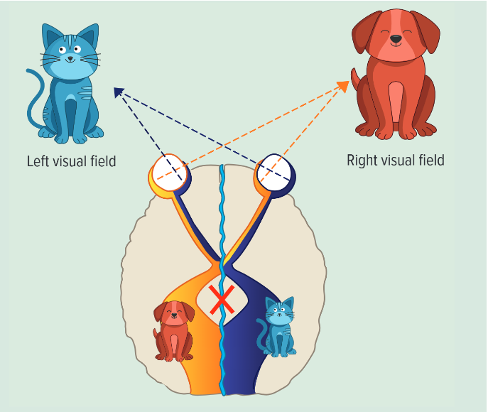
Participant fixates on a central point (to control visual input)
Stimuli presented briefly to one visual field at a time:
Left visual field → right hemisphere
Right visual field → left hemisphere
Presentation is fast → prevents eye movement, so info only goes to one hemisphere
what was the results of the split brain research?
if a dog was shown on the right visual field, they would say they saw a dog
if a cat was shown to the left visual field they would say they couldn’t see anything but they were able to draw the cat and then identify what they saw from what they drew
this is because info from the left visual field is processed by the right hemisphere which has no language centre so can’t respond verbally but can draw it
the left hemisphere which does have a language centre can’t say the cat as there was no way of communicating across the hemispheres as the corpus callosum was severed
what did we learn from the split brain study?
Work with split brained patients has discovered a number of differences between the 2 hemispheres e.g. the left hemisphere is responsible for speech and language, and the right hemisphere specialises in visual-spatial processing and facial recognition
However, split brain research has not shown that the brain is organised into discrete regions with specific sections responsible for specific tasks
Instead it suggests that the connectivity between the different regions is as important as the operation of different parts
what are 2 evaluation point against split brain study? (PET)
Point:
A limitation of split-brain research is low generalisability
Evidence:
Very small sample sizes (often 1–3 participants)
Participants often had epilepsy or incomplete separation of hemispheres
Control comparisons introduce extraneous variables → difficult to establish causality
Ethical issues → may make participants aware of differences
“Pure” cases without these issues are very rare
Therefore:
Findings cannot be confidently generalised to the wider population
Reduces validity of conclusions about hemispheric lateralisation
Point:
Split-brain research may oversimplify hemispheric lateralisation
Evidence:
In a normal brain, hemispheres constantly communicate via the corpus callosum
Brain plasticity allows functions to be shared or compensated across hemispheres
Therefore:
Findings from split-brain patients may not reflect typical brain functioning
Reduces validity of conclusions about lateralisation in the general population
what is an evaluation point to support split brain research?
Point:
A strength of split-brain research is that it has led to new insights about brain functioning
Evidence:
More recent research found split-brain patients can outperform controls on some tasks
e.g. faster at identifying the “odd one out” in visual arrays
Therefore:
Suggests separated hemispheres may process information independently and efficiently
Supports the idea that lateralisation can enhance certain cognitive abilitie
what are the 4 ways we can use to study/scan the brain?
post mortem examinations
fMRI (functional magnetic resonance imaging)
EEG (Electroencephalogram)
ERPs (Event related potentials)
what is a post mortem?
a way of examining the brains of people who have shown particular psychological abnormalities prior o their death to establish possible neurological case for this behaviour
includes examining brains of people who have died
what are post mortems used for?
Used to establish underlying neurobiology of a particular behaviour
Examine brain to look for structural differences that might explain behaviour
Made it possible to identify some of the brain structures involved in memory
Used to establish links between psychiatric disorders like schizophrenia and depression and underlying brain differences
who is the key case study for post mortems on?
Henry Molaison (H.M)
what happened in the key case study of Henry Molaison for post mortems?
he had surgery to relieve severe epilepsy - surgeon removed the hippocampus and surrounding structures
What were the findings when Henry Molaison was alive (post mortem key study)?
He had severe anterograde amnesia (inability to make new memories) and limited retrograde amnesia (losing memories prior to surgery)
Episodic and semantic memory heavily impacted
Procedural memory remained intact (e.g. learning how to draw in a mirror)
what were the findings after death of Henry Molaison (post mortem key study)?
Upon his death in 2008, his brain was preserved and sliced into 2,401 thin slices by Jacopo Annese at the Brain Observatory
Confirmed extent of the lesions, including removal of the hippocampus
Discovered a small portion of the posterior hippocampus remained
Identified a lesion in the orbitofrontal cortex
what was the conclusions following the post mortem of Henry Molaison’s brain (post mortem key study)?
Confirmed role of hippocampus in consolidating declarative memory
Provided 3D mapping of damaged structures
Medial temporal lobes, specifically the hippocampus, are crucial for memory formation
Helped distinguish between implicit and explicit memory systems
Led to the understanding that complex functions such as learning and memory are tied to discrete regions of the brain
what is a strength of post mortem examinations?
Provide detailed examination of anatomical structure and neurochemical aspects of the brain not possible with other scanning techniques (e.g. EEG, ERP and fMRI).
researchers can physically examine deep brain areas such as the hypothalamus and hippocampus, which cannot be accessed with non-invasive scanning methods.
This allows psychologists to gain insight into underlying biological mechanisms and form the basis for further research.
For example, Iverson found a higher concentration of dopamine in the limbic system of patients with schizophrenia, which prompted further research into the neural correlates of this disorder.
post-mortem studies have played a key role in understanding the origins of schizophrenia, identifying structural differences and changes in neurotransmitter systems
what are 2 disadvantages of post mortem examinations?
Post mortem research is retrospective so researchers cannot test hypotheses or follow up findings with P, which limits the depth of investigation
There could also be confounding factors such as age at death, drug treatments, cause of death and length of time between death and examination which may affect the condition of the brain - these make it hard to draw firm conclusions, reducing the internal validity of the findings
what is an fMRI?
Technique for measuring changes in brain activity while a person performs a task
Measures changes in blood flow and blood oxygenation in particular areas of the brain, which indicates increased neural activity
If an area becomes more active, there is an increased demand for oxygen
The brain responds by increasing blood flow, delivering oxygen in red blood cells
Researchers produce maps showing which areas are involved in mental activity
Can produce a 3D image by combining pictures
what is the key study of fMRI?
Used fMRI to study how amygdala activation relates to emotional memory
Found stronger amygdala activity correlates with better emotional memory recall
what are 2 strengths of fMRI?
it is more objective and reliable as it used quantitative data based on blood oxygen levels rather than subjective interpretation - increases scientific credibility of findings
non invasive - doesn’t require surgery and does not expose Ps to harmful radiation, making it safer more more ethical than some older brain scanning techniques
what are 2 limitations of fMRIs?
poor temporal resolution - temporal resolution of 1-4 secs, which is worse than EEG/ERPs (1-10 milliseconds), this means researchers cannot precisely determine the exact onset of brain activity reducing accuracy when studying rapid cognitive processes
focuses mainly on localised brain activity which may overlook the networked and interactive nature of the brain functioning, reducing the overall explanation of complex behaviours
what is a difference between fMRI and EEGs?
fMRI have a high spatial resolution whereas EEG do not
what is a similarity between fMRI and EEG?
fMRI and EEGs can both record your reaction to an event or stimuli
what is spatial resolution?
how accurately a brain imaging technique can show where activity is happening in the brain.
what is temporal resolution?
how accurately a brain imaging technique can show when activity is happening in the brain (how quickly it detects changes over time).
what is the difference between spatial resolution and temporal resolution?
Spatial resolution is about location of brain activity, whereas temporal resolution is about the timing of brain activity
what is one similarity of ERPs and EEGs?
Both EEG and ERPs measure electrical activity in the brain using electrodes placed on the scalp.
what is one difference between ERPs and EEGs?
EEG records overall, continuous brain activity, whereas ERPs are specific responses to particular stimuli or events.
what is an EEG (Electroencephalogram)
Method of recording changes in electrical activity of the brain using electrodes attached to the scalp
Electrodes detect small electrical changes from activity of brain cells
Graphed signals over time produce an EEG
what are EEGs used for?
Used to detect epilepsy and diagnose disorders that influence brain activity (e.g. Alzheimer’s)
what are the 4 basic brain wave patterns EEGs pick up and what do they mean?
alpha = relaxed
beta = alert/active
theta/delta = sleep (deeper = delta)
how are EEGs done?
Around 20 metal discs placed on scalp
Records electrical signals as wavy lines on a computer
Activation procedures include closing eyes, hyperventilation, or exposure to flashing light
Routine EEG lasts 20–30 minutes (sleep-deprived or video EEGs last longer)
what are 2 strengths of EEGs?
high temporal resolution - measures brain activity in milliseconds which allows researchers to accurately match brain activity to specific tasks or stimuli in real time
non-invasive and low cost - does not require surgery and is relatively inexpensive compared to fMRI, makes it more accessible and ethical
what are 2 limitations of EEGs?
poor spatial resolution - electrical signals may be picked up by neighbouring electrodes, so it is difficult to pinpoint the exact source of the activity
limited depth - cannot record activity from deep brain structures because inserting electrodes into a living brain would be too invasive and unethical and so it limits the completeness of the data collected
What are ERPs (Event-Related Potentials)?
Very small voltage changes triggered by specific events or stimuli
Use raw EEG data to investigate cognitive processing of an event
Require many presentations of a stimulus
Responses are averaged together (‘averaging’)
Extraneous neural activity is cancelled out, allowing stimulus-linked activity to stand out
Reduces background neural ‘noise’
what are sensory and cognitive ERPs?
First 100 milliseconds = sensory ERPs (initial response to physical characteristics of stimulus)
After 100 milliseconds = cognitive ERPs (information processing and evaluation)
Latency = time between stimulus and response
Use same equipment as EEG but focus on activity related to a specific stimulus
what are 2 strengths of ERPs?
high temporal resolution - can detect brains activity within milliseconds, makes ERPs particularly useful for studying rapid cognitive processes
can measure covert processing - does not require a behavioural response from Ps, allows researcher to study unconscious or automatic processing
what are 2 limitations of ERPs?
small signals may require many trials - ERPs are very small and difficult to distinguish from background activity, researchers must present stimulus many times which limits the types of research question that can realistically be investigated
restricted to neocortex - only strong voltage changes across the scalp can be recorded, activity occurring deep within the brain cannot be measured, which limits the overall understanding of brain functioning
What is the location of the motor cortex and it’s function in the brain?
responsible for the generation of voluntary motor movements
frontal lobe
in both hemispheres
diff parts of motor cortex control different parts of body - regions arranged logically e.g. region that controls foot is near region that controls the leg
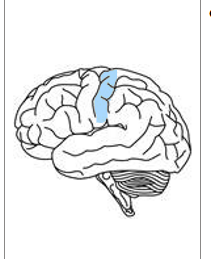
what is the location of the somatosensory area and function in the brain?
detects sensory events arising from different regions of the body
in parietal lobe
uses sensory information
in both hemispheres, receives info from opposite sides
Using sensory information from the skin, the somatosensory cortex produces sensation on touch, pressure, pain and temperature, which it then localizes to specific body regions.
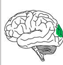
What is the location of the visual cortex and it’s function in the brain?
Visual cortex processes visual information from the eyes
Enables object recognition, motion detection, and spatial awareness
Primary visual cortex is in the occipital lobe
Visual processing starts in the retina (light hits photoreceptors)
Signals are sent from the retina to the brain via the optic nerve
Most signals go to the thalamus, which acts as a relay station to the visual cortex
A small number of signals go to areas controlling circadian rhythms
Visual information is contralateral:
Right hemisphere processes left visual field
Left hemisphere processes right visual field
The visual cortex has different areas for processing different features (e.g. colour, shape, movement)
What is the location of the auditory cortex and it’s function in the brain?
crucial for processing sound, language comprehension, and integrating auditory information with other functions
in temporal lobe on both sides of brain
Sound is converted to nerve impulses in the cochlea
Impulses travel via the auditory nerve to the brain
Pathway: brain stem (basic decoding) → thalamus (relay) → auditory cortex
In the auditory cortex, sound is recognised and interpreted, leading to a response
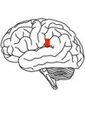
What is the location of Broca’s area and it’s function in the brain?
Crucial for speech production and language production
named after Paul Broca
in left hemisphere of brain
what was the experiment that discovered Broca’s area?
done by Paul Broca
treated a patient who was referred to as Tan as that is all he could say
he could understand but couldn’t speak or write
Broca later studied eight more patients with similar language difficulties and damage (lesions) in the left frontal hemisphere.
Patients with damage in the right hemisphere did not show the same language problems.
This led Broca (1865) to identify a ‘language centre’ in the posterior part of the left frontal lobe.
Broca’s area is important for speech production.
However, research shows Broca’s area is also active during non-language cognitive tasks.
Another researcher also discovered 2 regions of Broca's area, one selectively language, the other responding to many demanding tasks
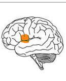
What is the location of Wernicke’s area and it’s function in the brain?
primarily responsible for language comprehension - helps us understand both spoken and written words by interpreting the meaning of words and sentences
Discovered by carl Wernicke, shortly after Broca identified the speech production area
Patients with damage to Wernicke's area could speak but couldn't understand language (opposite to Broca's)
Wernicke proposed language involves separate motor regions (in Broca's area) for speech production and sensory regions (in Wernicke's area) for language comprehension
Motor region is near areas controlling mouth, tongue and vocal cords
Sensory regions is near areas for auditory and visual input
Input is sent to Wernicke's areas where it is recognised as language and given meaning

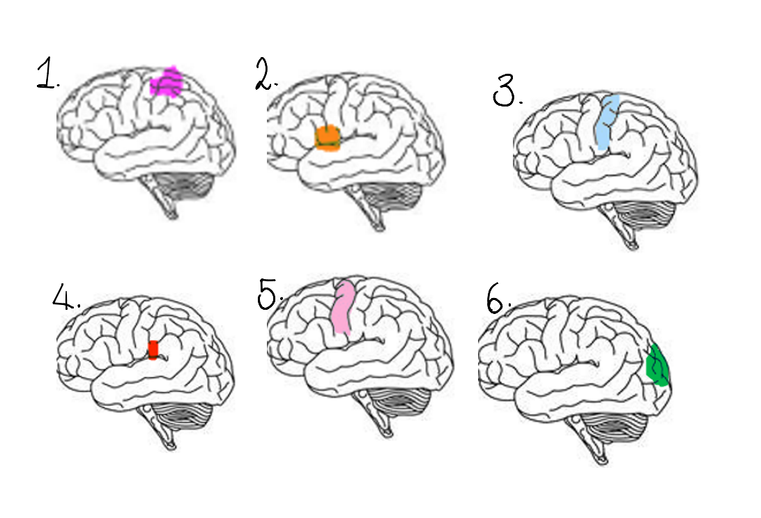
what are the names of the areas highlighted?
Wernicke’s area
Broca’s area
Somatosensory
Auditory
Motor
Visual

what are 2 evaluation points against localisation of function in the brain? (PET)
P – Equipotentiality theory challenges localisation of function.
E – Some researchers argue that while basic motor and sensory functions are localised, higher mental functions are not. Instead, intact areas of the cortex can take over functions after brain damage, meaning the effects depend on the extent of damage rather than its location.
T – Therefore, this suggests cognitive abilities are not confined to specific brain areas, challenging the idea of strict localisation.
P – The importance of communication between brain regions challenges localisation.
E – Carl Wernicke proposed that although brain areas have specialised functions, they must interact to work effectively. For example, a case showed reading ability was lost due to damage between the visual cortex and Wernicke’s area, not the areas themselves.
T – Therefore, complex behaviours like language and movement rely on networks of brain regions working together, rather than isolated localised areas.
what are 2 evaluation points to support localisation of function of the brain? (PET)
P – Localisation of language varies between individuals.
E – Patterns of activation during language tasks differ between people; one researcher found activity in the right temporal lobe as well as left frontal, temporal and occipital lobes when reading. Other studies show gender differences, with women having proportionally larger Broca’s and Wernicke’s areas.
T – Therefore, language functions are not localised in the same way in all individuals, challenging strict localisation.
P – Language production is not limited to Broca’s area alone.
E – Re-examination of Broca’s patients using MRI showed damage extended beyond Broca’s area, suggesting other regions contribute to speech; damage to Broca’s area alone usually causes only temporary disruption.
T – Therefore, language involves multiple brain regions working together, rather than a single localised area.
Briefly explain the Phineas gage example of localisation of the brain
Survived a severe brain injury where a iron rod was blasted through his skull during an explosion while preparing rock for railway construction
Rod destroyed part of his frontal lobe
He survived
Before the accident he was described as responsible, hardworking and well liked
After the accident there were many personality and behavioural changes such as: becoming impulsive and aggressive, poor decision making, difficulty controlling behaviour and acting in socially inappropriate ways
Provides early evidence that different areas of the brain have different functions
Damage to frontal lobe is linked to personality, decision making and behavioural control
Supports the idea of localisation in the brain
However, some later reports say his personality change may have been less extreme than originally reported
Also is only a single case study so can't be generalised to everyone
what is the flight or fight response?
A sequence of activity within the body that is triggered when the body prepares itself for defending or attacking (flight) or running away to safety (flight)
Involves a change in the nervous system and secretion of hormones
what is an acute stressor?
a stress which is sudden and short term