BFUN: Labs 1-4
1/275
There's no tags or description
Looks like no tags are added yet.
Name | Mastery | Learn | Test | Matching | Spaced | Call with Kai |
|---|
No analytics yet
Send a link to your students to track their progress
276 Terms
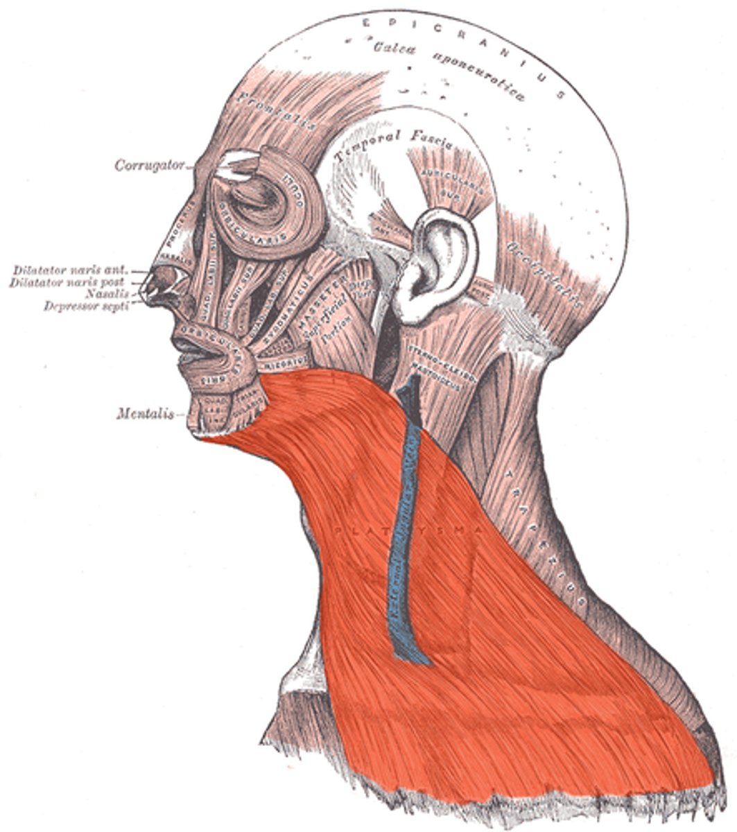
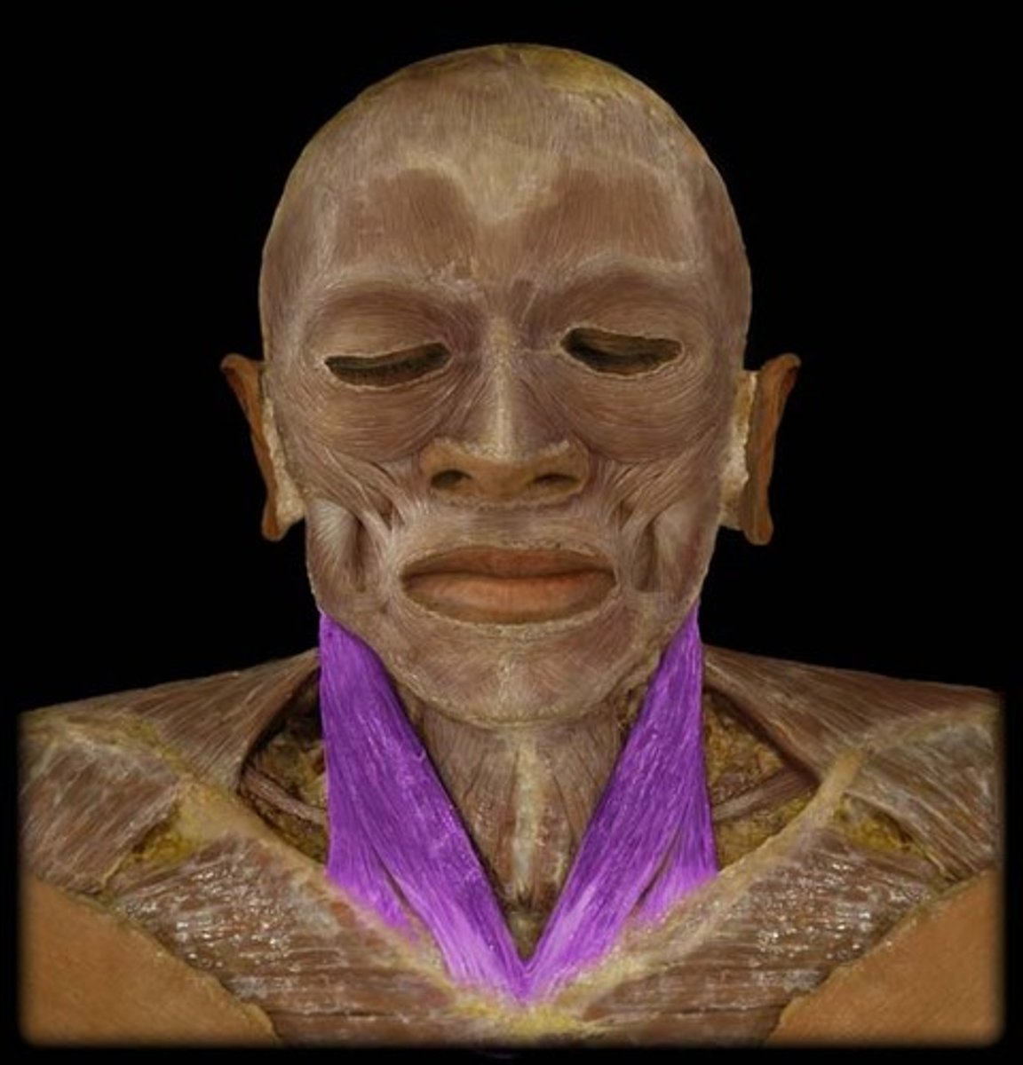
Platysma
Note: Thin, Located in Subcutaneous Neck Tissue
Name the Muscle
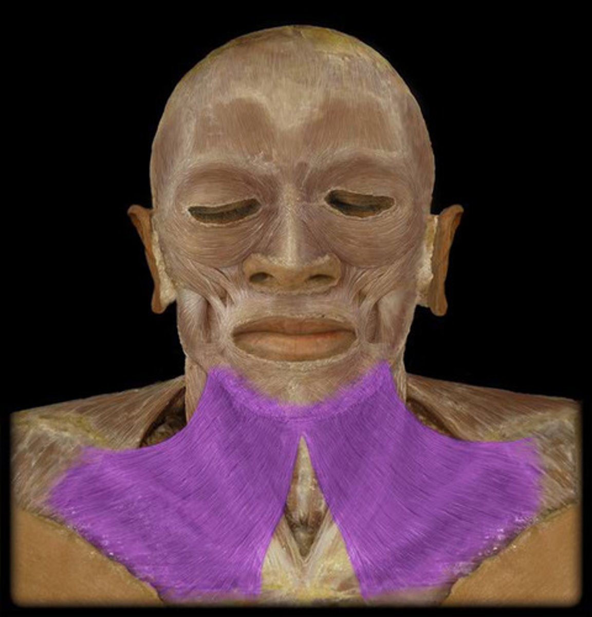
Platsyma
Name the Muscle
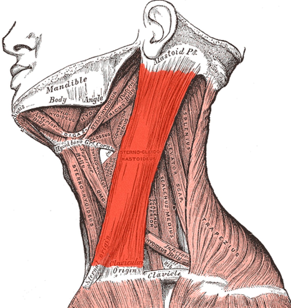
Sternocleidomastoid
Note: Inserts into Sternam & Clavicle, Originates in Mastoid
Name the Muscle
Sternocleidomastoid
Name the Muscle
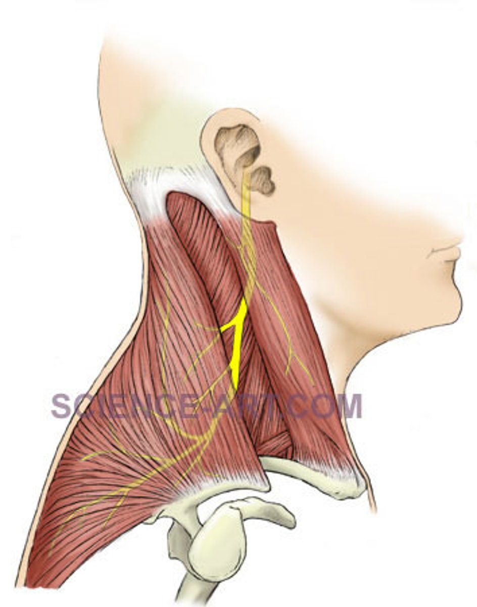
Accessory
Note: Innervates Trapezius & Sternocleidomastoid
Name the Nerve
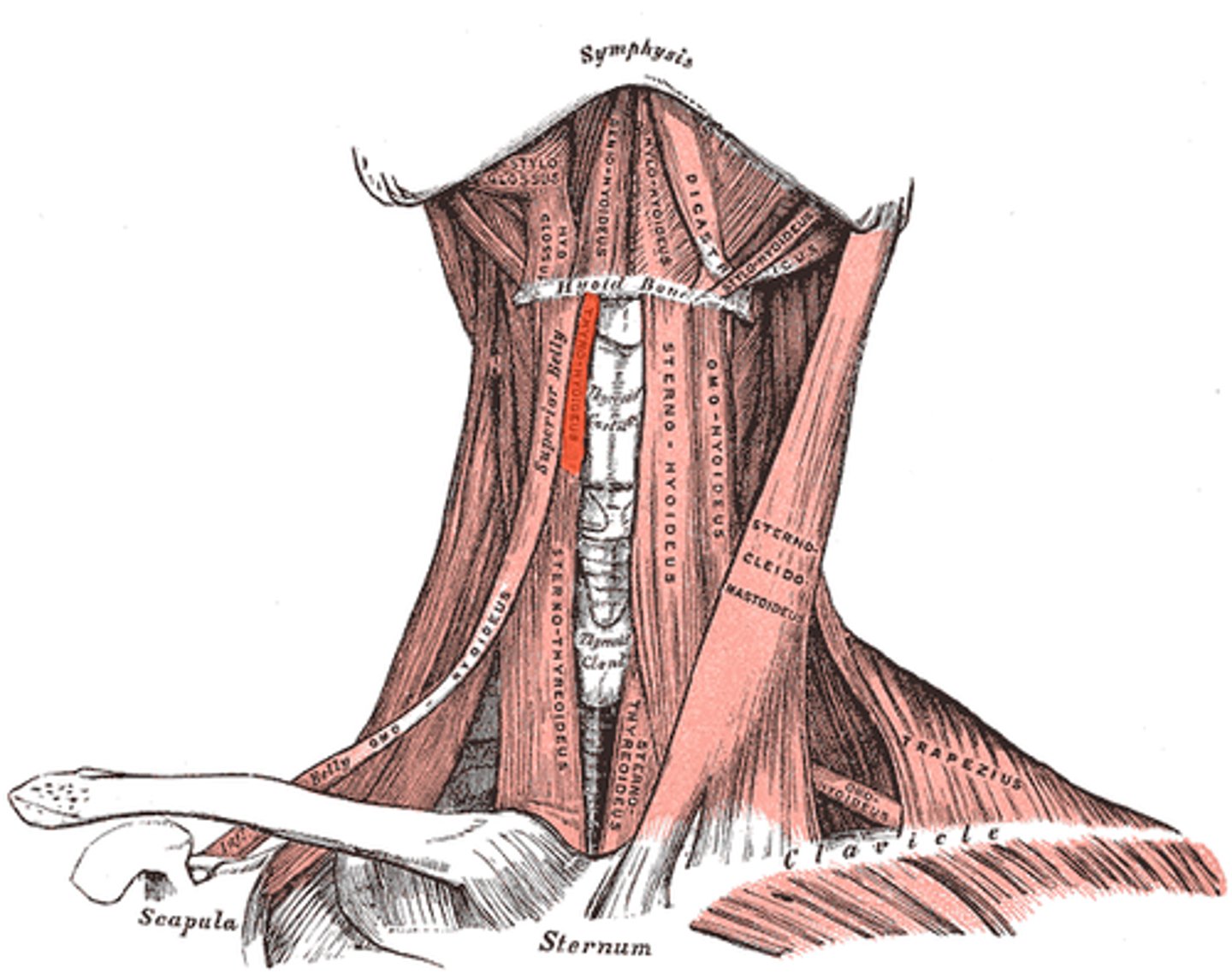
Omohyoid (Infrahyoid Muscle)
Note: Inserts into Upper Scapula, has Superior & Inferior Belly Separated by Intermediate Tendon, Connects to Hyoid Bone
Name the Muscle
Omohyoid
Name the Muscle
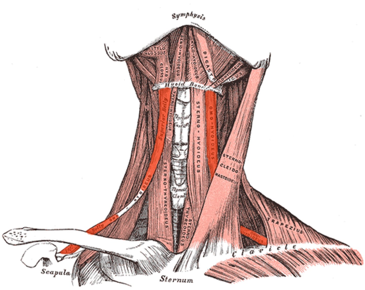
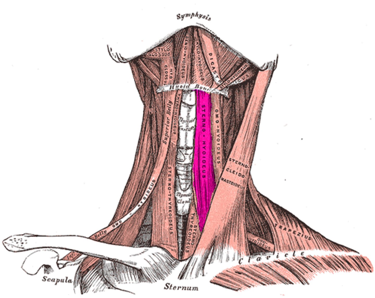
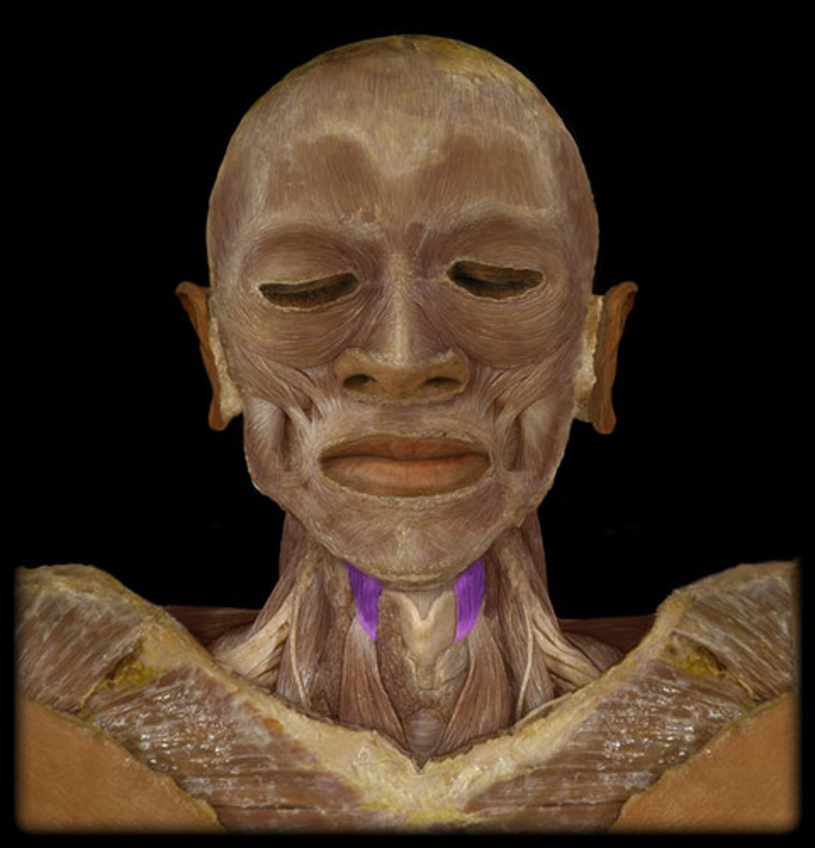
Sternohyoid (Infrahyoid Muscle)
Note: Connects Sternum & Hyoid Bone
Name the Muscle
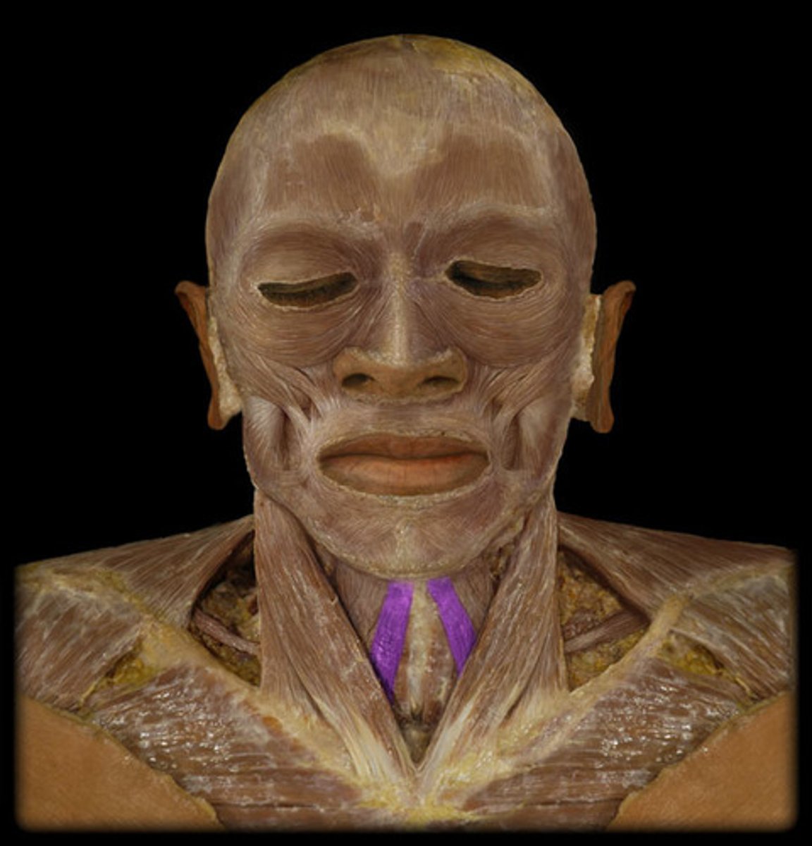
Sternohyoid
Name the Muscle
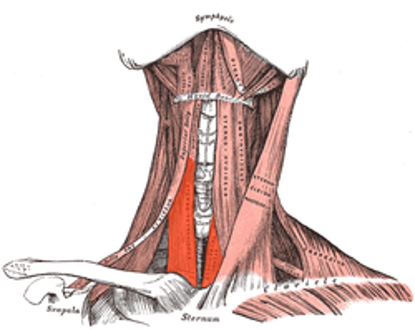
Sternothyroid (Infrahyoid Muscle)
Note: Connects Sternum & Thyroid Bone
Name the Muscle
Sternothyroid
Name the Muscle
Thyrohyoid (Infrathyroid Muscle)
Note: Connects Thyroid Cartilage & Hyoid Bone
Name the Muscle
Thyrohyoid
Name the Muscle
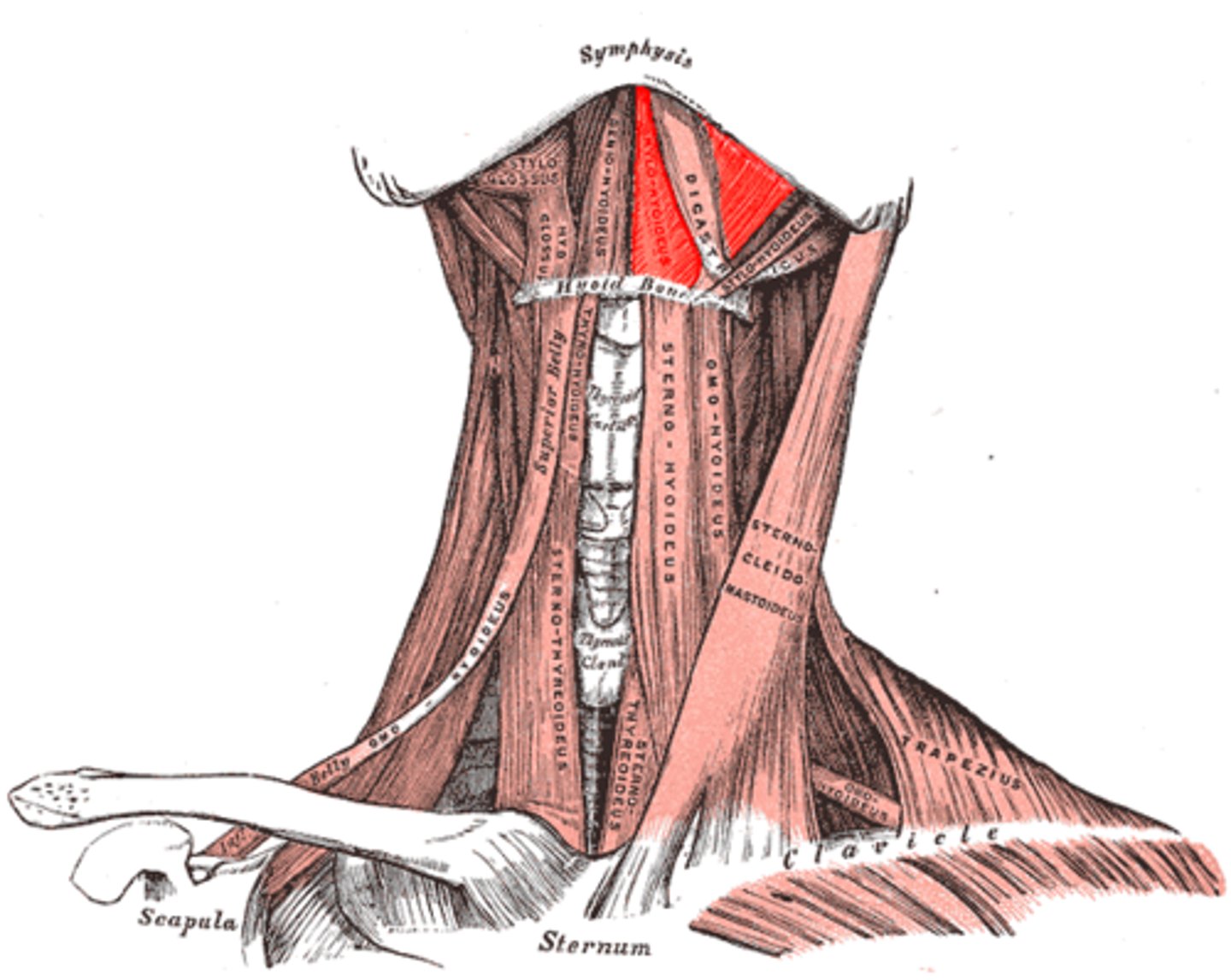
Digastric (Suprahyoid Muscle)
Note: Has anterior (innervated by V3, mandibular) and posterior belly (innverated by facial nerve) separated by an intermediate tendon
Name the Muscle
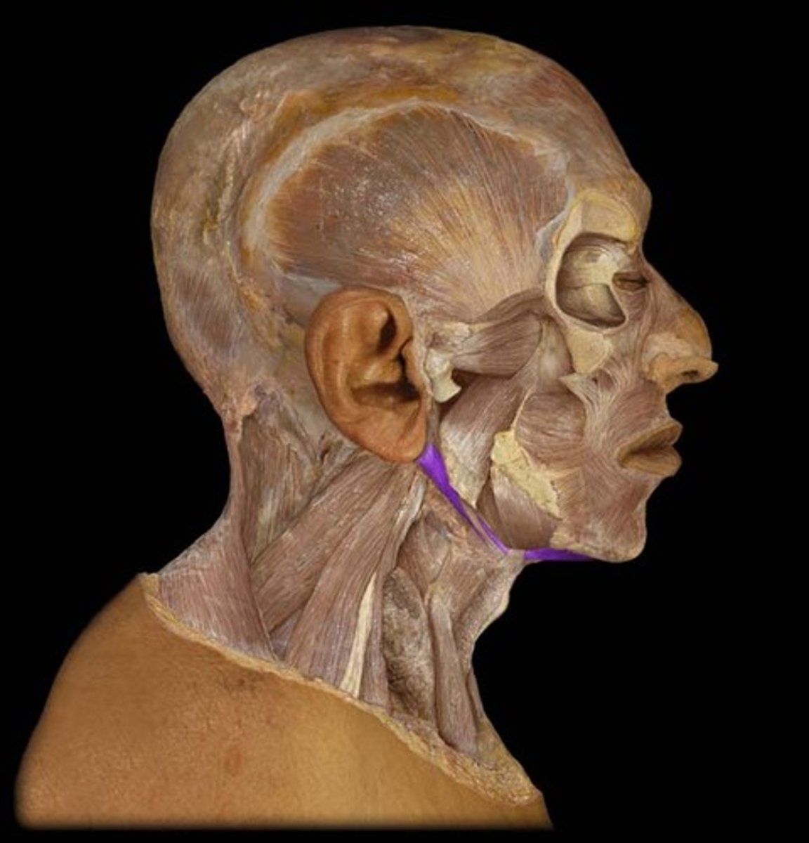
Digastric
Name the Muscle
Mylohyoid (Suprahyoid Muscle)
Note: forms floor of mouth, visible deep to anterior digastric belly
Name the Muscle
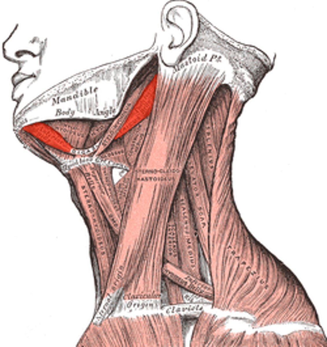
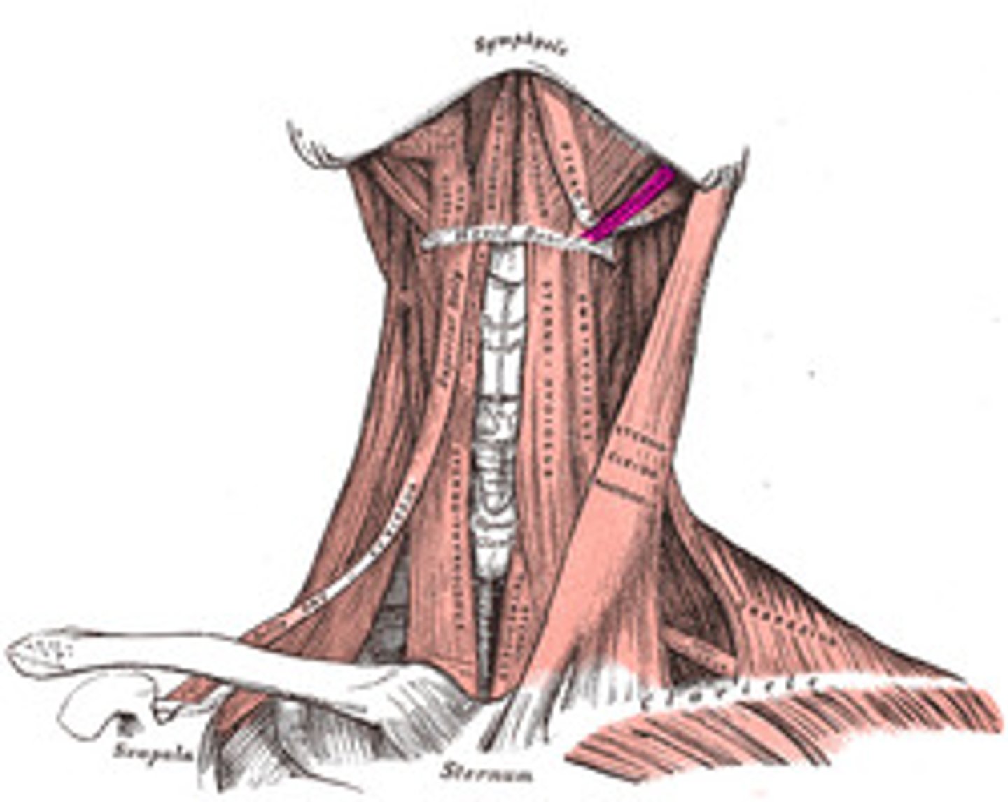
Stylohyoid (Suprahyoid Muscle)
Note: On Superior Surface of Posterior Digastric Belly
Name the Muscle
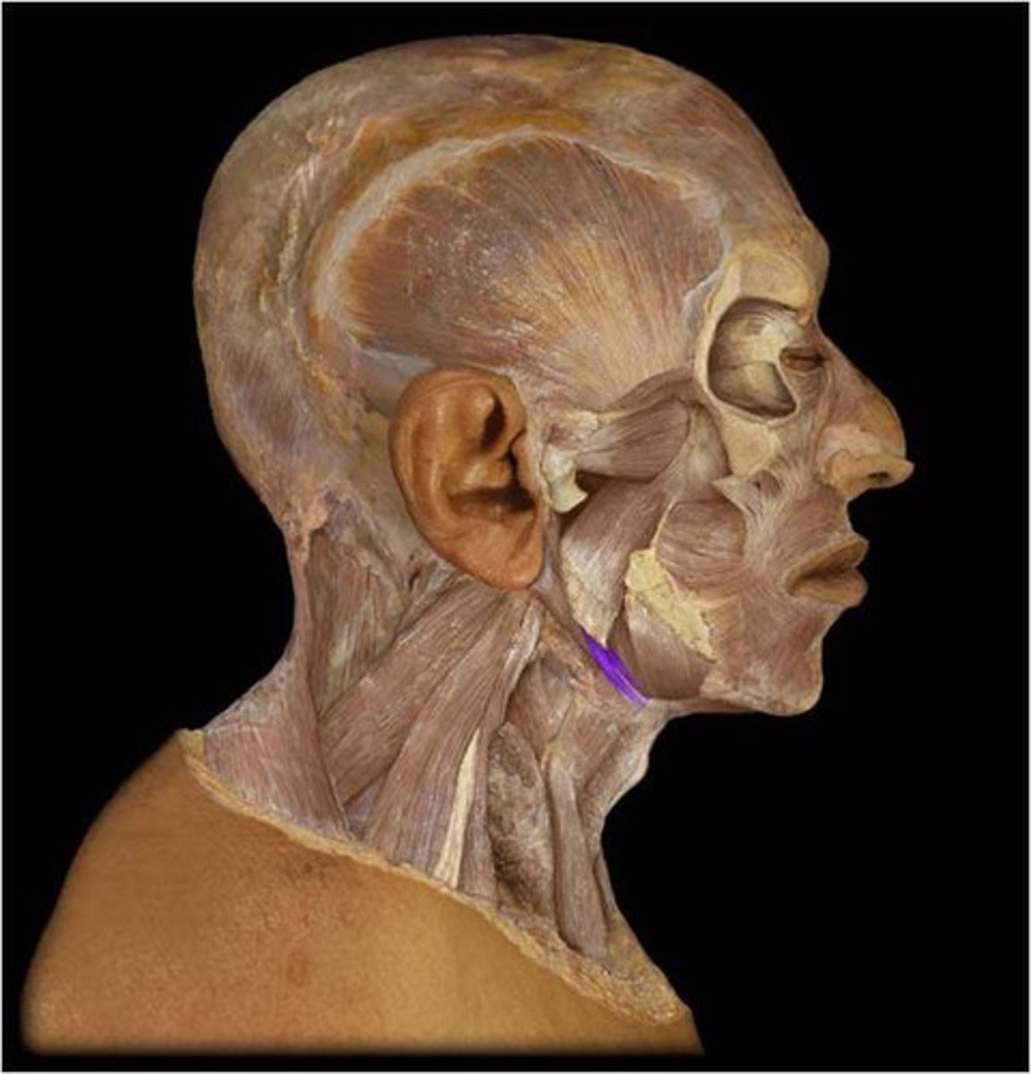
Stylohyoid
Name the Muscle
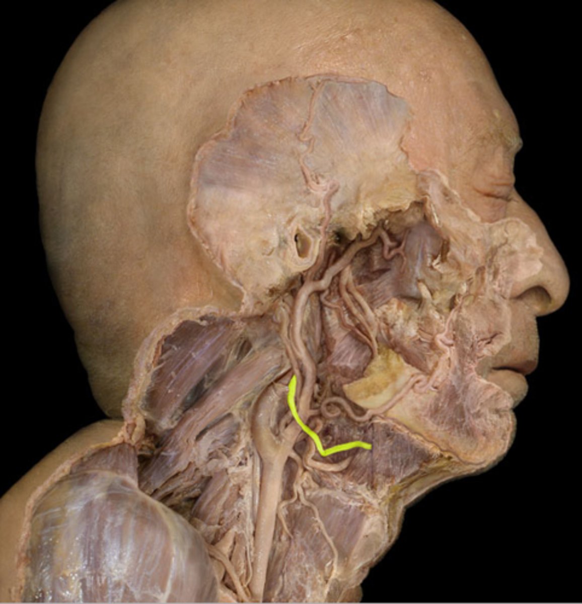
Hypoglossal
Note: Immediately Medial to Intermediate Digastric Tendon
Name the Nerve
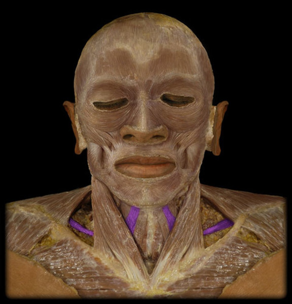
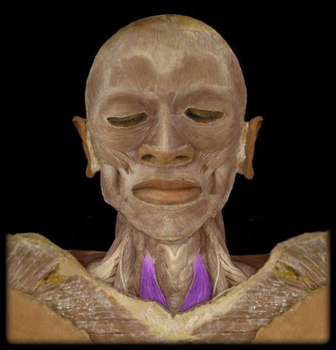
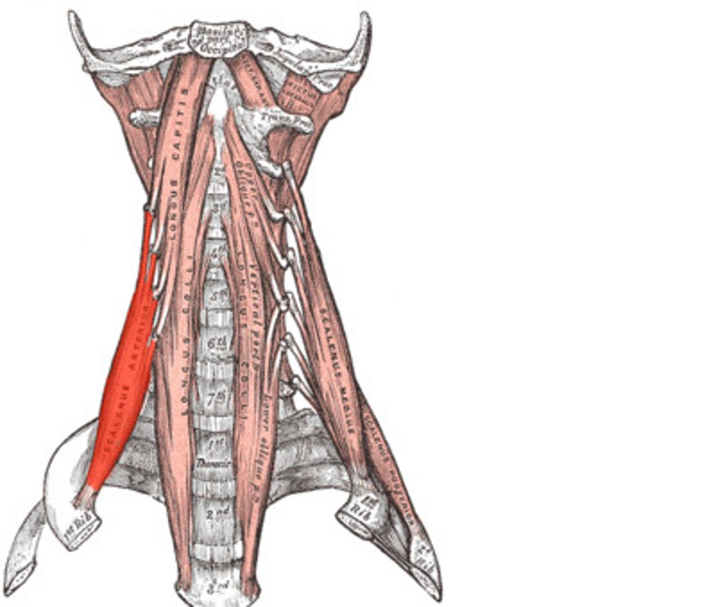
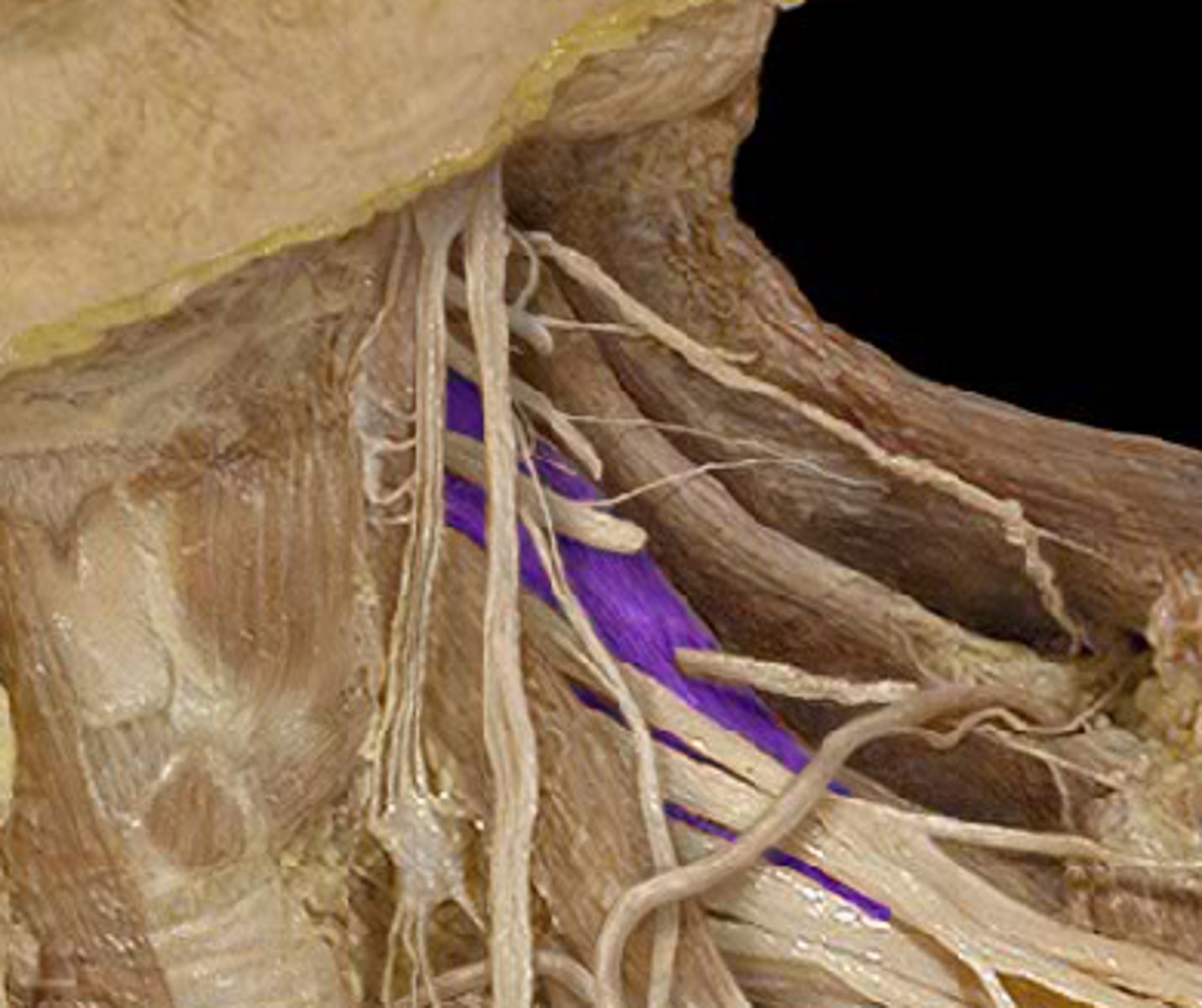
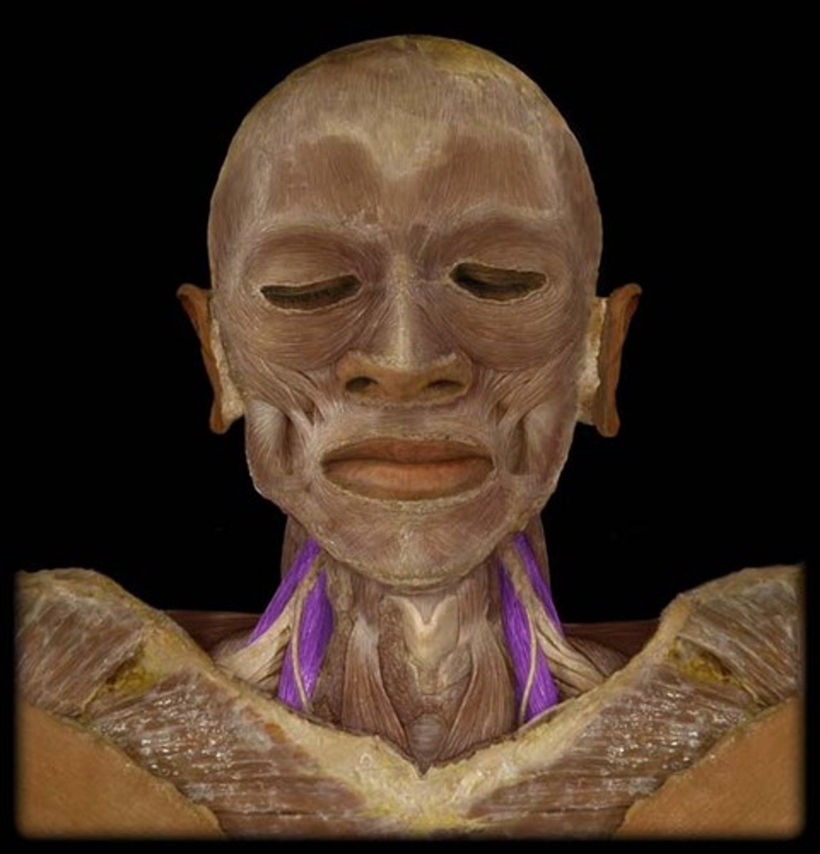
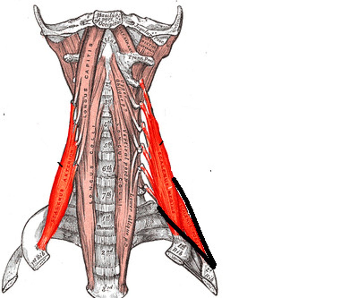
Anterior Scalene
Note: anterior to brachial artery & brachial plexus trunks
Name the Muscles (the 2 purple ones close to the front)

Anterior Scalene
Name the Muscle
Middle Scalene
Note: posterior to brachial artery & brachial plexus trunks
Name the Muscle
Middle Scalene
Name the Muscle
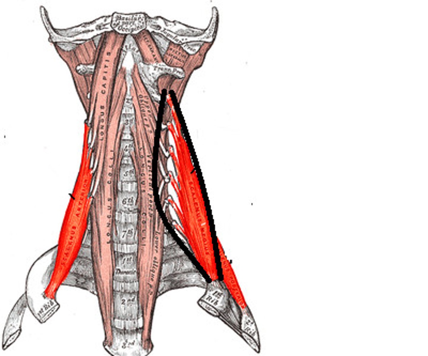
Posterior Scalene
Note: inserts into 1st rib, NOT 2nd rib (unlike anterior and middle)
Name the Muscles (the 2 purple ones close to the back)
Posterior Scalene
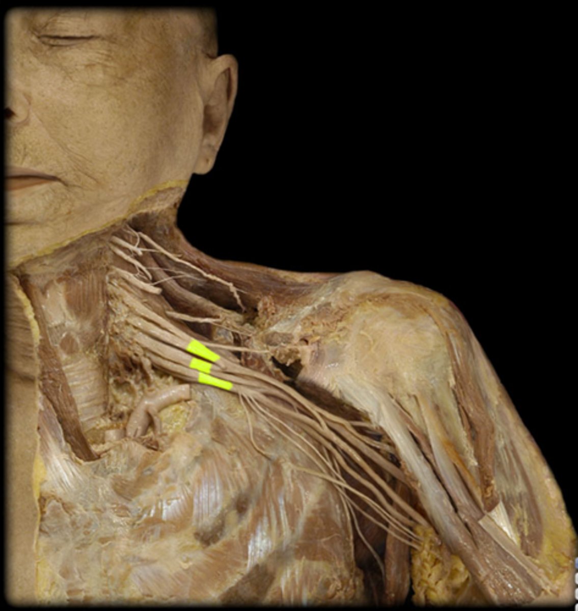
Name the Muscle
Roots of Brachial Plexus
Note: C5-T1
Name the Nerves
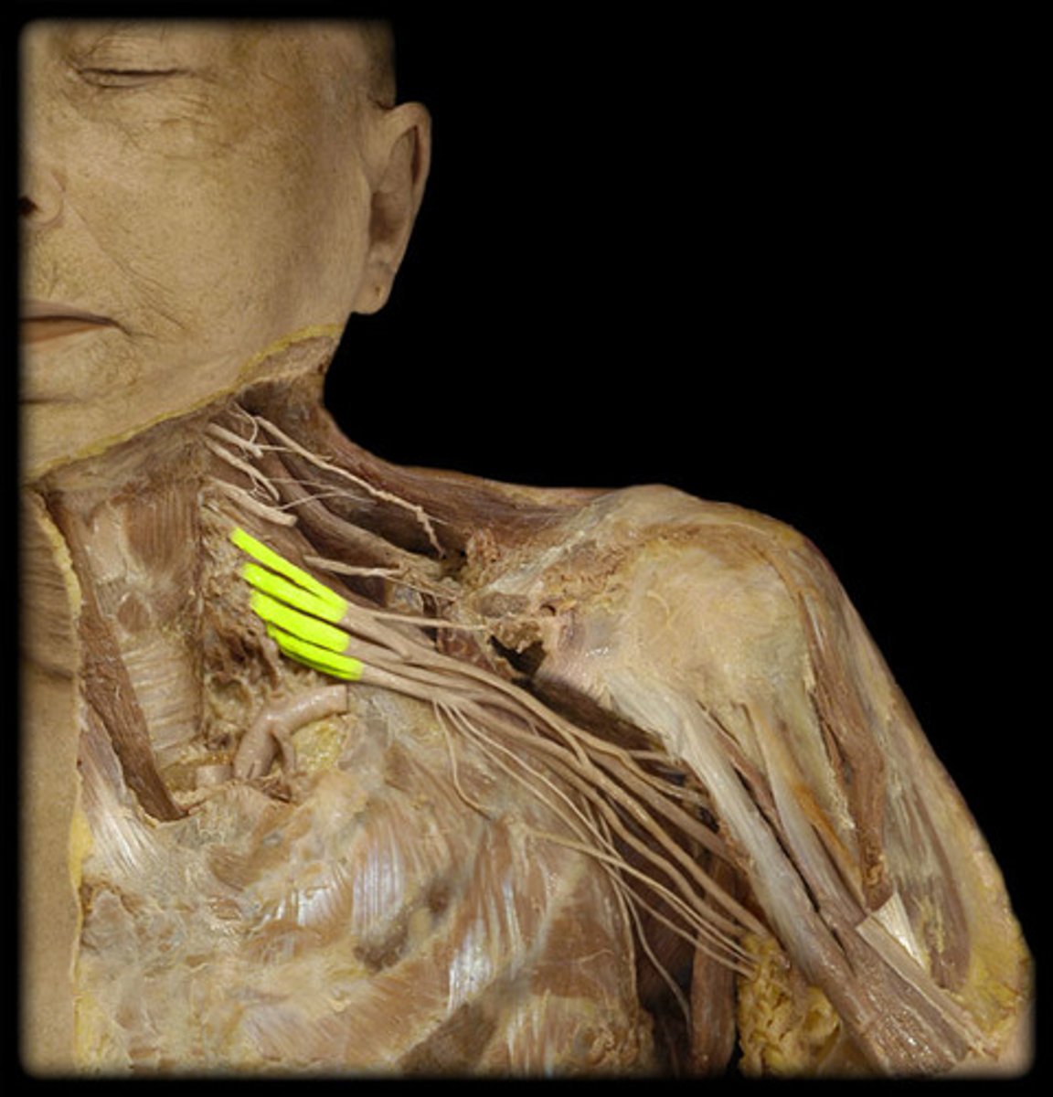
Trunks of Brachial Plexus
Note: remember the superior (C5-C6), middle (C7), and inferior (C8-T1)
Name the Nerves
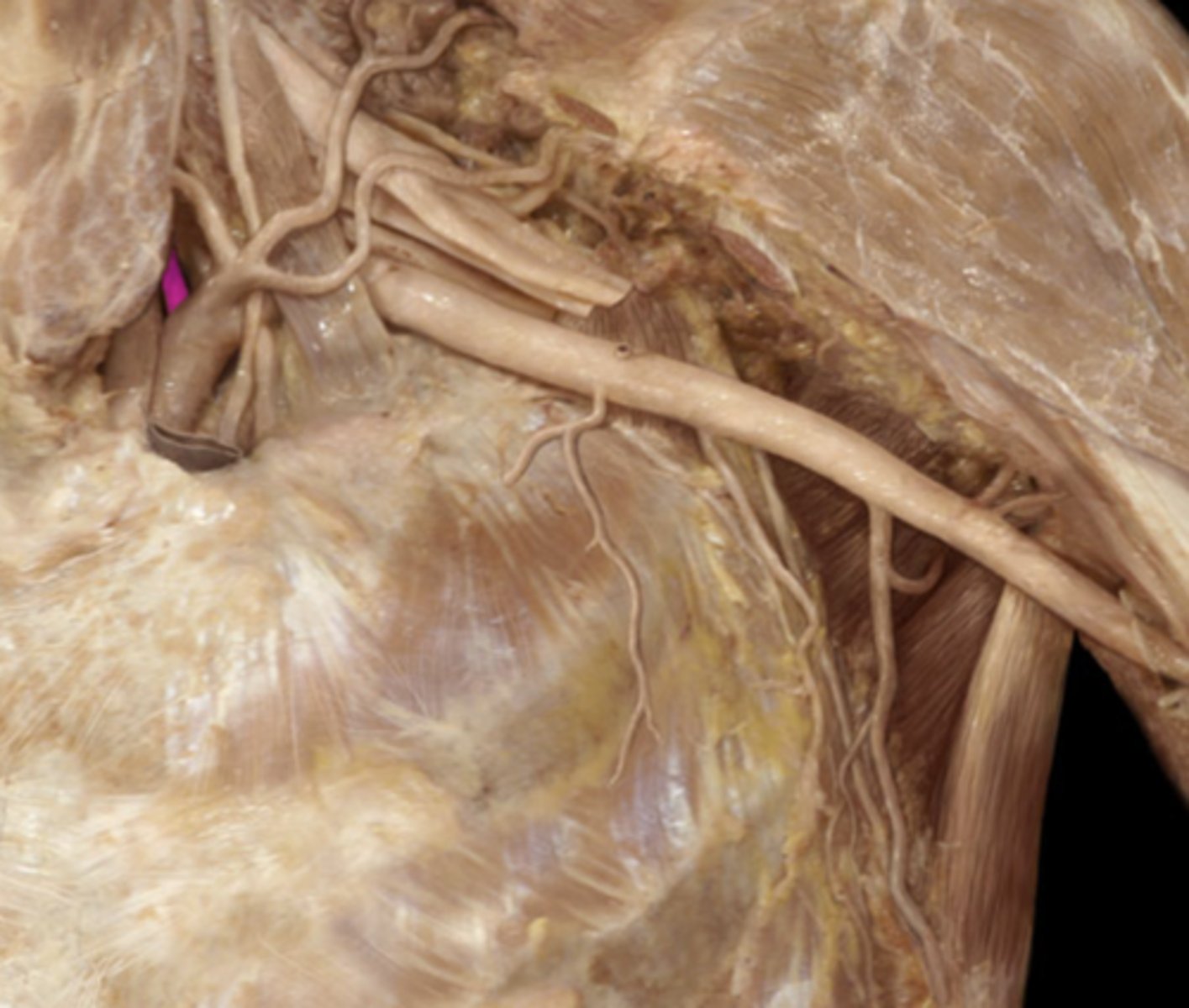
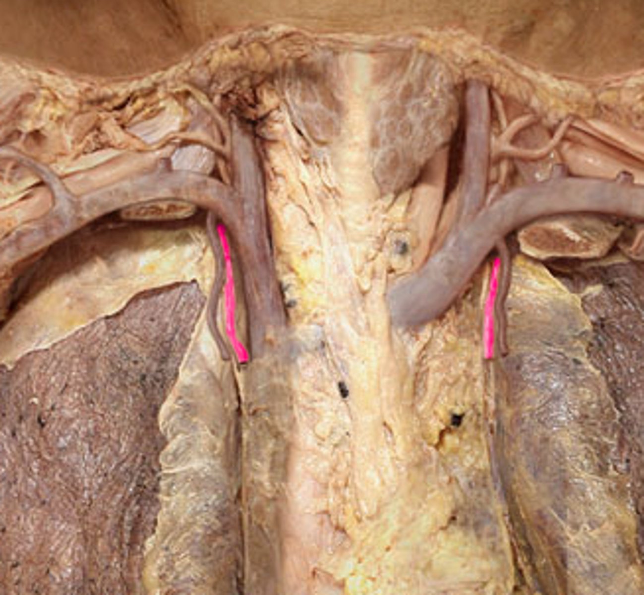
Left Subclavian
Note: travels between anterior and middle scalene muscles
Name the Artery
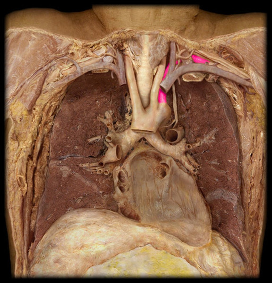
Right Subclavian
Note: travels between anterior and middle scalene muscles
Name the Artery
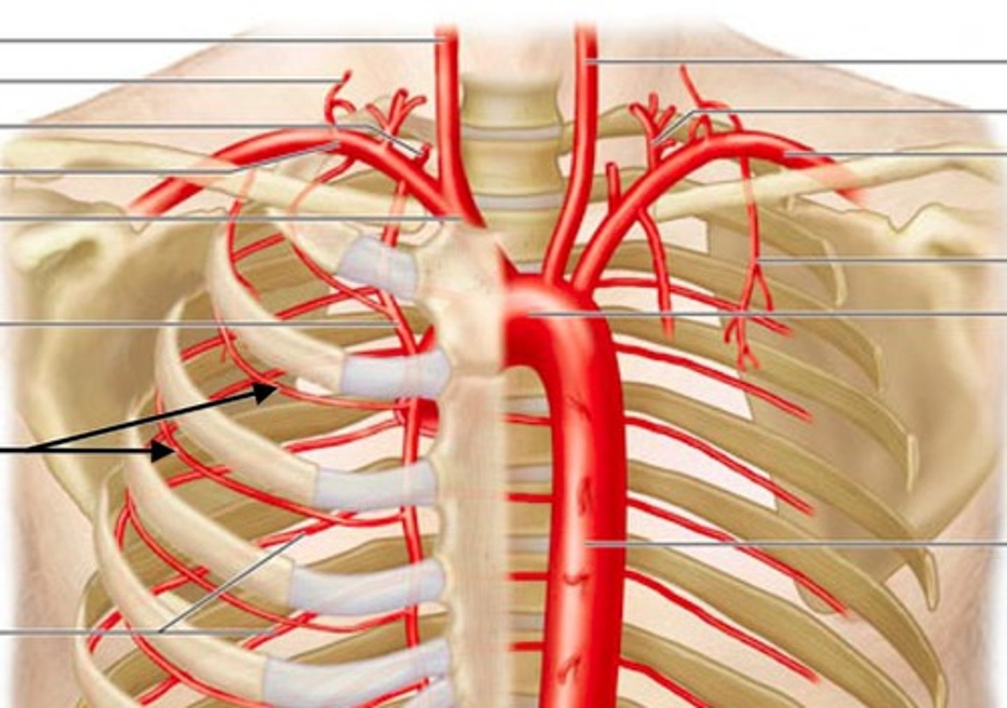
Subclavian
Note: travels anterior to anterior scalene muscles
Name the Vein
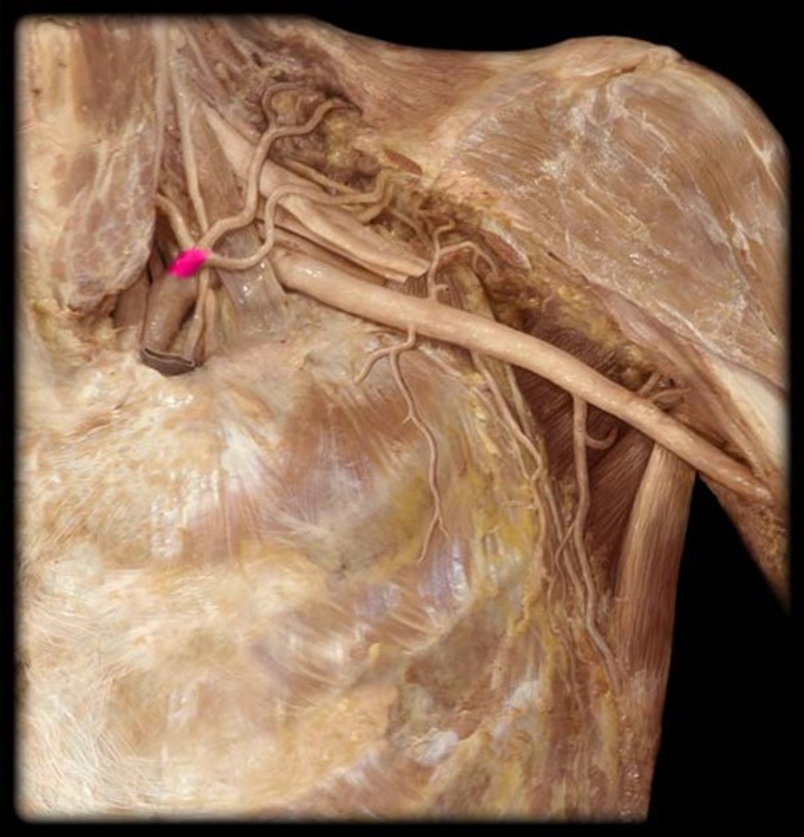
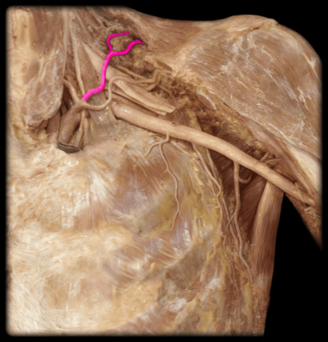
Thyrocervical Trunk
Note: second branch of subclavian after vertebral
Name the Arterial Structure
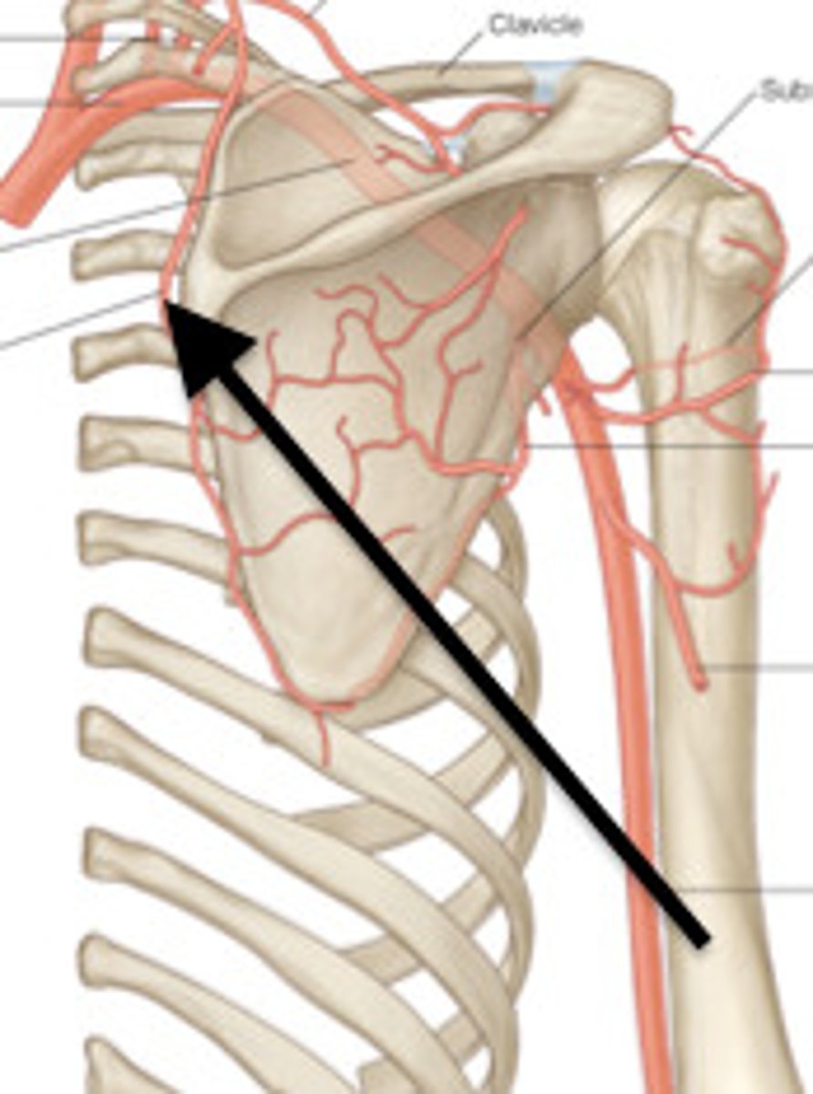
Suprascapular
Note: branch of thyrocervical trunk
Name the Artery
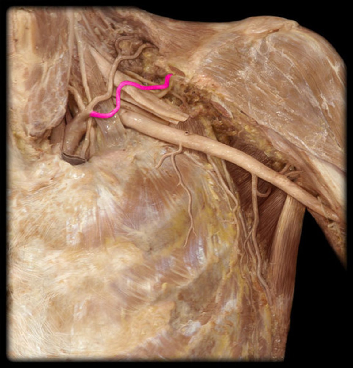
Transverse Cervical
Note: branch of thyrocervical trunk
Name the Artery
Inferior Thyroid
Note: branch of thyrocervical trunk
Name the Artery

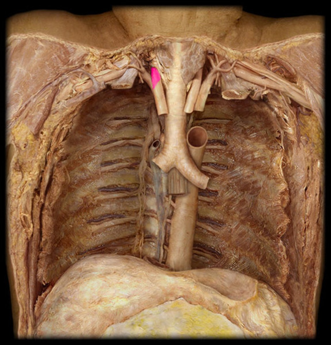
Vertebral
Note: first branch of subclavian
Name the Artery
Dorsal Scapular
Note: either deep branch of transverse cervical OR directly comes off subclavian, then travels between trunks of brachial plexus en route to rhomboids & levator scapulae
Name the Artery (Sorry, Can't Find a Better Pic)
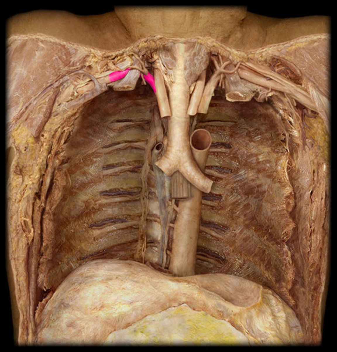
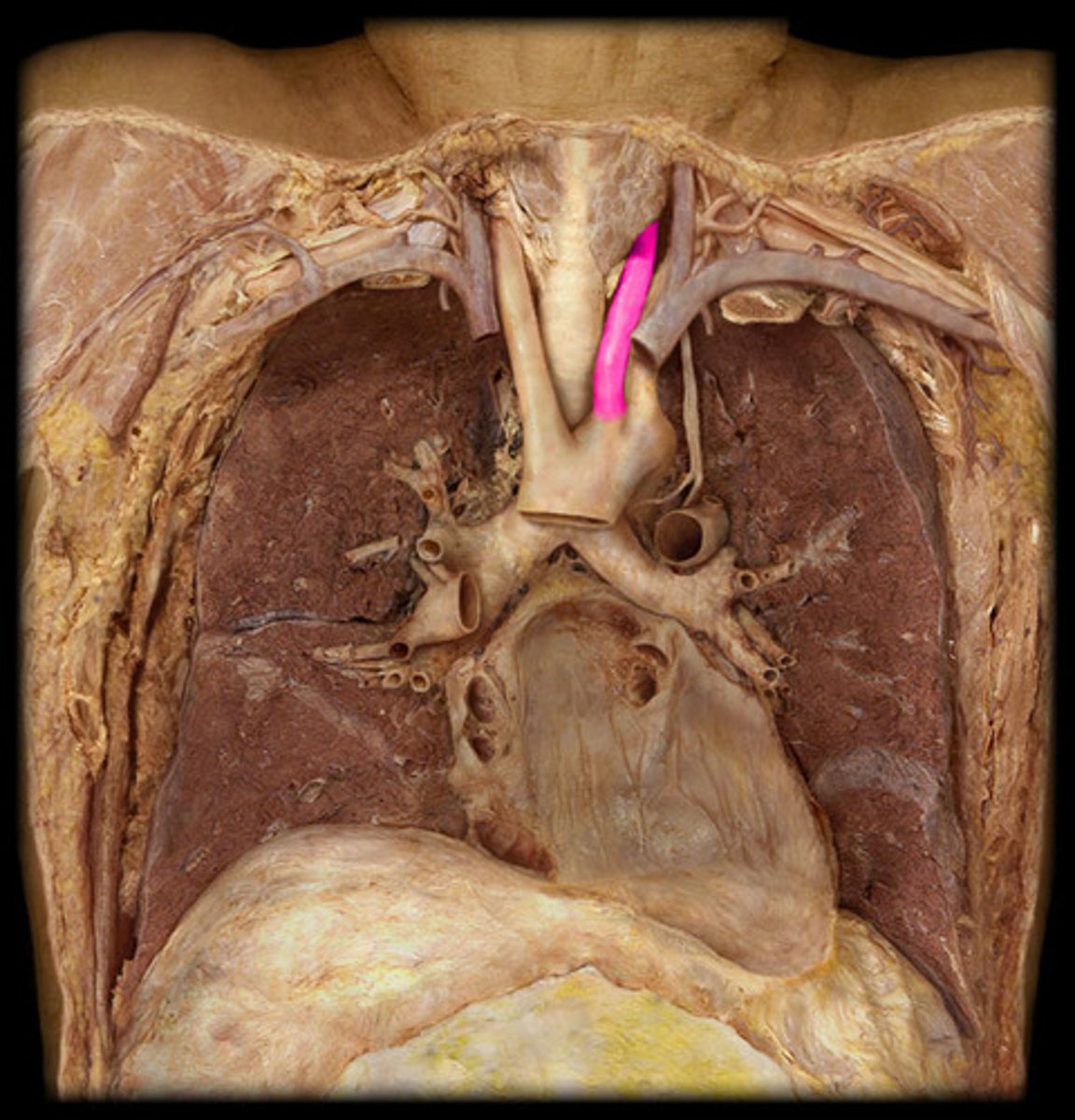
Left Common Carotid
Name the Artery
Right Common Carotid
Name the Artery
Carotid Sinus
Note: dilation of most proximal portion of internal carotid (just before it merges with external into common), acts as baroreceptor
Name the Structure
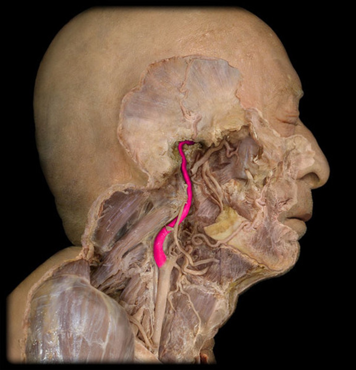
Internal Carotid
Name the Artery
External Carotid
Name the Artery

Superior Thyroid
Note: branch of external carotid
Name the Artery

Lingual
Note: branch of external carotid
Name the Artery

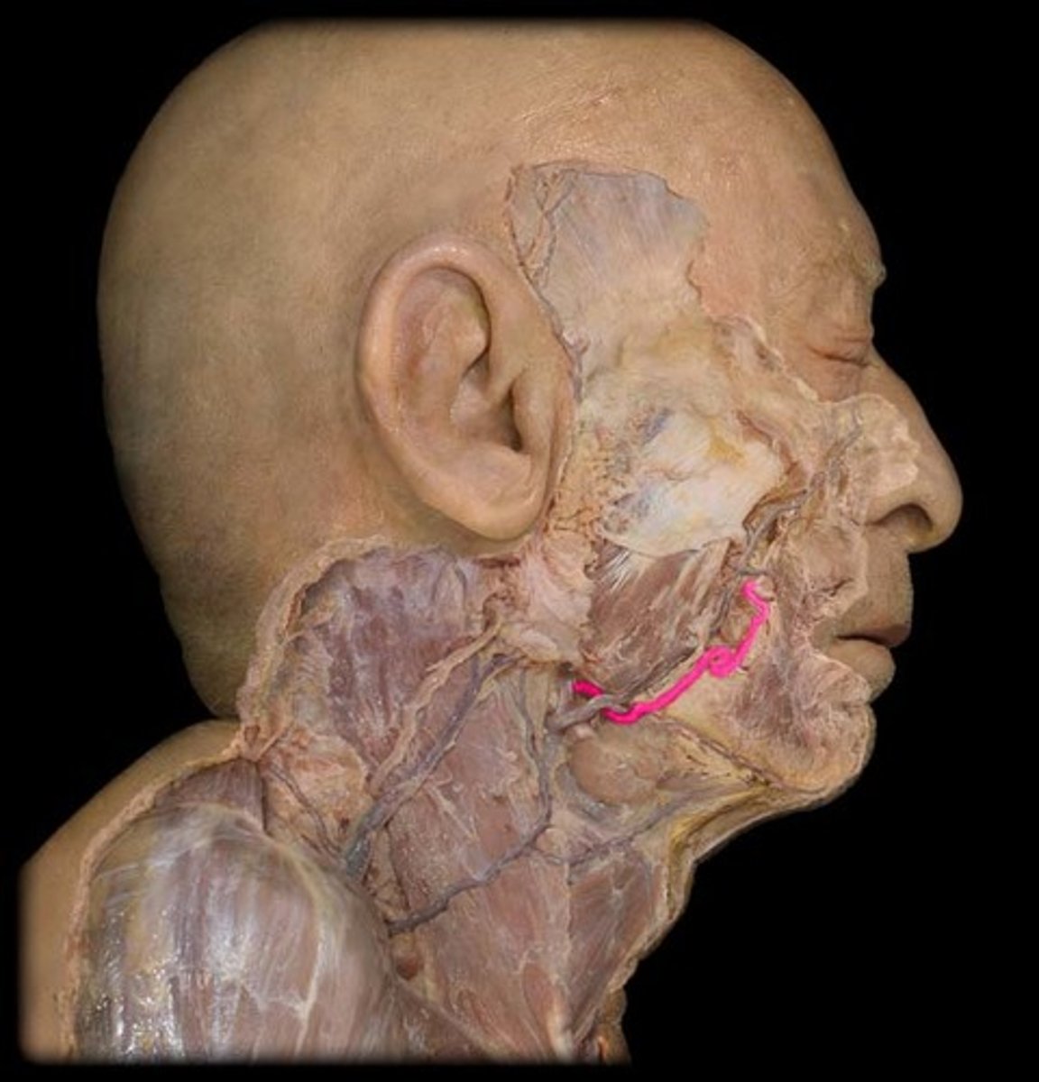
Facial
Note: branch of external carotid, cross inferior margin of mandible
Name the Artery
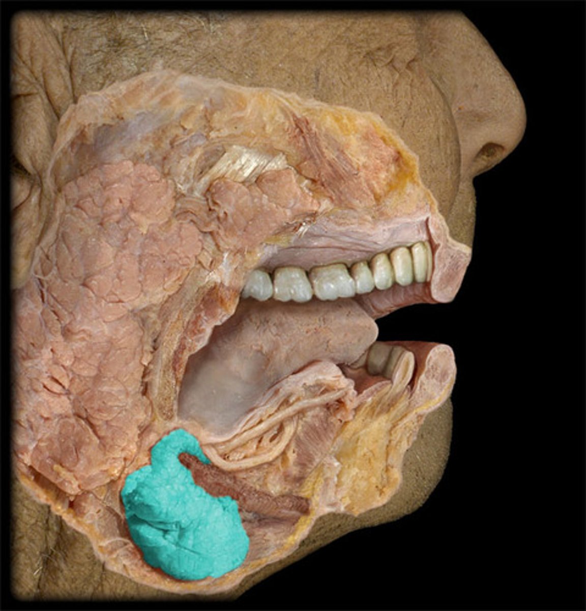
Submandibular Gland
Name the Artery
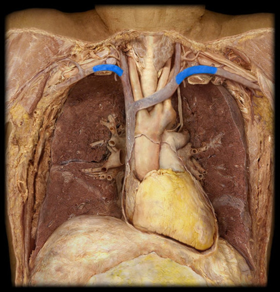
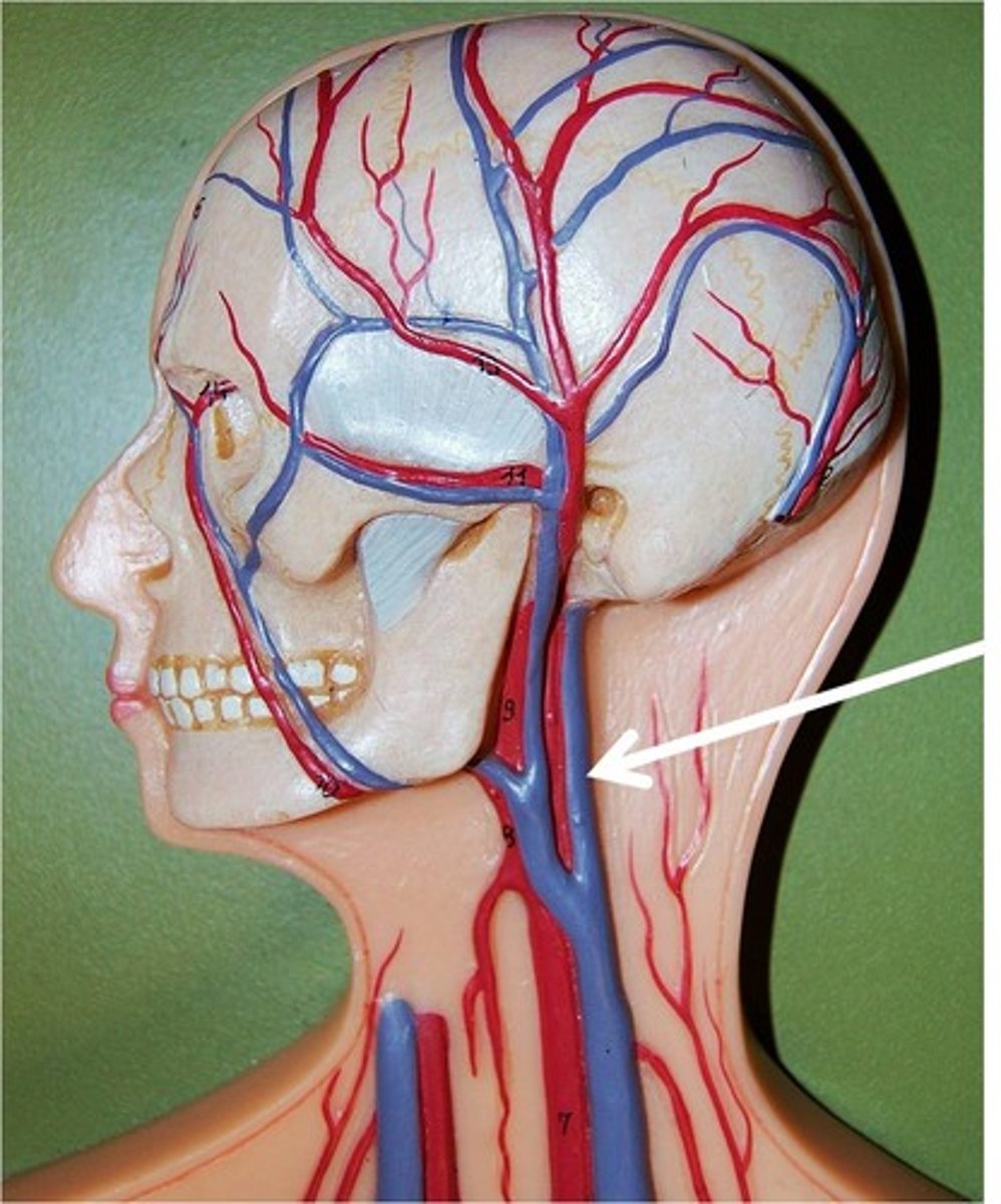
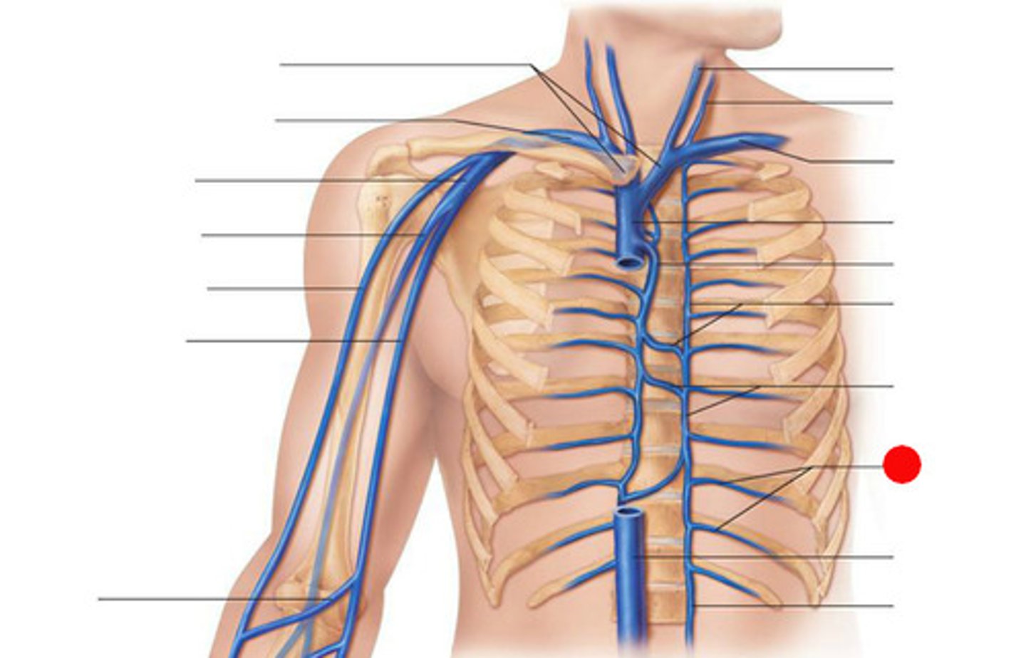
Internal Jugular
Note: branch of brachiocephalic vein (other branch is subclavian vein)
Name the Vein
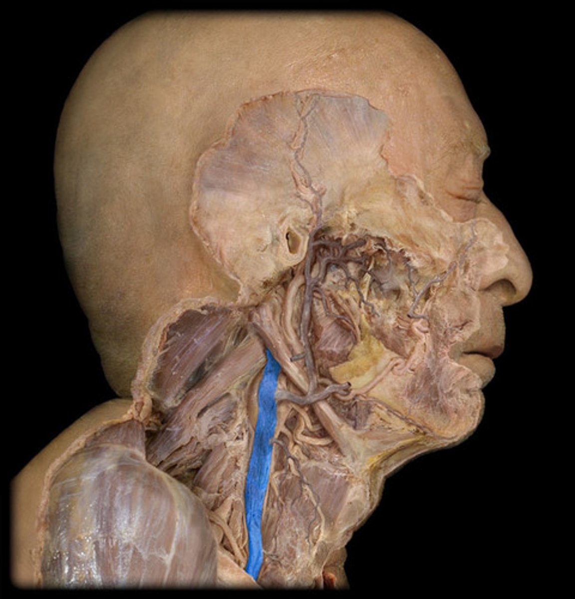
Internal Jugular
Name the Vein
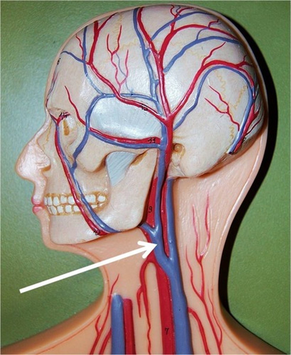
External Jugular
Note: branch of subclavian vein
Name the Vein
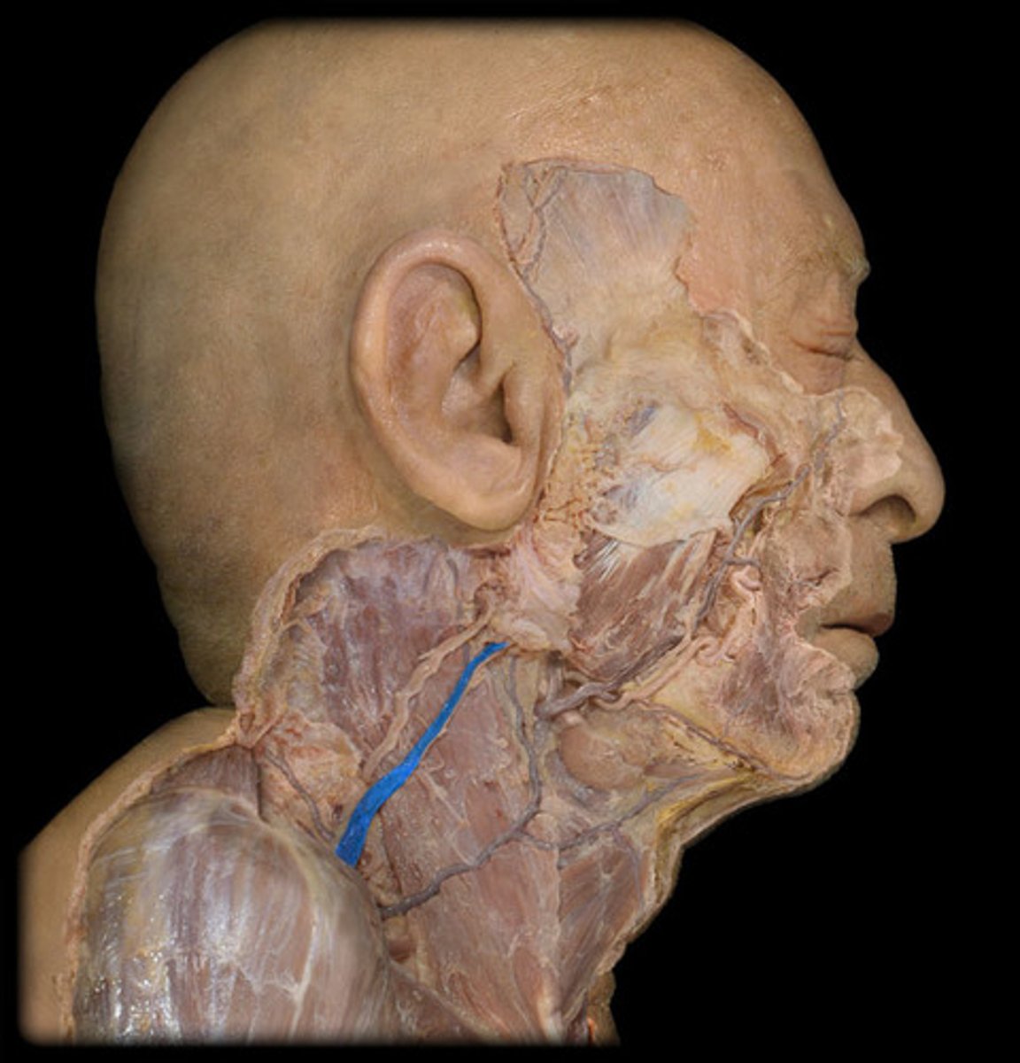
External Jugular
Name the Vein
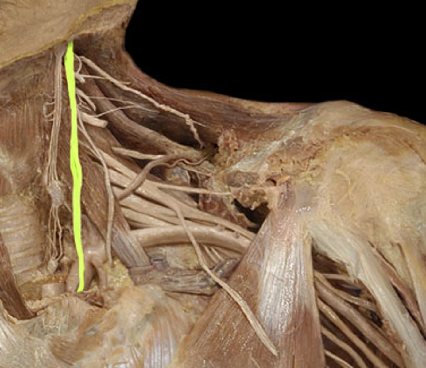
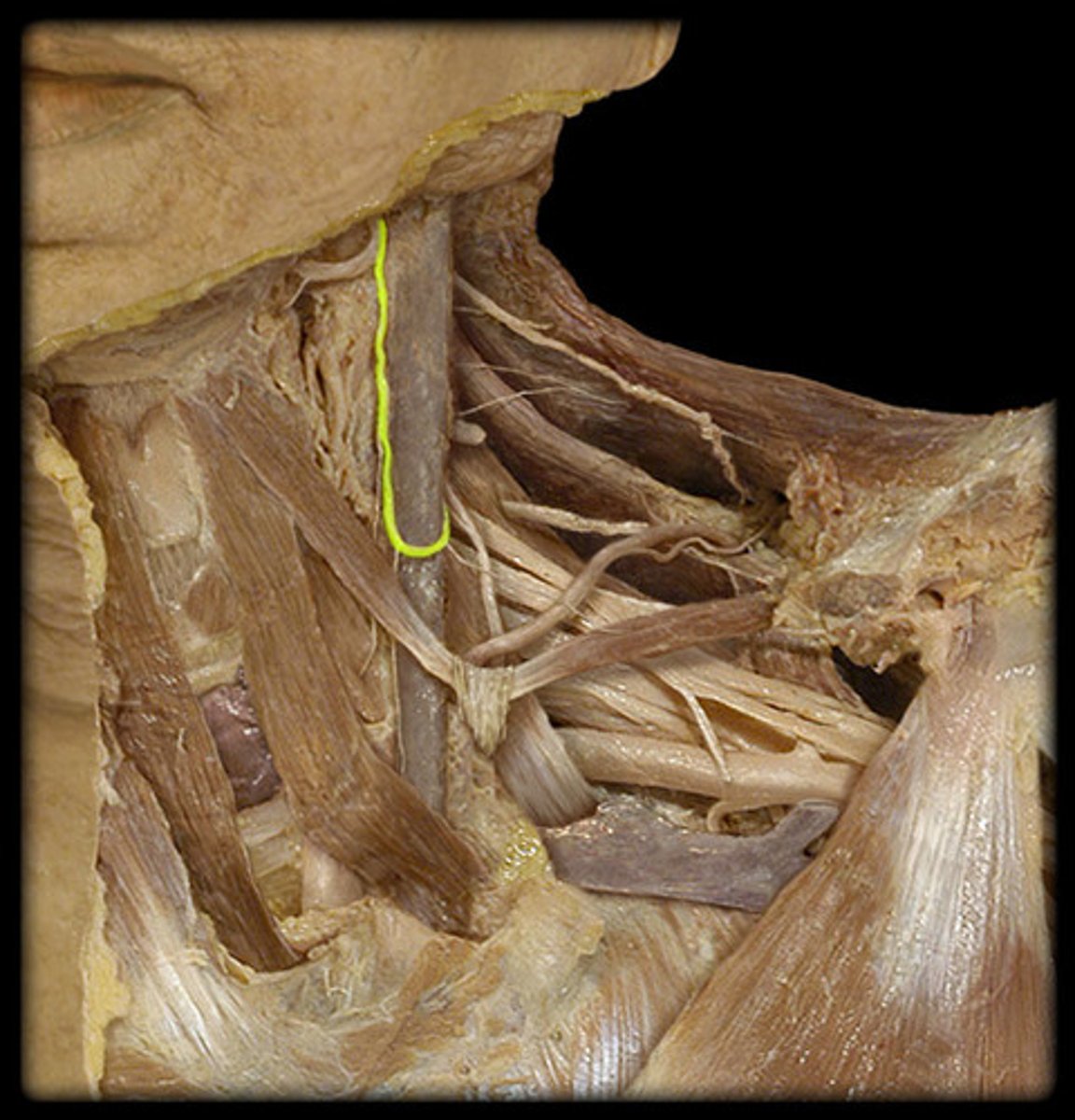
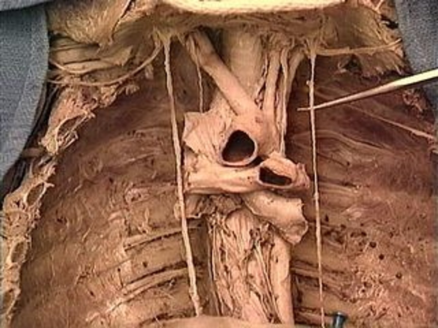
Vagus Nerve
Note: tucked in carotid sheath between common carotid and internal jugular vein
Name the Nerve
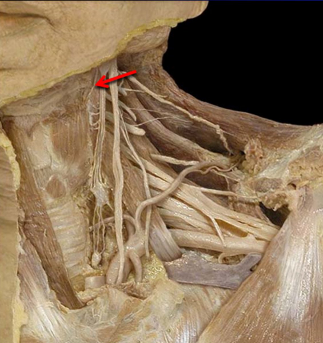
Ansa Cervicalis
Note: innervates 3/4 infrahyoid muscles (not thyrohyoid), on anterior surface of carotid sheath, has superior (off C1) and inferior roots (off C3) connected by loop
Name the Nerve
Nerve to Thyrohyoid
Name the Nerve
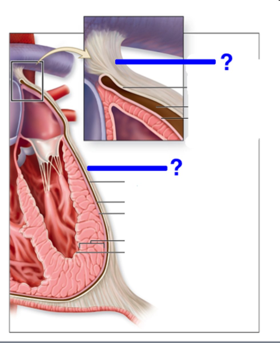
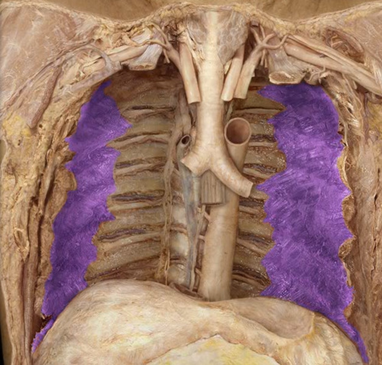
Parietal Pleura
Note: lines inside of pleural cavity, broken further down into costal/diaphragmatic/mediastinal portions
Label Layer 2

Visceral Pleura
Note: lines lung, cannot be distinguished from lung itself
Label Layer 1
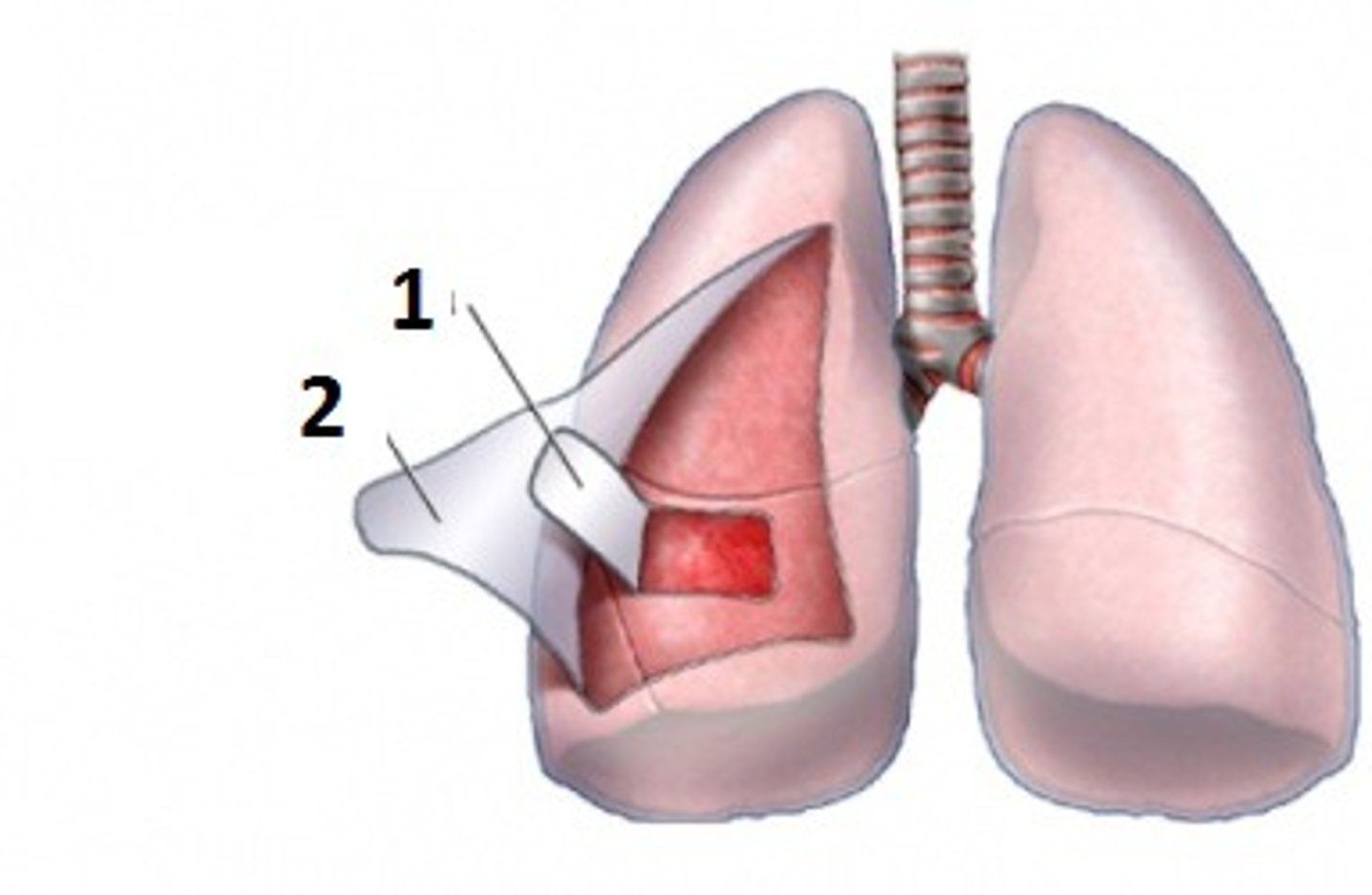
Pleural Cavity
Name the Space
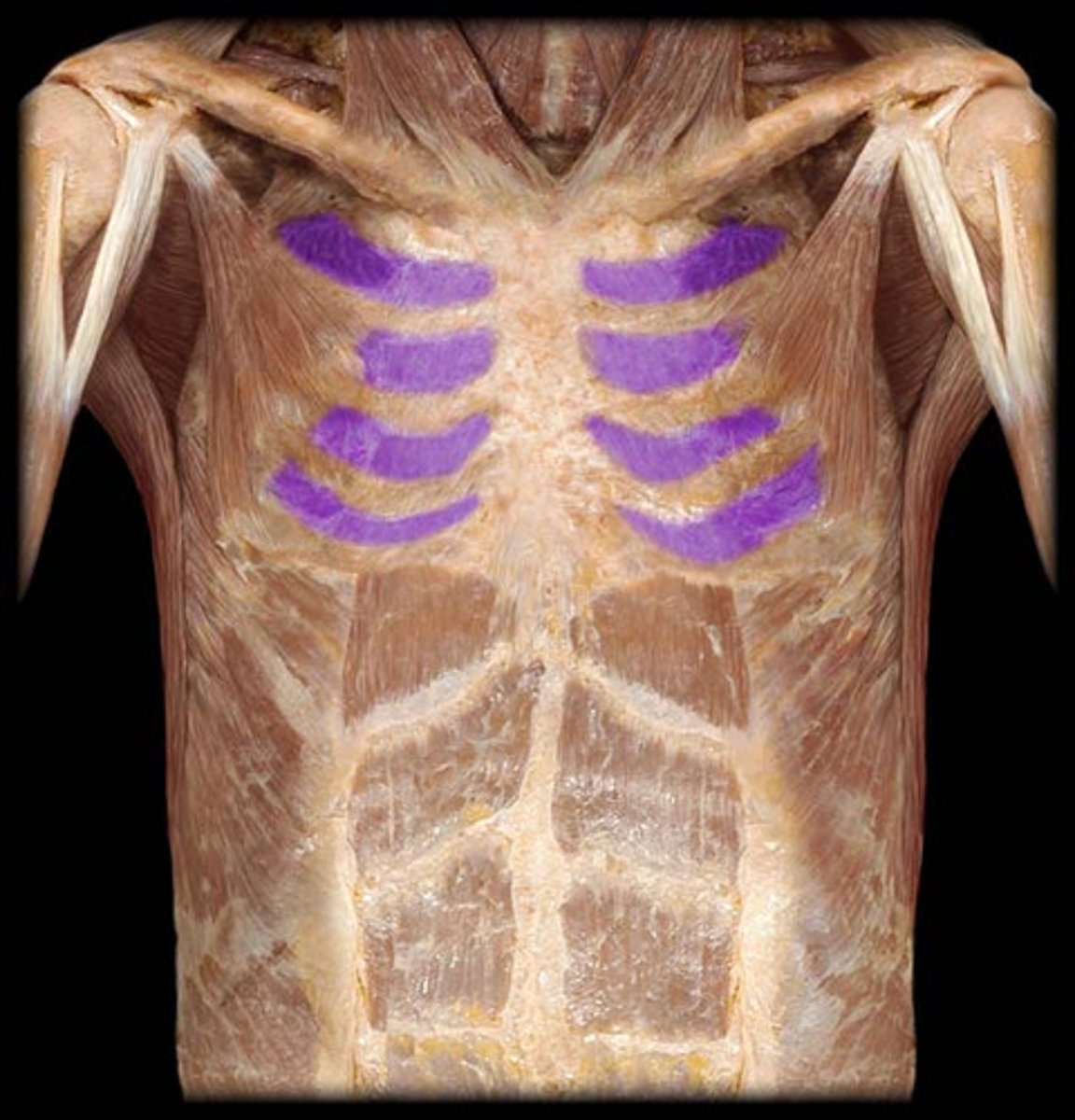
External Intercostal
Note: slanted inferomedially (slant goes down and to the middle of the body)
Name the Muscles
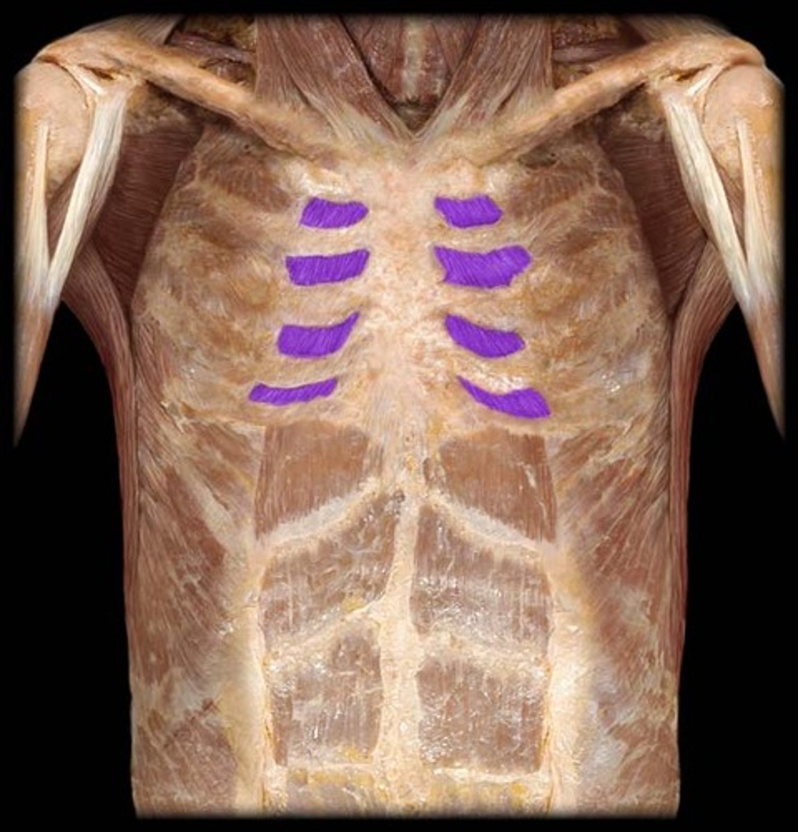
Internal Intercostal
Note: deep to innermost muscles, perpendicular to external
Name the Muscles
Innermost Intercostal
Name the Muscles
Tranverse Thoracis
Name Muscle Group 2

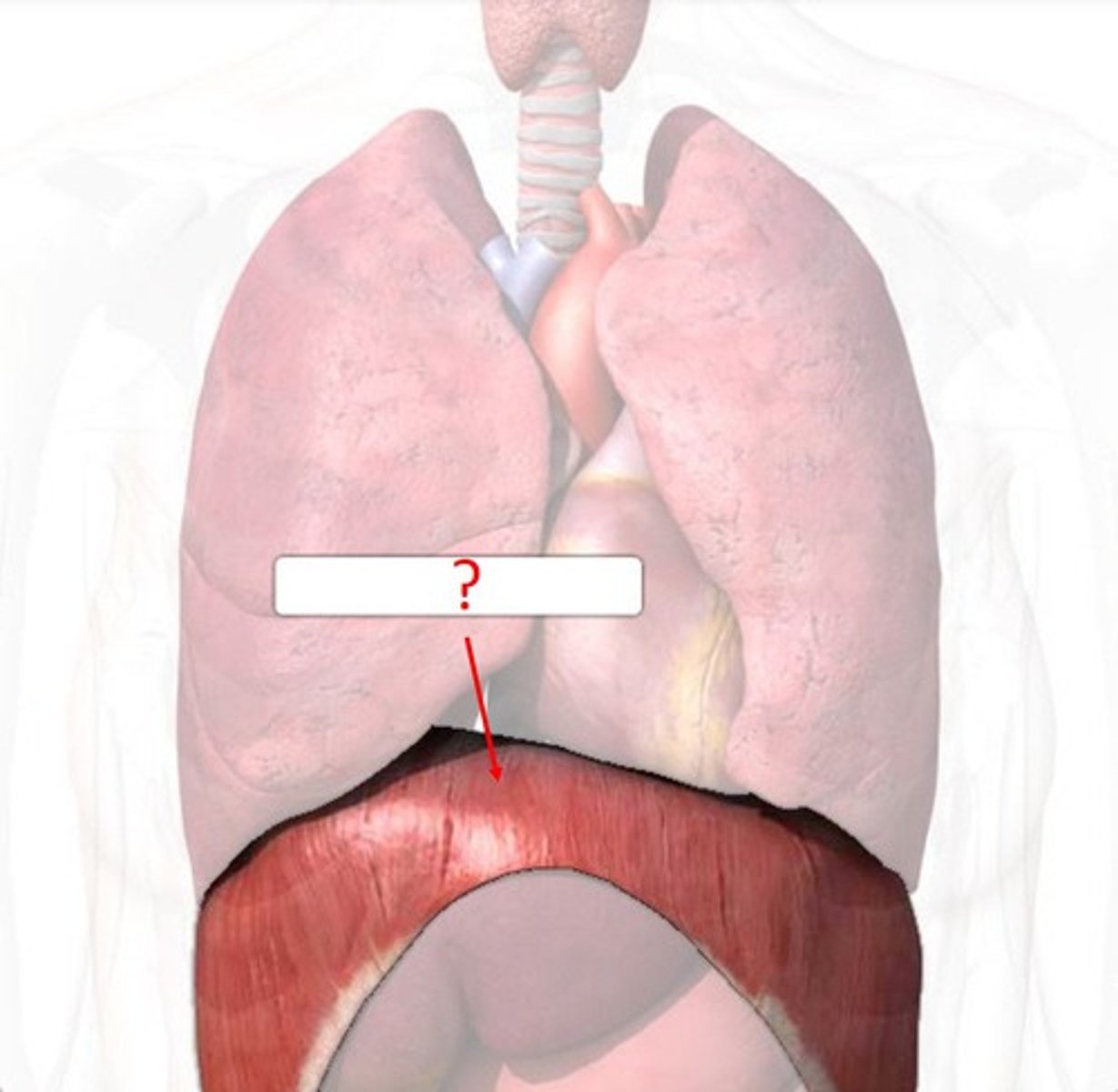
Diaphragm
Name the Structure
Phrenic
Note: kinda hard to distinguish from vagus nerve & sympathetic trunk in distal part, each half innervates ipsilateral diaphragm, runs along anterior scalenes, roots are C3-C5
Name the Nerve
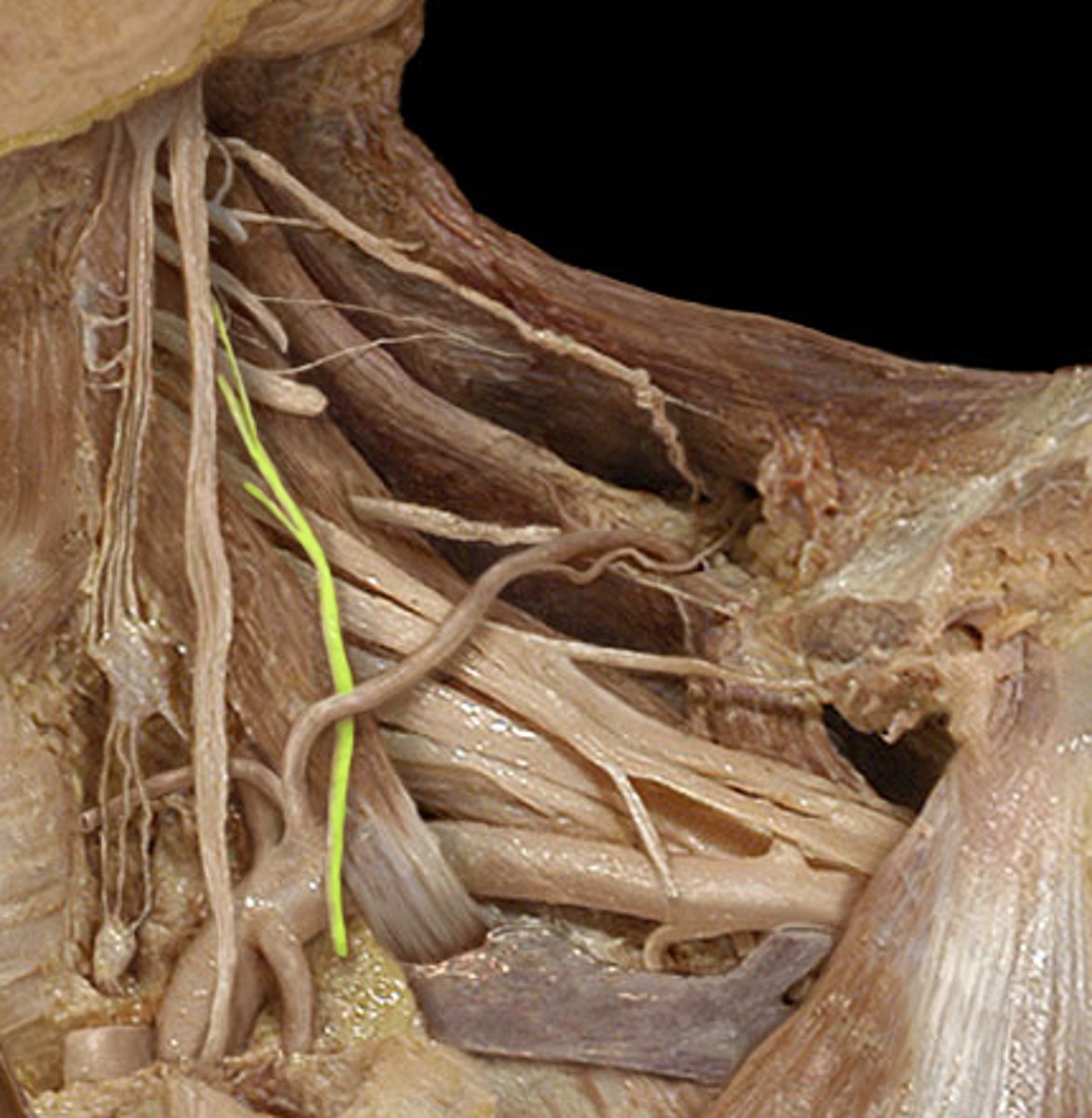
Phrenic
Name the Nerve
Internal Thoracic
Note: branch of subclavian artery and runs along posterior surface of anterior thoracic wall, deep to transverse thoracis
Name the Artery
Internal Thoracic Vein
Note: drains into subclavian vein and runs along posterior surface of anterior thoracic wall, deep to transverse thoracis
Name the Vein
Anterior Intercostal
Name the Arteries (apologies for not finding a better picture, they are quite hard to find since they are very tiny vessels)
Anterior Intercostal
Name the Veins (apologies for not finding a better picture, they are quite hard to find since they are very tiny vessels)
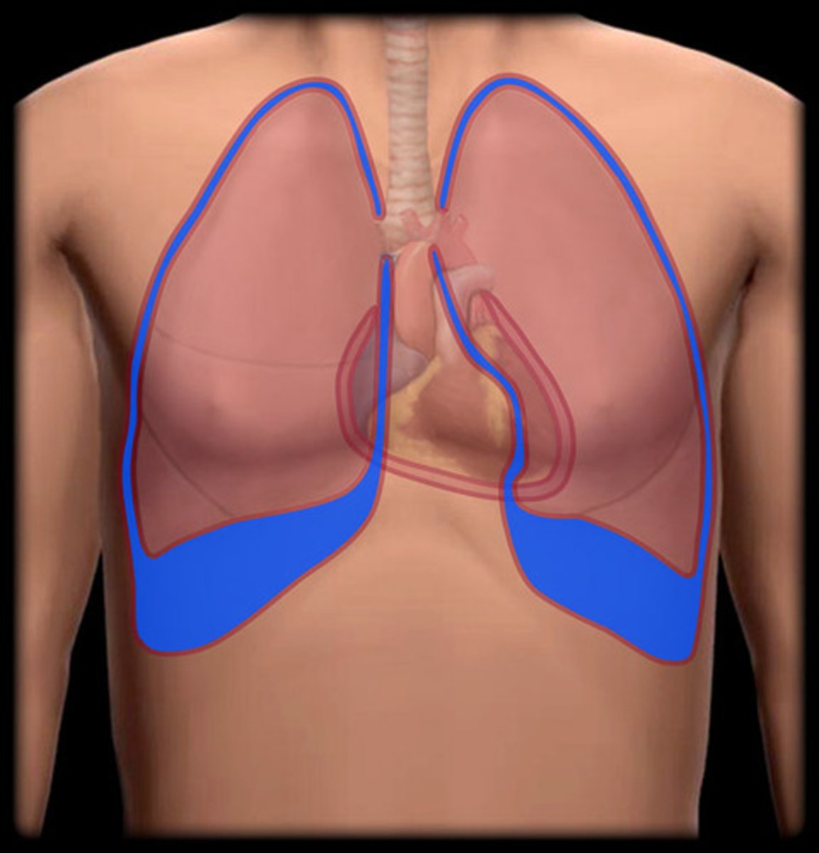
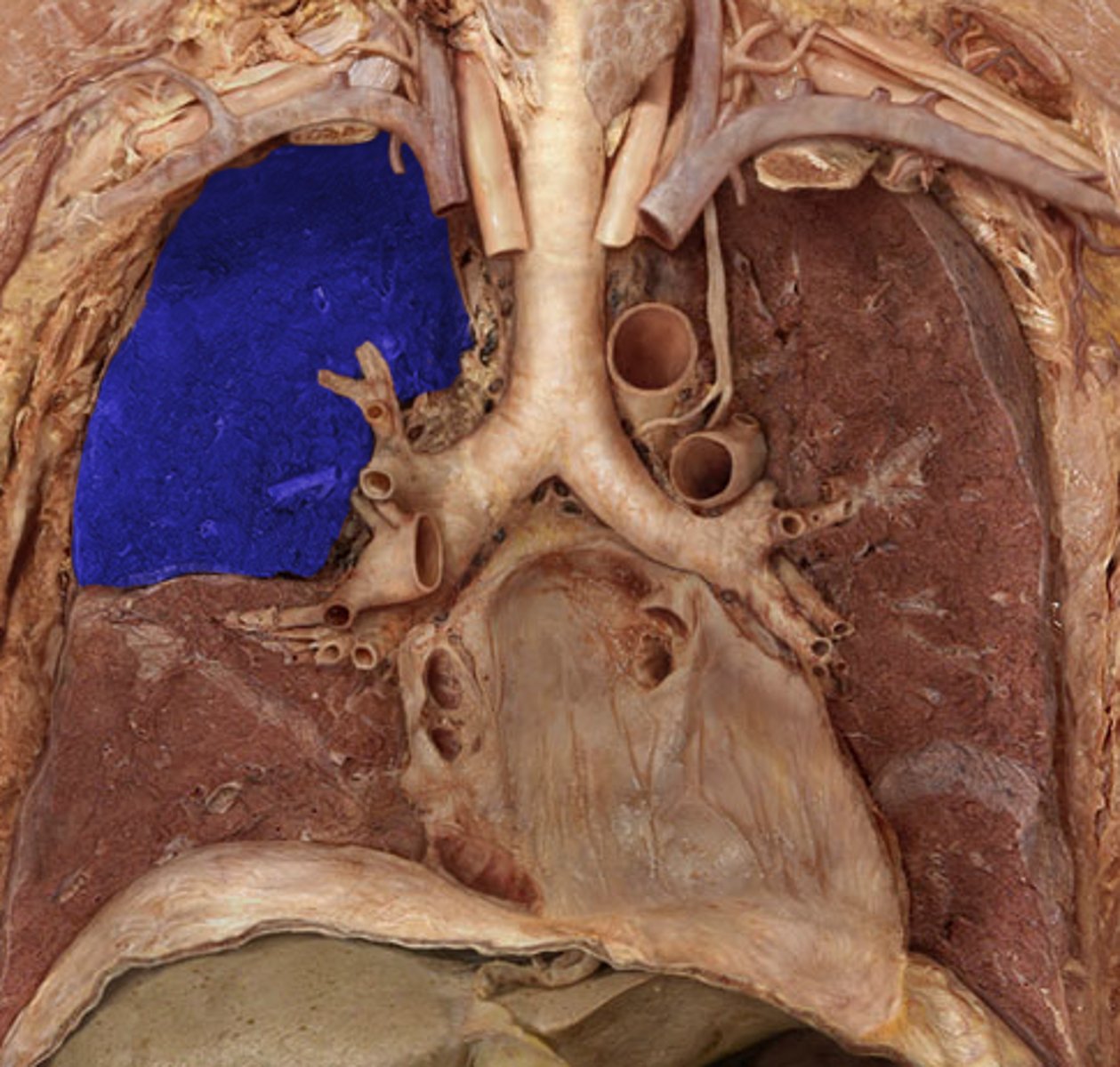
Costomediastinal Recess
Label Space A
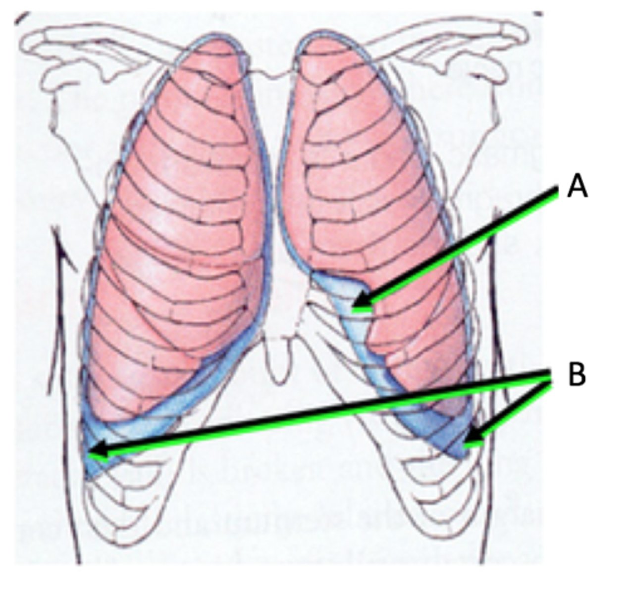
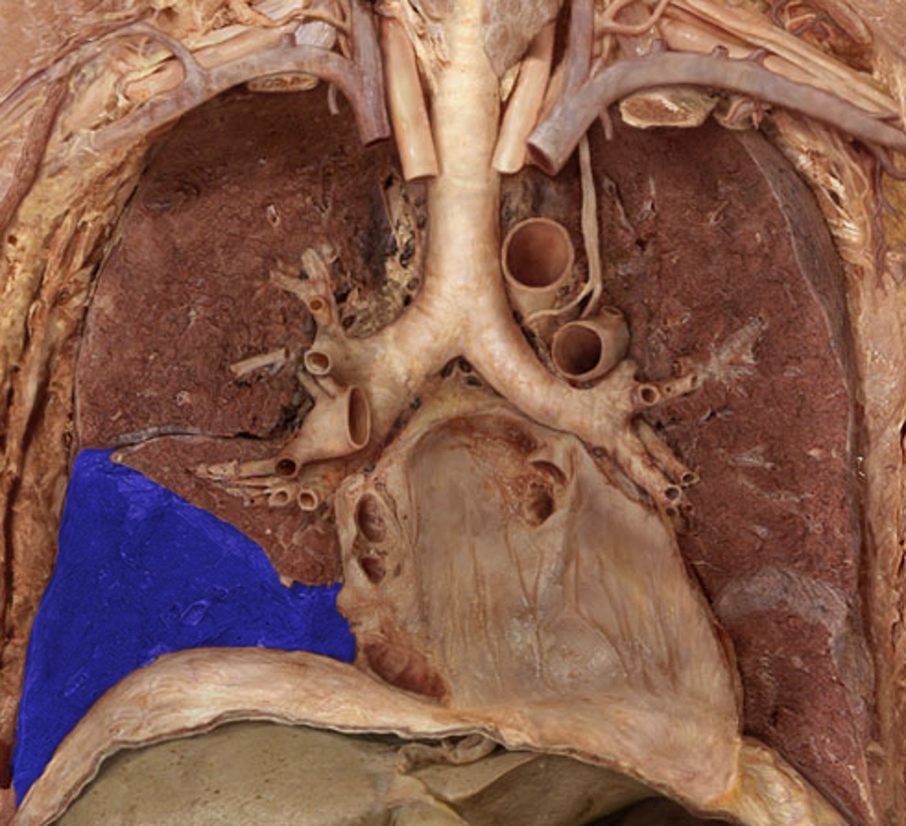
Costodiaphragmatic Recess
Label Space B

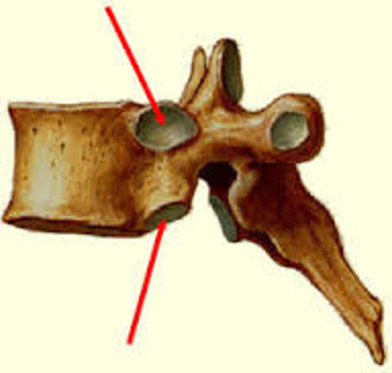
Costal Demifacet
Note: superior and inferior components, articulates with same # rib's head, specific to thoracic vertebrae
Label the Boney Structure
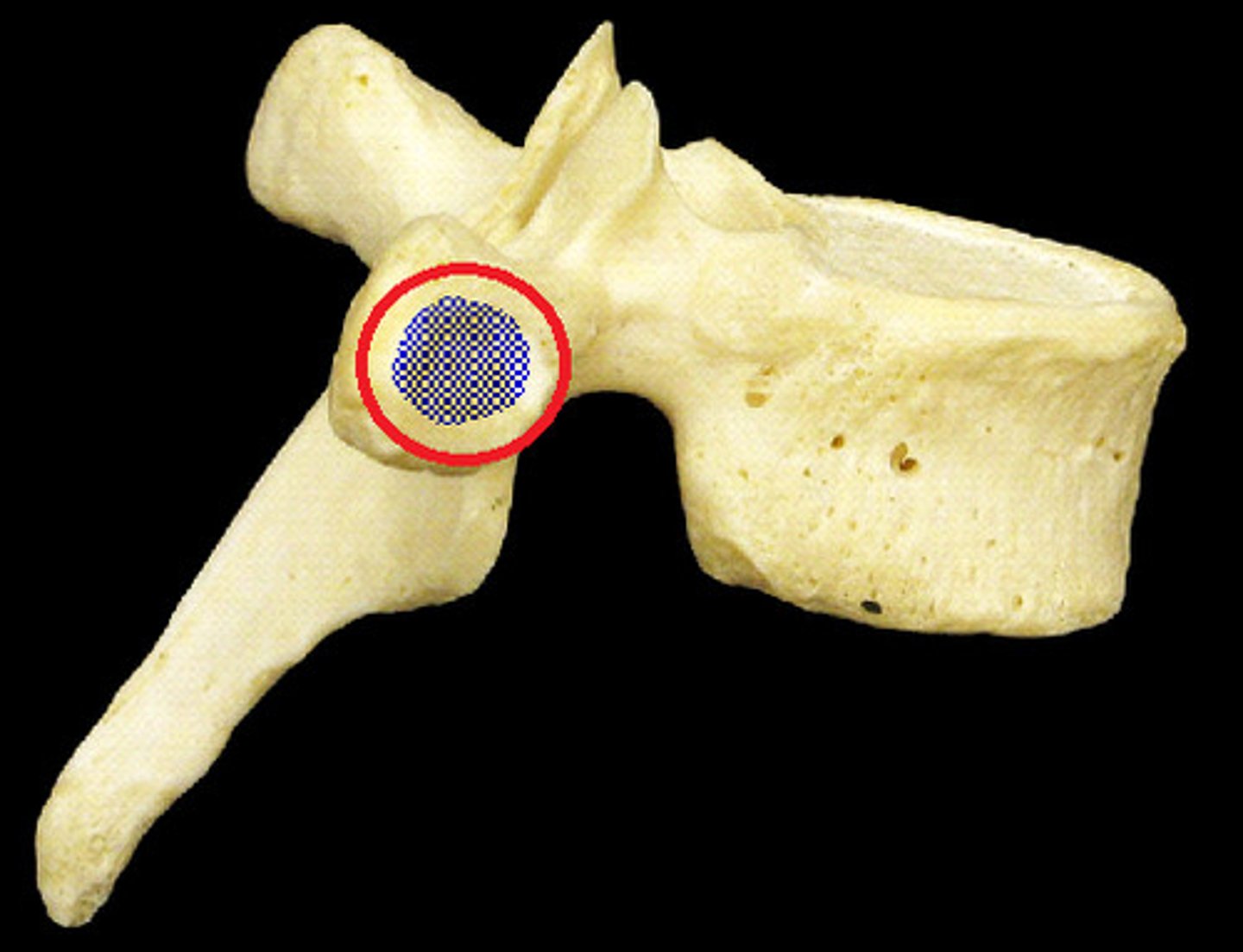
Transverse Costal Facet
Note: articulates with same # rib's tubercle, specific to thoracic vertebrae
Name the Boney Structure
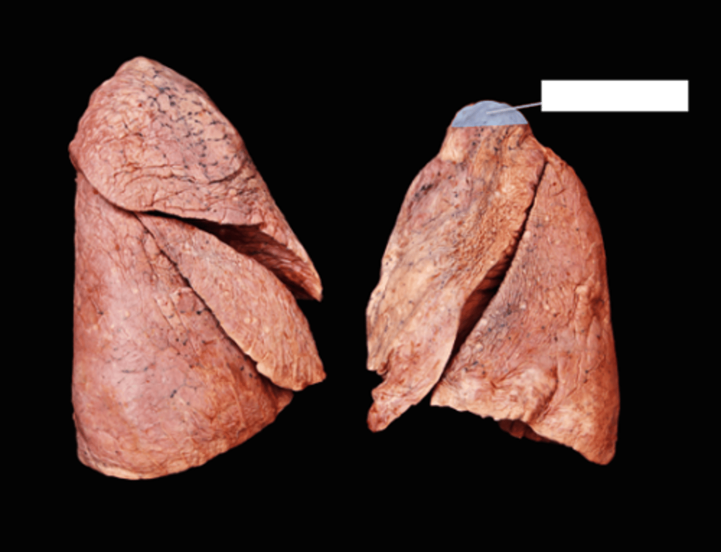
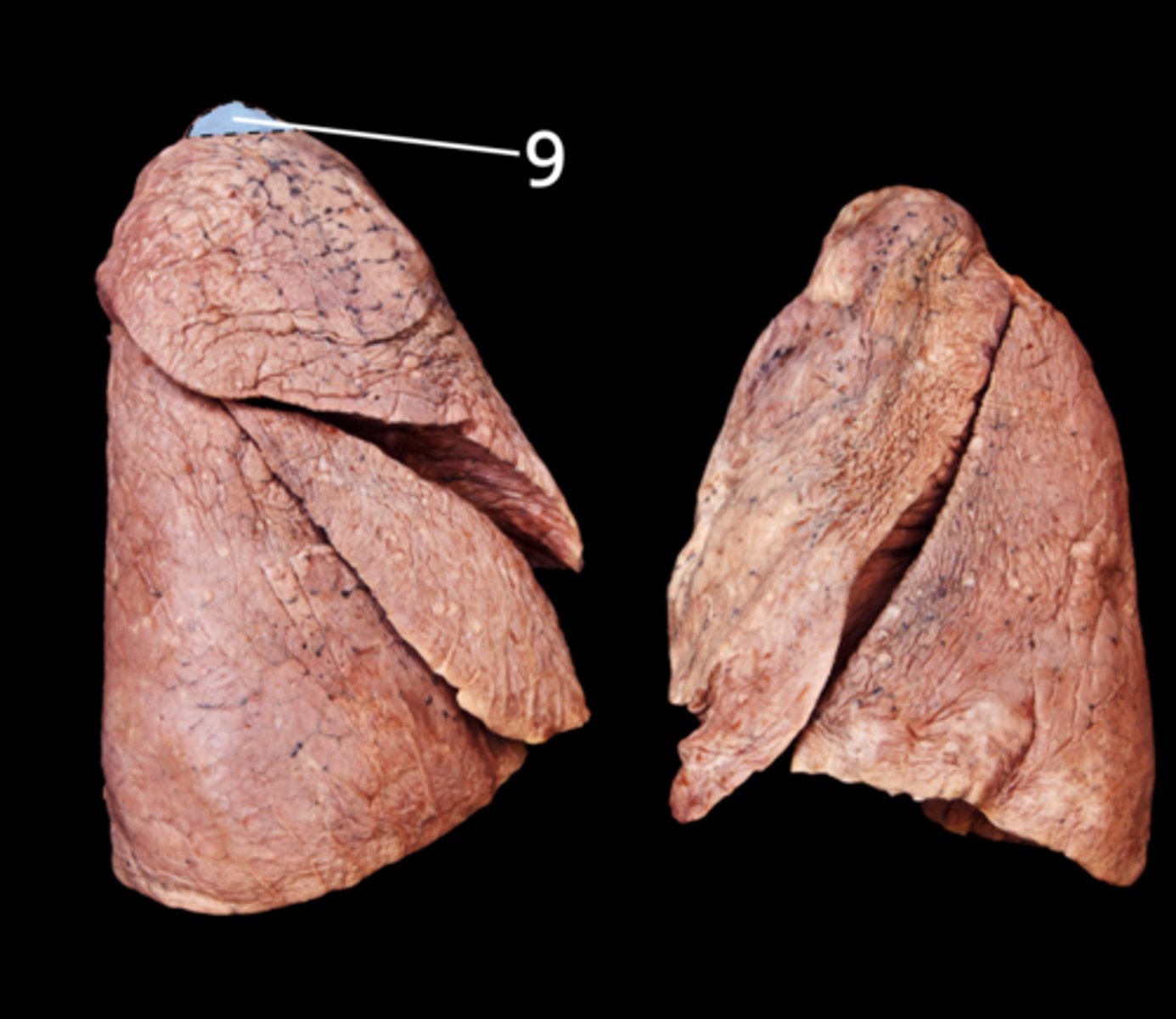
Left Apex
Label the Lung Segment
Left Superior Lobe
Label the Lung Segment
Left Inferior Lobe
Label the Lung Segment
Left Oblique Fissure
Note: separates superior and inferior lobes
Label the Lung Segment
Lingula
Label the Lung Segment
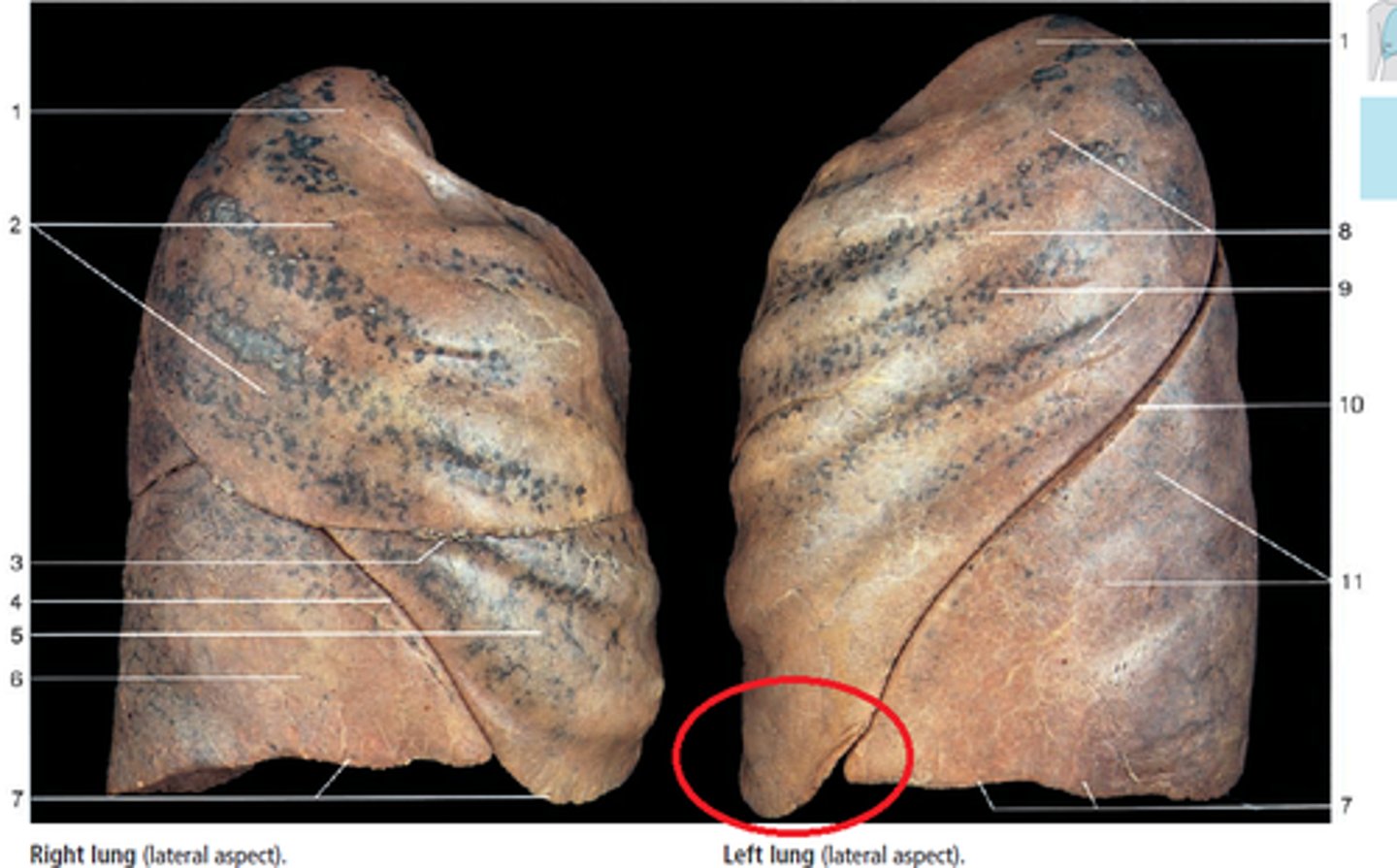
Cardiac Notch
Label the Notch
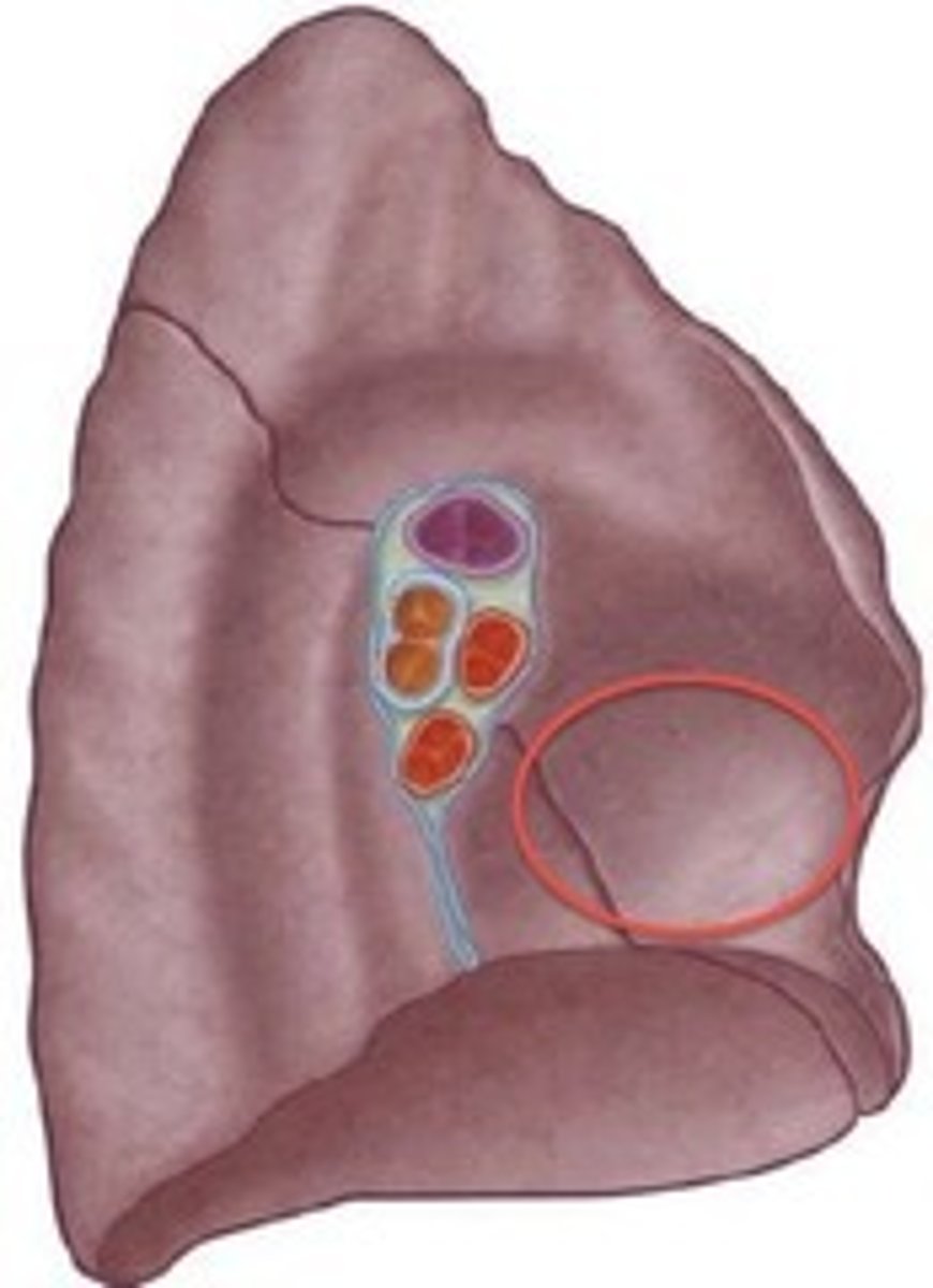
Cardiac Impression
Label the Space
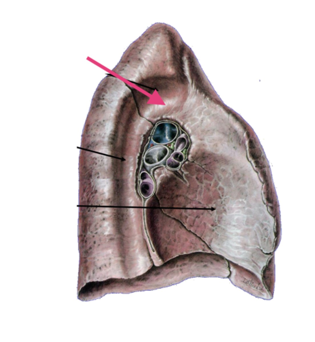
Groove for Arch of Aorta
Note: superior to left lung root
Label the Space
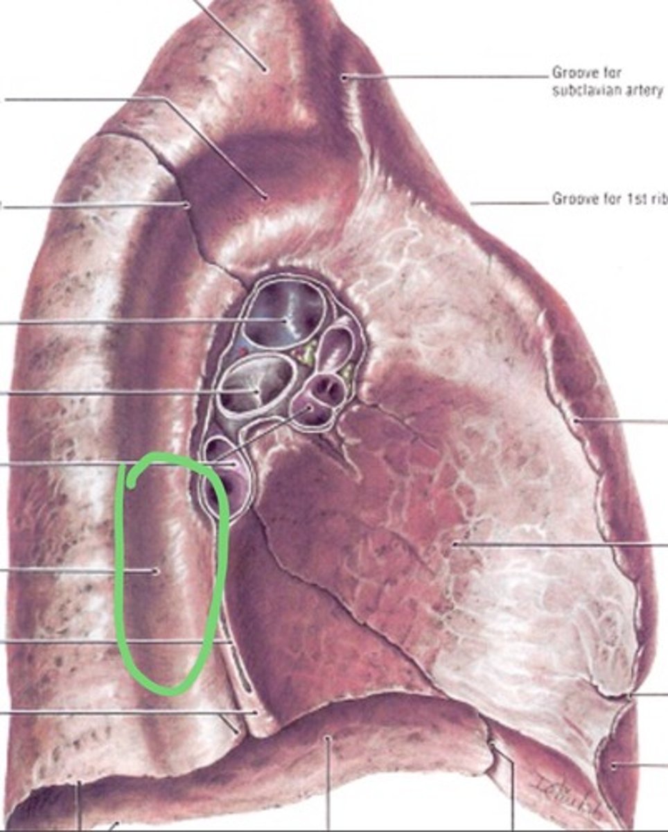
Groove for Thoracic Aorta
Note: posterior to left lung root
Label the Space
Left Diaphragmatic Surface
Label the Surface
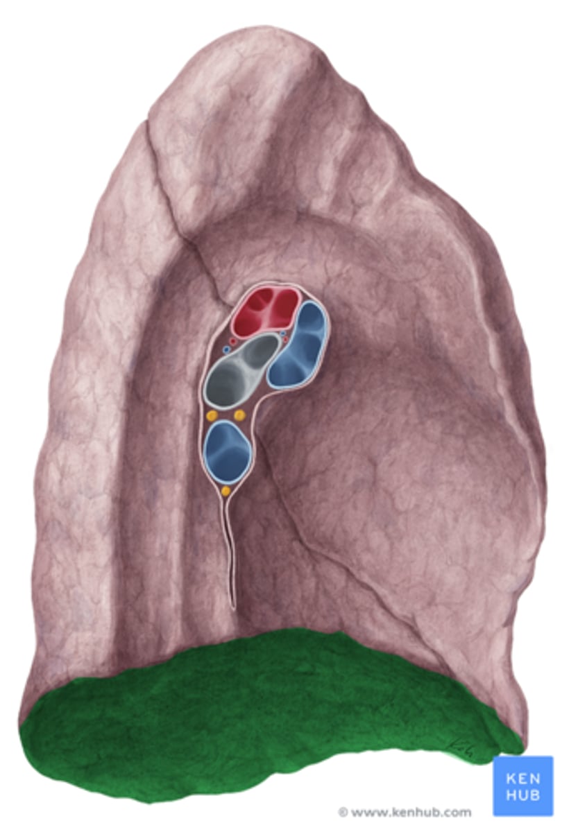
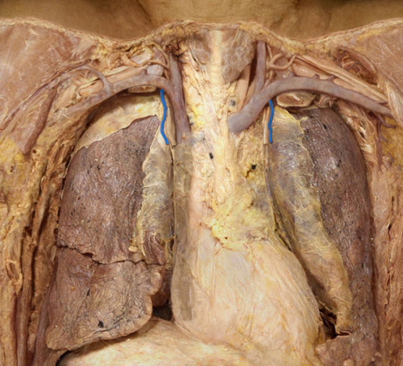
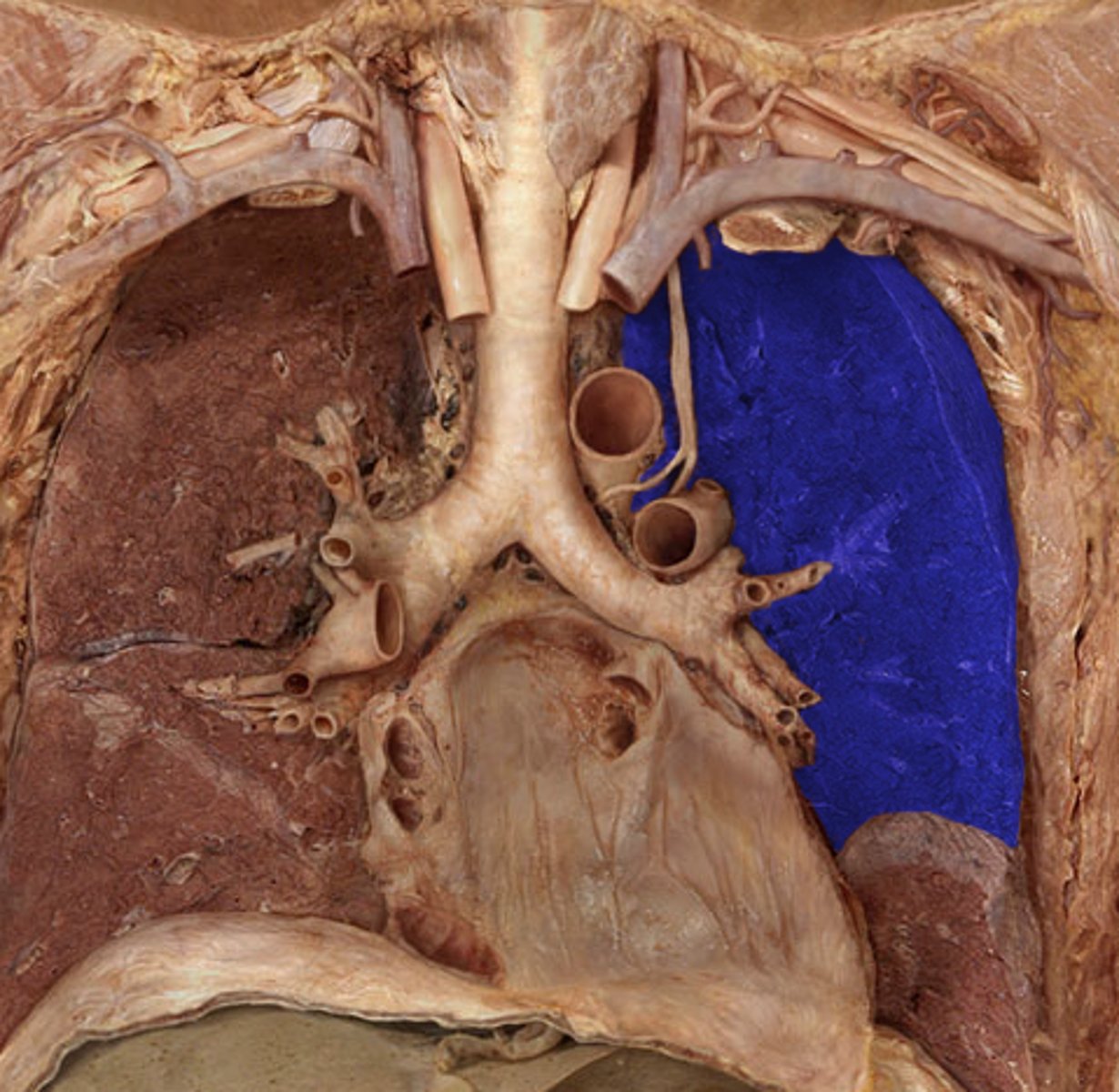
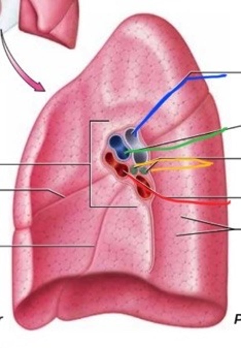
Left Main Bronchus (Primary)
Label the Segment
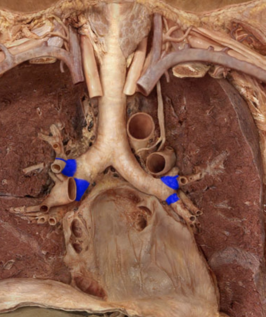
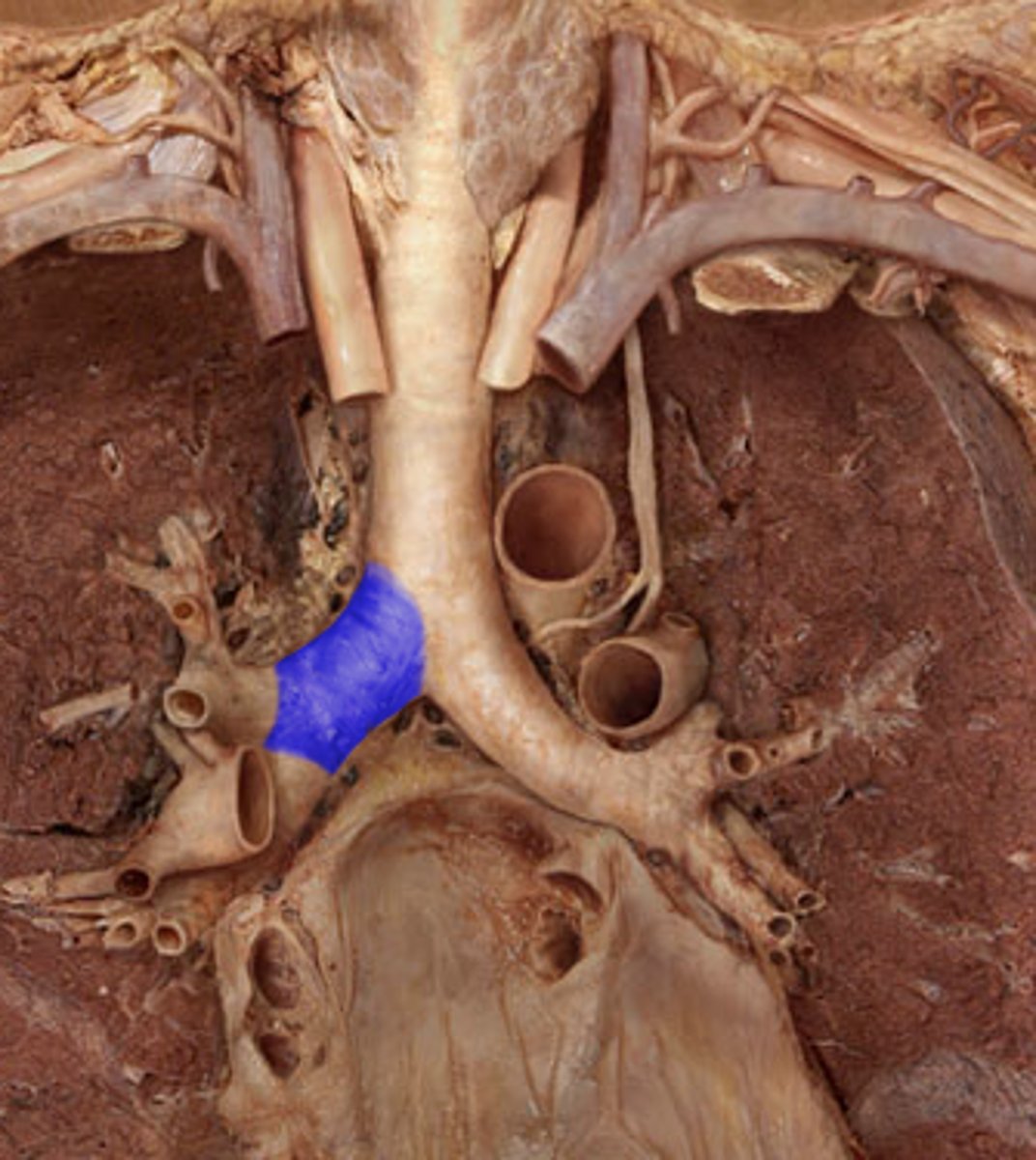
Left Lobar Bonchi (Secondary)
Note: has 2 bronchi...superior & inferior
Label the Blue Segment on the Left Side of Chest
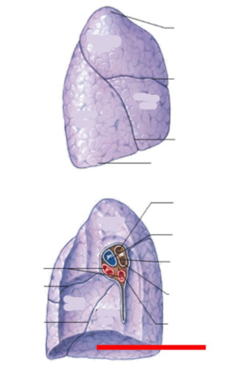
Left Pulmonary Artery
Note: usually superior to pulmonary veins in lung root
Label the Artery
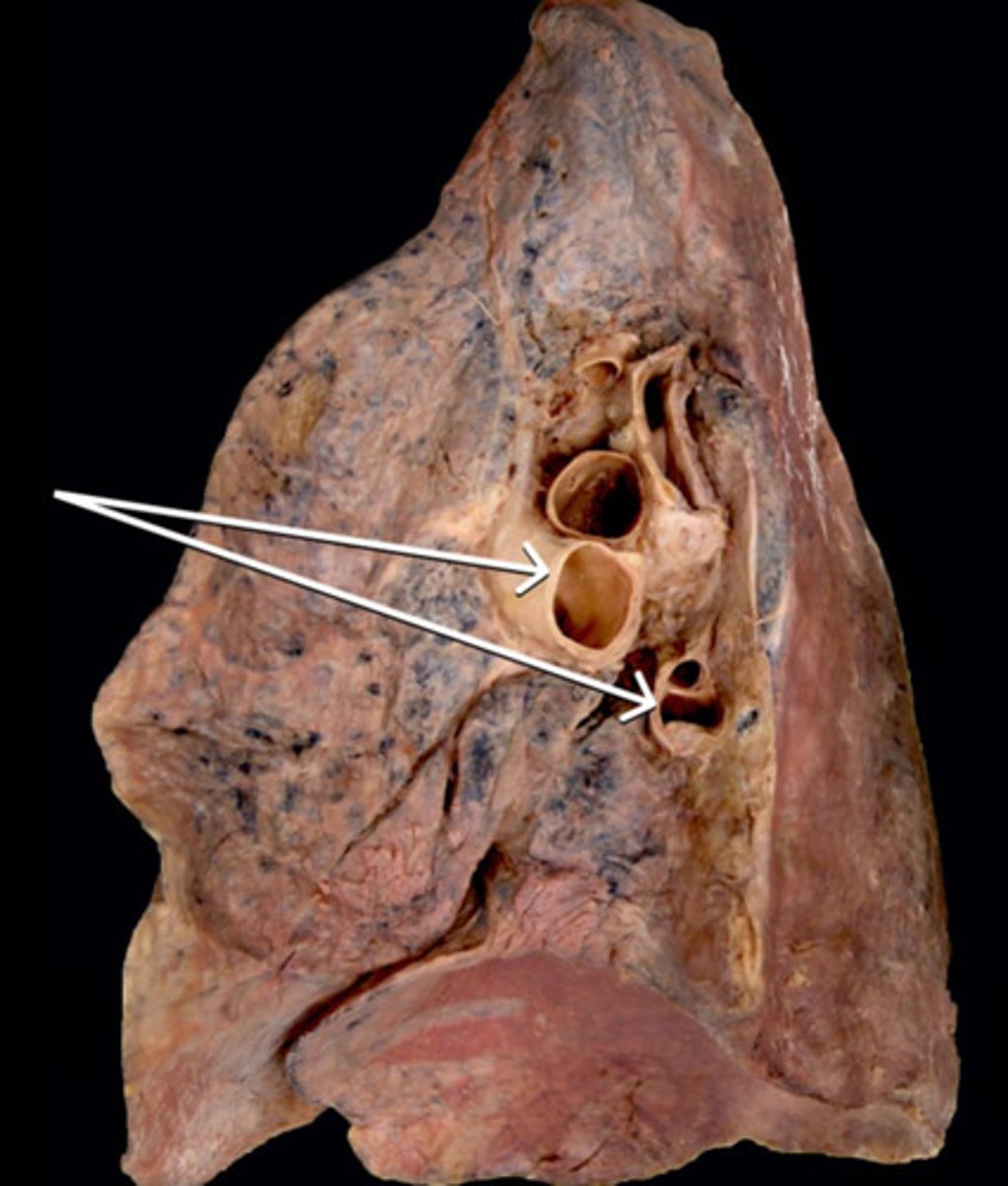
Left Pulmonary Veins
Note: contains superior & inferior veins
Label the Veins (Yellow Arrows)
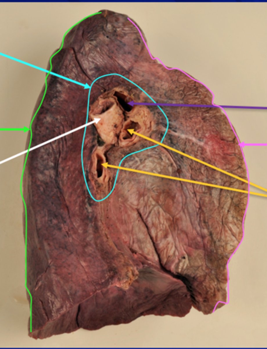
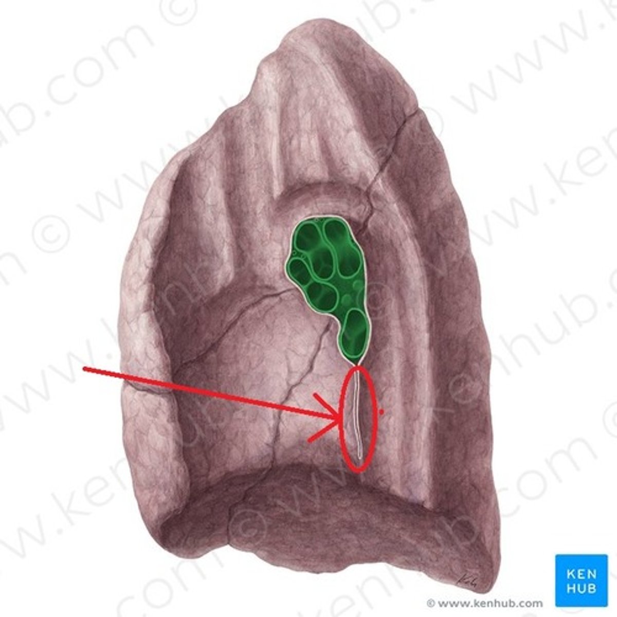
Left Hilar Lymph Nodes
Name This Structure
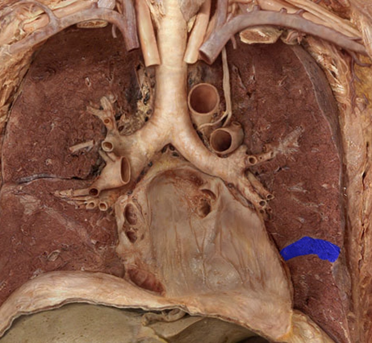
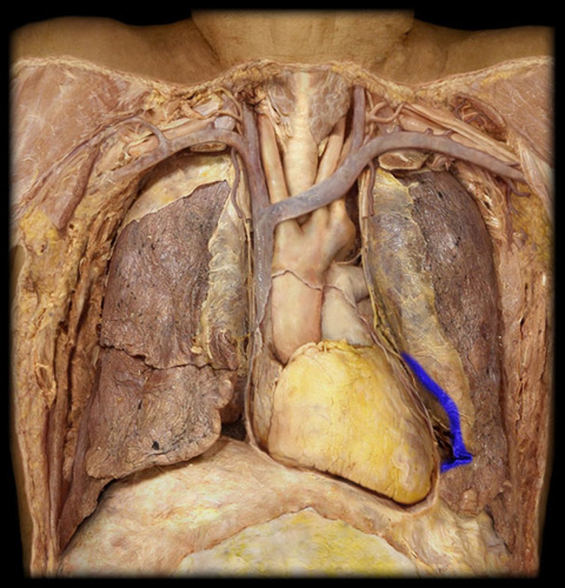
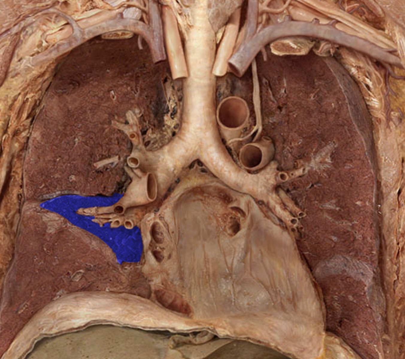
Left Pulmonary Ligament
Name The Green Arrow
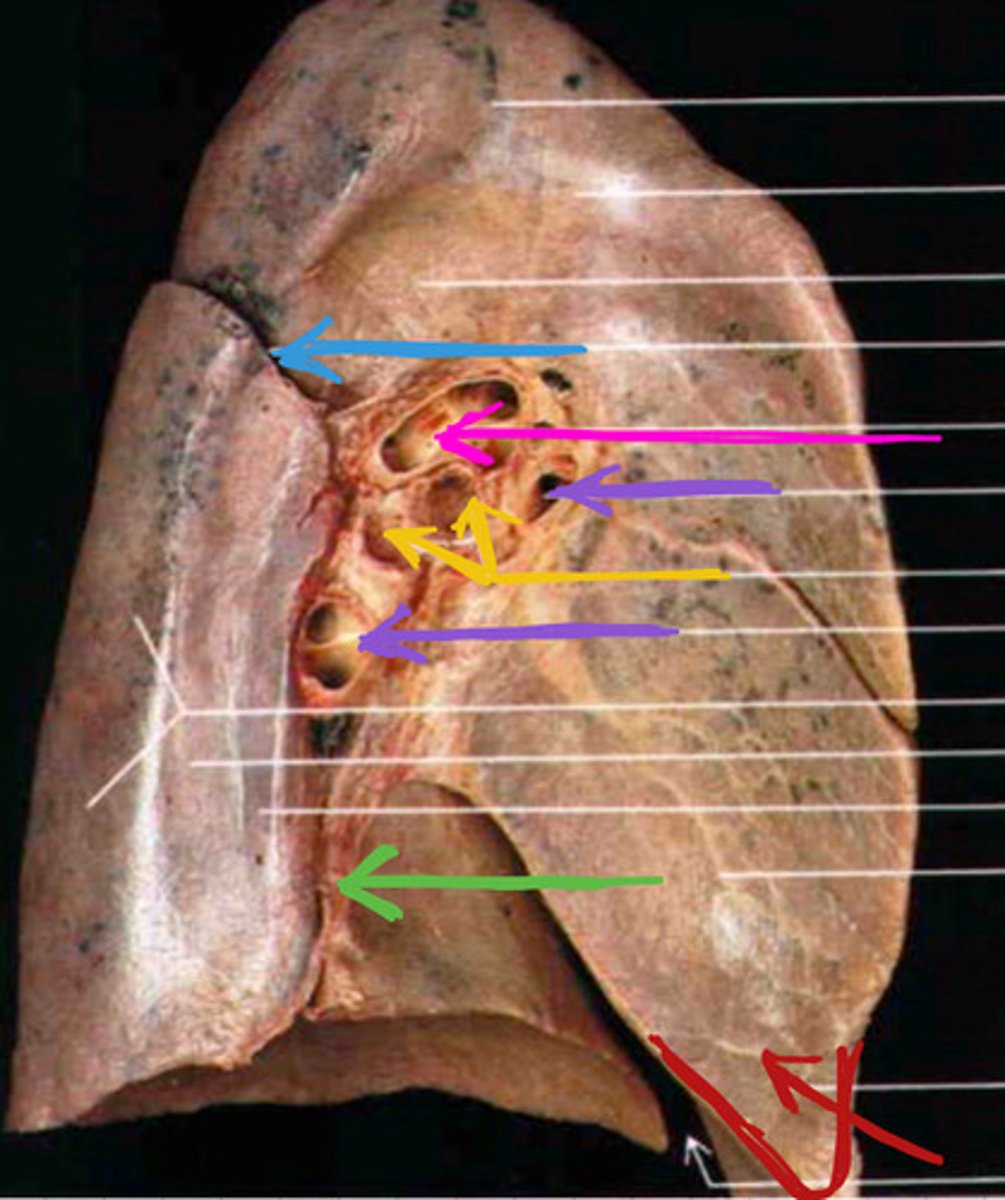
Right Apex
Label the Lung Segment
Right Superior Lobe
Label the Lung Segment
Right Middle Lobe
Label the Lung Segment
Right Inferior Lobe
Label the Lung Segment
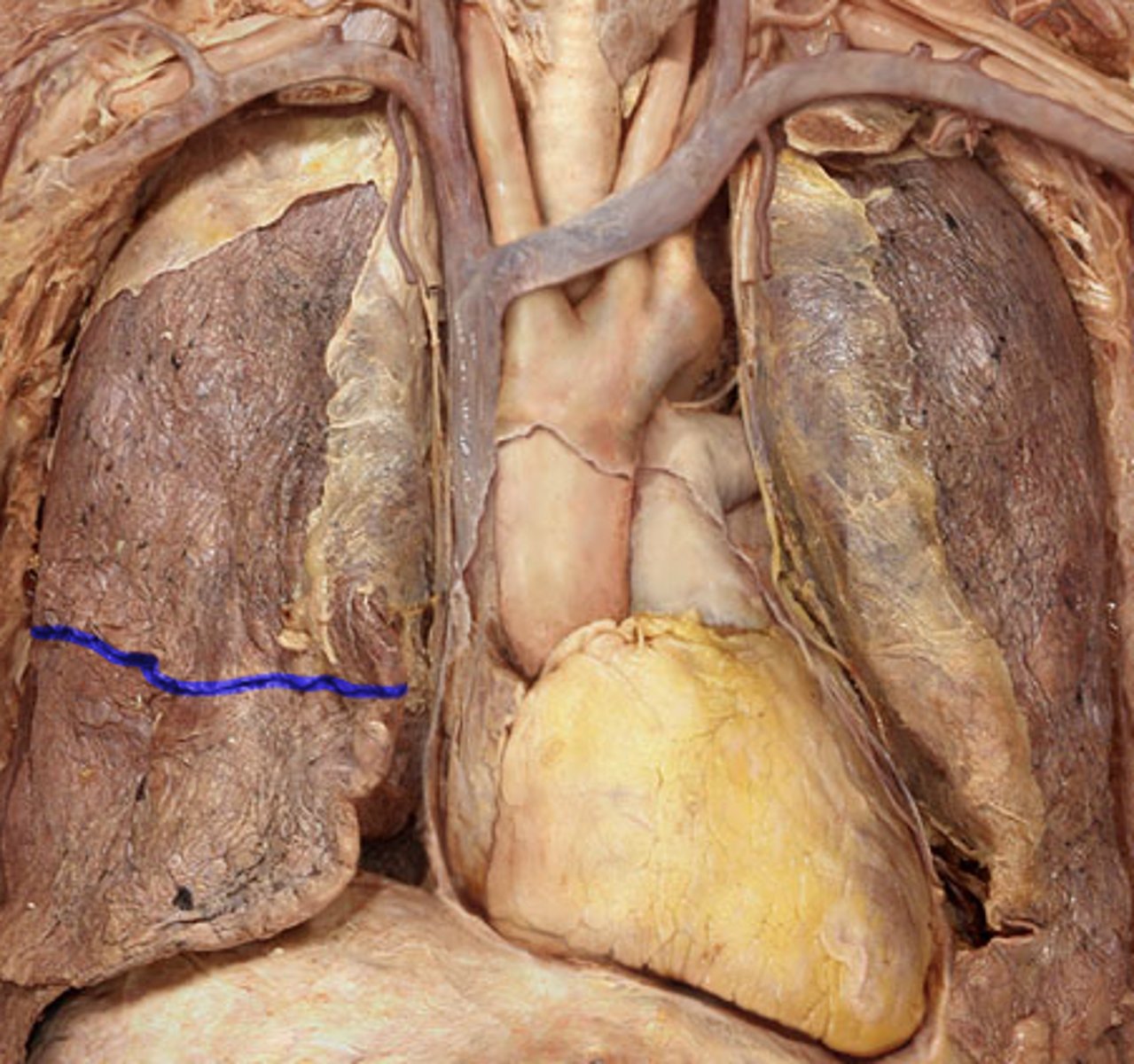
Right Oblique Fissure
Note: separates inferior lobe from other 2 lobes
Label the Lung Segment
Right Horizontal Fissure
Note: separates superior from middle lobe
Label the Lung Segment
Right Lung Diaphragmatic Surface
Label the Surface
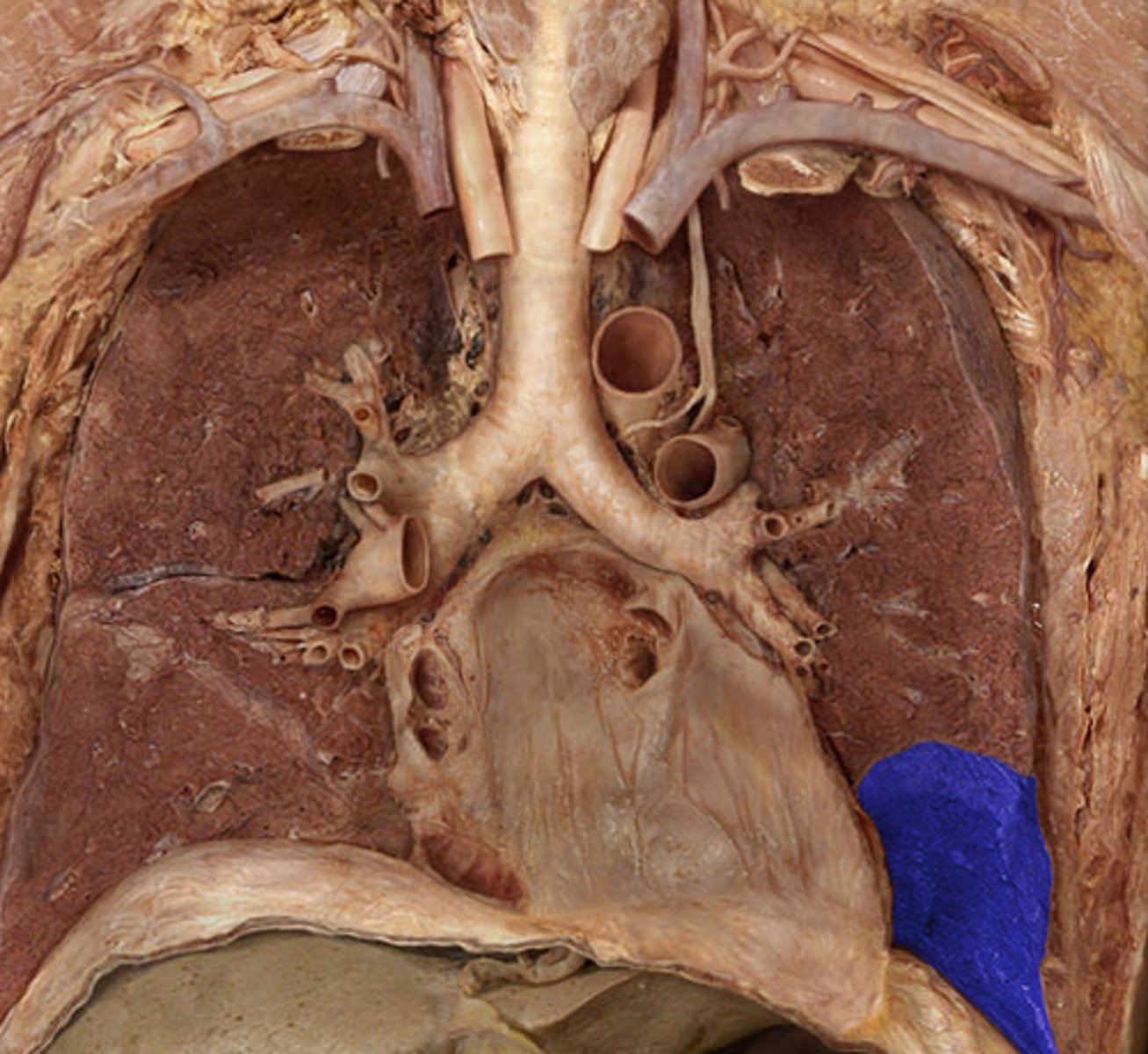
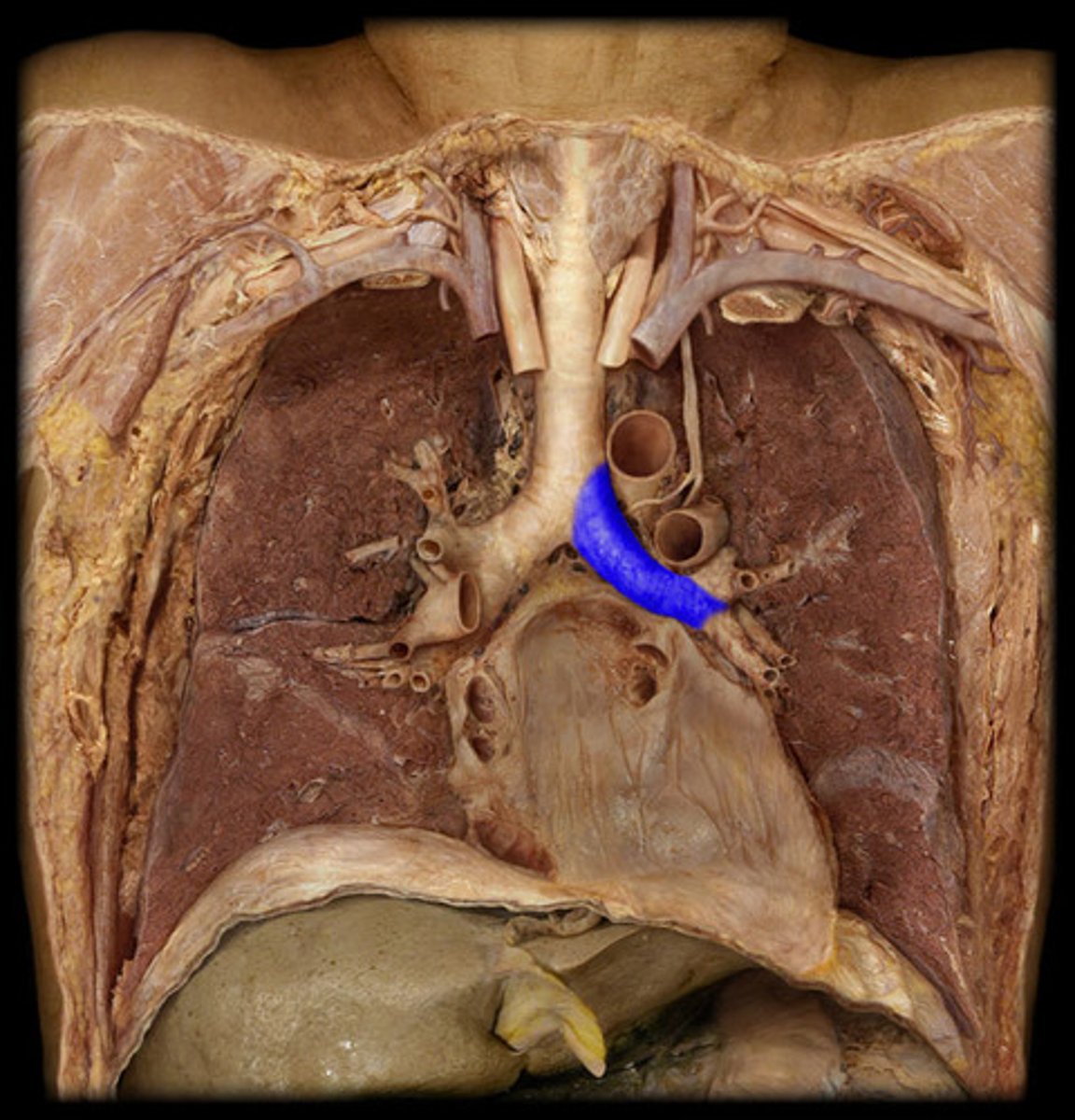
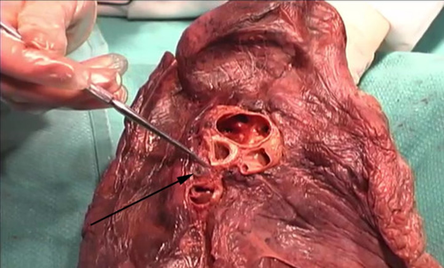
Right Main Bronchus (Primary)
Label the Segment
Right Lobar Bronchi (Secondary)
Note: has 3 bronchi...superior, middle, & inferior
Label the Blue Segment on the Right Side of Chest

Right Pulmonary Artery
Note: usually superior to pulmonary veins in lung root
Name the Artery
Right Pulmonary Veins
Note: contains superior & inferior veins
Name the Veins
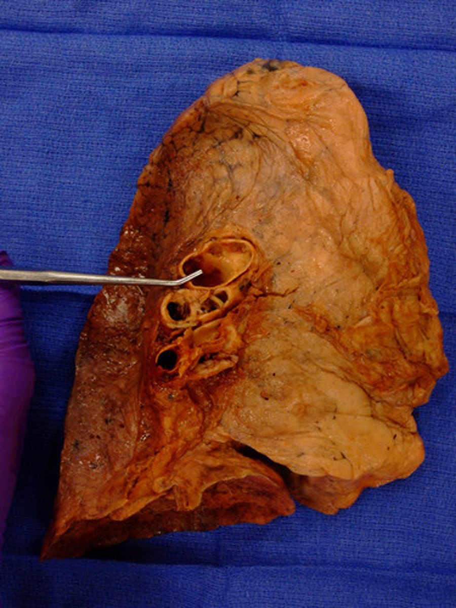
Right Hilar Lymph Nodes
Name This Structure (Yellow Arrow)
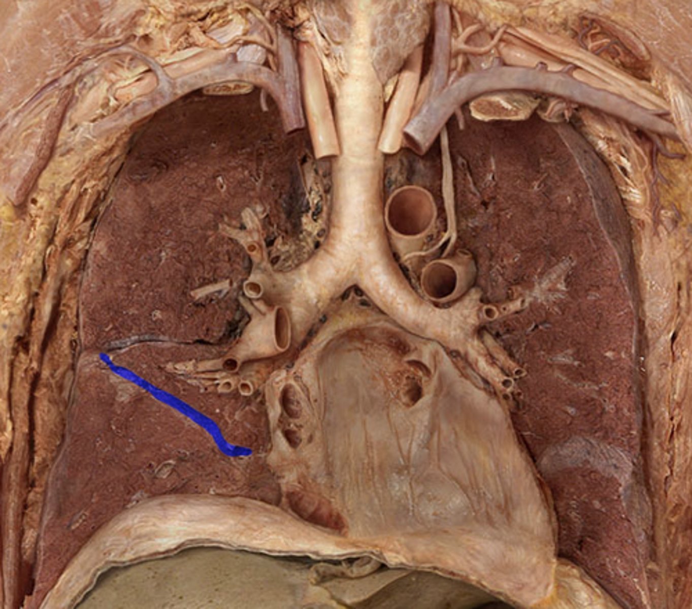
Right Pulmonary Ligament
Name The Arrow
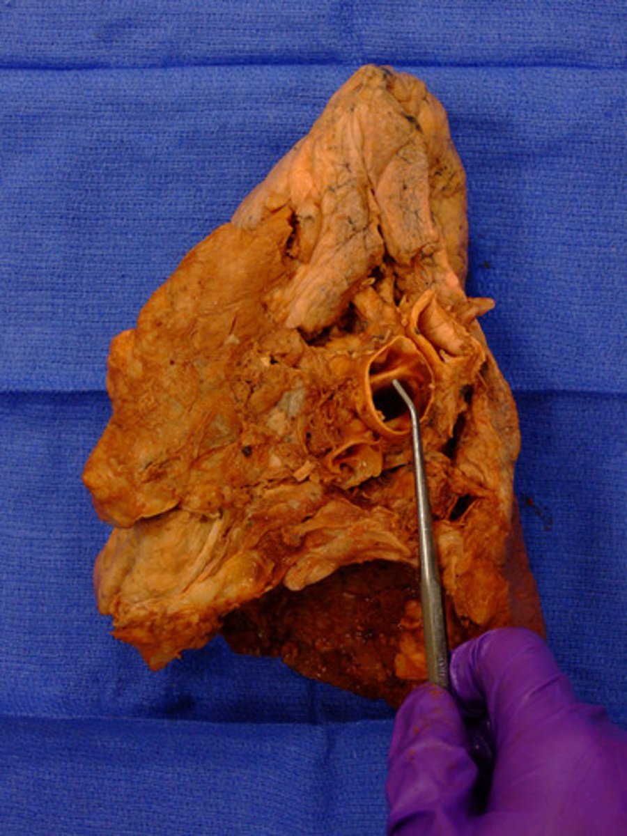
Fibrous Pericardium
Name the Layer