Dysphagia Overview & CSE
 Call Kai
 Learn
Learn Practice Test
 Spaced Repetition
 Match
Match Flashcards
 Knowt Play
1/238

There's no tags or description
Looks like no tags are added yet.
Name | Mastery | Learn | Test | Matching | Spaced | Call with Kai | Chat |
|---|
No analytics yet
Send a link to your students to track their progress
239 Terms
What is dysphagia?
A swallowing disorder involving the oral cavity, pharynx, esophagus, or gastroesophageal junction.
What is deglutition?
The entire act of moving food from the mouth until it enters the stomach.
What is feeding?
Placement and manipulation of food in the mouth before the swallow, including oral transit.
What is mastication?
Chewing; it is a voluntary act.
What is a bolus?
A formed mass of food or liquid ready to be swallowed.
What is penetration?
When material enters the laryngeal vestibule but does not pass through the vocal folds.
What is aspiration?
When material passes through the vocal folds into the lower airway.
What is residue?
Material remaining in the oral cavity, pharynx, or esophagus after a swallow.
What are the four phases of swallowing?
Oral preparatory, oral transit/transport, pharyngeal, and esophageal.
Which phases are voluntary and which are reflexive?
The oral preparatory and oral transit phases are primarily voluntary. The pharyngeal and esophageal phases are primarily involuntary/reflexive.
What are the two major divisions of the skull?
The cranium and the facial skeleton.
What is the function of the cranium?
It houses and protects the brain.
What is the function of the facial skeleton?
It forms the framework for mastication, speech, respiration, special senses, and facial expression.
What are the six cranial bones?
Frontal, parietal, temporal, occipital, sphenoid, and ethmoid.
What are the facial bones?
Mandible, maxilla, palatine, vomer, zygomatic, nasal, and inferior nasal conchae.
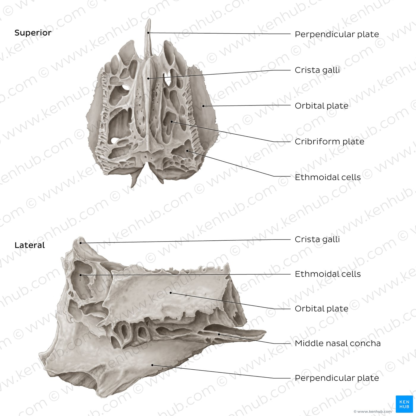
What are important landmarks of the ethmoid bone?
Cribriform plate, perpendicular plate, crista galli, and middle nasal conchae.
What are important features of the frontal bone?
It forms the anterior part of the brain case; its vertical plate contributes to the forehead and its horizontal plate contributes to the roof of the orbit and nasal cavities.
What do the parietal bones form?
Most of the rounded roof of the cranium.
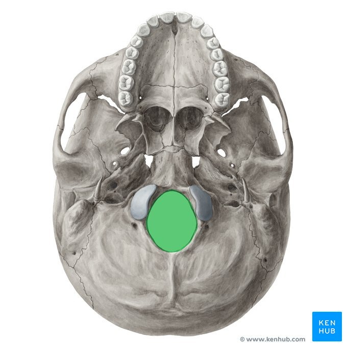
What is an important landmark of the occipital bone?
The foramen magnum. The occipital condyles articulate with the first cervical vertebra. The Foramen magnum is the largest opening at the base of the skull that connects the brain inside the head to the spinal cord in the neck
What is important about the temporal bone?
It forms much of the lateral base and sides of the brain case. Its petrous portion houses the organs of hearing and equilibrium, and it contains the mastoid and styloid processes.
What is the sphenoid bone?
A bone at the base of the skull located behind the ethmoid and in front of the foramen magnum. Important landmarks include the body, greater wings, lesser wings, and medial and lateral pterygoid plates.

What are important landmarks of the mandible?
Mental symphysis, body, ramus, coronoid process, condyle/condyloid process, and mental spines.

What is the maxilla?
The paired bone forming the upper jaw. It contributes to the roof of the mouth, nasal cavity, and floor of the orbit.
What are the four processes of the maxilla?
Palatine, zygomatic, frontal, and alveolar processes.

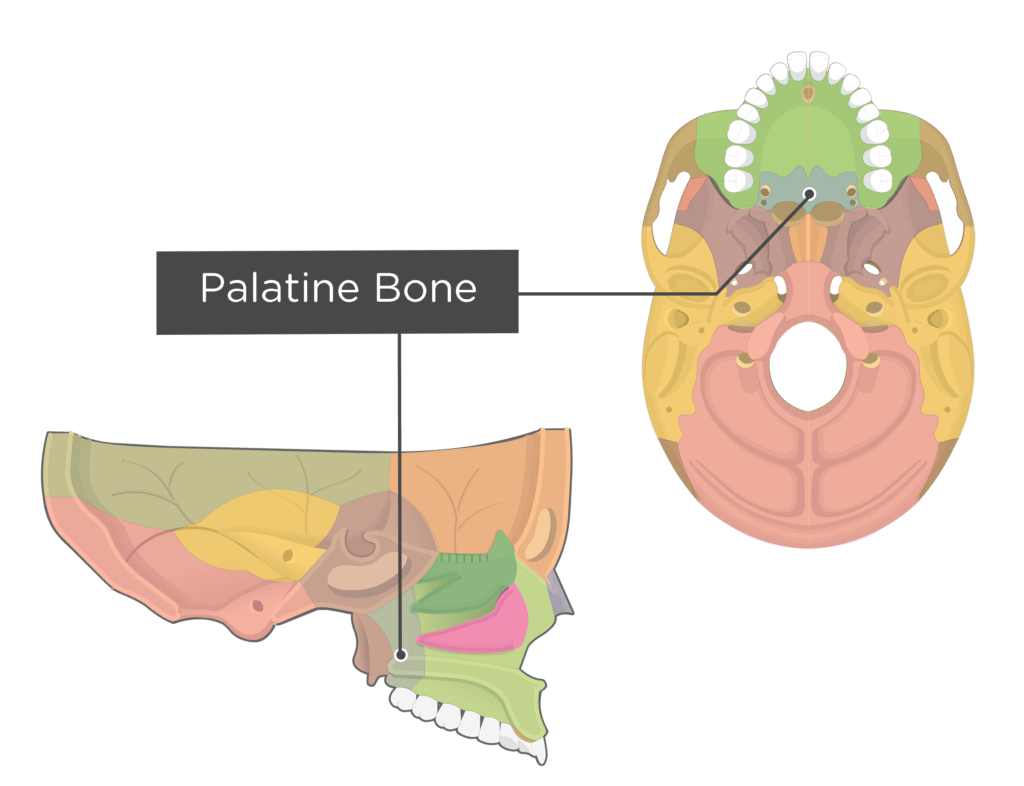
What do the palatine bones contribute to?
The floor and lateral wall of the nasal cavity, the posterior one-fourth of the hard palate, and the floor of the orbit.
What does the vomer form?
The inferior half of the bony nasal septum

What are the two major portions of the tongue?
The oral portion and the pharyngeal portion.
What does the oral portion of the tongue include?
The tip, blade, and dorsum.
Where does the pharyngeal portion of the tongue begin?
At the circumvallate papillae.

Where does the tongue base begin?
At the hyoid bone.
What are the three extrinsic tongue muscles?
Genioglossus, hyoglossus, and styloglossus
What does the genioglossus do?
Protrudes and depresses the tongue. It also helps prevent posterior displacement of the tongue.
What does the hyoglossus do?
Depresses and retracts the tongue.
What does the styloglossus do?
Retracts and elevates the tongue.
What does the palatoglossus do?
Elevates the tongue base and approximates it to the soft palate.
Which cranial nerve innervates the tongue muscles?
Cranial nerve XII, the hypoglossal nerve, innervates all intrinsic and extrinsic tongue muscles except the palatoglossus.
What nerve innervates the palatoglossus?
Cranial nerve X, the vagus nerve.
What are the four major muscles of mastication?
Masseter, temporalis, medial pterygoid, and lateral pterygoid.
What does the masseter do?
Elevates and protracts the mandible.
What does the temporalis do?
Elevates and retracts the mandible
What does bilateral contraction of the medial pterygoid do?
Elevates, closes, and protrudes the mandible
What does unilateral contraction of the medial pterygoid do?
Swings the mandible to the opposite side.
What does bilateral contraction of the lateral pterygoid do?
Protrudes and depresses the mandible.
What does unilateral contraction of the lateral pterygoid do?
Moves the mandible medially.
What cranial nerve innervates the muscles of mastication?
Cranial nerve V, the trigeminal nerve.
What does orbicularis oris do?
Closes the mouth, purses the lips, and rounds the lips.
What does levator labii superioris do?
Lifts and everts the upper lip.
What do the zygomaticus major and minor do?
Pull the corners of the mouth upward and backward for smiling.
What does risorius do?
Pulls the corners of the mouth laterally.
What does depressor labii inferioris do?
Pulls the lower lip downward and outward.
What does depressor anguli oris do?
Pulls the corners of the mouth downward for frowning.
What does mentalis do?
Wrinkles the chin and pushes the lower lip upward or outward.
What does buccinator do?
Compresses the cheek against the teeth to help with chewing and prevents food from collecting in the cheek.
What cranial nerve innervates the facial muscles involved in lip and cheek movement?
Cranial nerve VII, the facial nerve.
What are the four major suprahyoid muscles?
Digastric, mylohyoid, geniohyoid, and stylohyoid.
What does the anterior belly of the digastric do?
Elevates and stabilizes the hyoid during swallowing. It also helps depress the mandible for jaw opening.
What does the posterior belly of the digastric do?
Elevates and retracts the hyoid bone.
What do the two bellies of the digastric do together?
Lift the tongue base and hyoid and help depress the mandible for jaw opening, chewing, and speech.
What does the mylohyoid do during swallowing?
Elevates the base of the tongue and moves the hyoid anterosuperiorly. It can also depress the mandible and elevate the floor of the oral cavity when the hyoid is fixed.
What does the geniohyoid do?
Pulls the hyoid up and forward during swallowing, helping bolus flow and pharyngeal clearing.
What does the stylohyoid do?
Elevates and retracts the hyoid.
What nerve innervates the anterior belly of the digastric?
Cranial nerve V, the trigeminal nerve.
What nerve innervates the posterior belly of the digastric?
Cranial nerve VII, the facial nerve.
What nerve innervates the mylohyoid?
Cranial nerve V, the trigeminal nerve.
What nerve innervates the stylohyoid?
Cranial nerve VII, the facial nerve.
What nerve innervates the geniohyoid?
Cranial nerve XII, the hypoglossal nerve
What are the important soft palate/pharyngeal muscles?
Tensor veli palatini, levator veli palatini, palatopharyngeus, salpingopharyngeus, and stylopharyngeus.
What does tensor veli palatini do?
Tenses the soft palate and opens the auditory tube.
What does levator veli palatini do?
Elevates the soft palate.
What does palatopharyngeus do?
Elevates and shortens the pharynx, pulls the pharyngeal walls superiorly, anteriorly, and medially during swallowing, and helps close the nasopharynx from the oropharynx.
What does salpingopharyngeus do?
Raises the pharynx and larynx during swallowing and draws the pharyngeal walls upward laterally.
What does stylopharyngeus do?
Elevates the pharynx and larynx.
Which three muscles elevate the soft palate and tongue base during swallowing?
Levator veli palatini, palatoglossus, and styloglossus.
What nerve innervates tensor veli palatini?
Cranial nerve V, the trigeminal nerve.
What nerve innervates levator veli palatini?
Cranial nerve X, the vagus nerve, through its pharyngeal branches.
What nerve innervates palatopharyngeus?
Cranial nerve X, the vagus nerve, through its pharyngeal branches.
What nerve innervates salpingopharyngeus?
Cranial nerve X, the vagus nerve, through its pharyngeal branches.
What nerve innervates stylopharyngeus?
Cranial nerve IX, the glossopharyngeal nerve.
What are the three pharyngeal constrictors?
Superior, middle, and inferior pharyngeal constrictors
What is the function of the pharyngeal constrictors during swallowing?
They contract behind the bolus and generate positive pressure to propel the bolus through the pharynx.
What nerve innervates the pharyngeal constrictors?
Cranial nerve X, the vagus nerve, through the pharyngeal plexus.
What are the muscles responsible for shortening and elevating the pharynx?
Stylopharyngeus, salpingopharyngeus, and palatopharyngeus.
What structure is primarily responsible for closing the airway during a swallow?
The larynx.
What structures make up the laryngeal skeletal framework?
The hyoid bone and the thyroid, cricoid, arytenoid, corniculate, cuneiform, and epiglottic cartilages.
Which laryngeal cartilages are paired?
Arytenoid, corniculate, and cuneiform cartilages.

What do the cuneiform and corniculate cartilages do?
They reinforce and support the aryepiglottic folds and may help the arytenoids move.
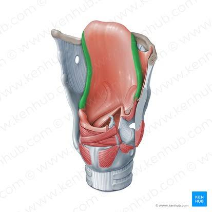
What is the aditus laryngis?
The superior opening of the laryngeal vestibule.
What forms the boundaries of the aditus laryngis?
The epiglottis anteriorly, aryepiglottic folds laterally, and the arytenoid apex posteriorly.
What is the pyriform sinus?
A deep depression located lateral to the larynx.
What is the laryngeal vestibule?
The space below the aditus laryngis and above the ventricular/false vocal folds.

What is the laryngeal ventricle?
The space just below the ventricular/false vocal folds.

What is the glottis?
The region involving the vocal folds and the space between them.
What are the five major intrinsic laryngeal muscles?
Thyroarytenoid, cricothyroid, posterior cricoarytenoid, lateral cricoarytenoid, and interarytenoids.
What does the thyroarytenoid do?
Shortens and adducts the vocal folds.
What does the cricothyroid do?
Lengthens and tenses the vocal folds and adducts them.
What happens when the pars recta of the cricothyroid contracts?
It reduces the distance between the cricoid arch and thyroid lamina, lengthening and tensing the vocal folds.
What does the pars obliqua of the cricothyroid do?
Slides the thyroid cartilage forward.
What does the posterior cricoarytenoid do?
Abducts the vocal folds. It is the muscle that opens the vocal folds.
What does the lateral cricoarytenoid do?
It is the primary adductor of the vocal folds
What do the interarytenoids do?
Adduct the posterior vocal folds.