forearm bones and muscles
 Call Kai
 Learn
Learn Practice Test
 Spaced Repetition
 Match
Match Flashcards
 Knowt Play
1/47

There's no tags or description
Looks like no tags are added yet.
Name | Mastery | Learn | Test | Matching | Spaced | Call with Kai | Chat |
|---|
No analytics yet
Send a link to your students to track their progress
48 Terms
head of radius
articulates with capitulum of distal end of humerus
strong annular ligament hodls head of radius in contact with radial notch of ulna
interosseous border
thin layer of tissue that holds radius and ulna together and separates forearm in anter/posterior compartments
sharp edge spanning distal ½ of radius
attachment site for interosseous membrane
ulnar notch
a shallow notch located on the medial surface of the distal end of the radius
articulates with the head of the ulna
lister’s tubercle
aka distal tubercle of the radius
“bump” located on the distal and dorsal end of radius
acts as a pulley for the tendon of extensor pollicis longus
articular facets of radius
distal end of radius has two facets:
scaphoid
lunate
forearm joints
superior and inferior radioulnar → both are pivot synovial
radiocarpal → condyloid synovial
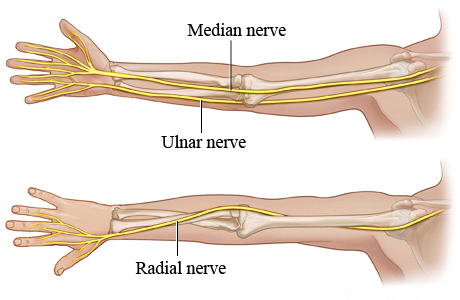
forearm nerve muscle innervations/where they run
ulnar - innervates 4th/5th digits
radial - innervates posterior
median - innervates 1-3 digits
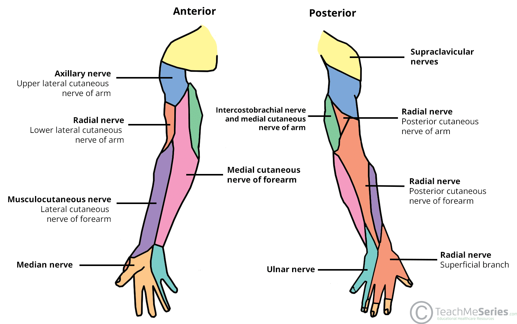
forearm nerve - sensory innervations
ulnar - innervates 4th/5th digits
radial - innervates posterior
median - innervates 1-3 digits
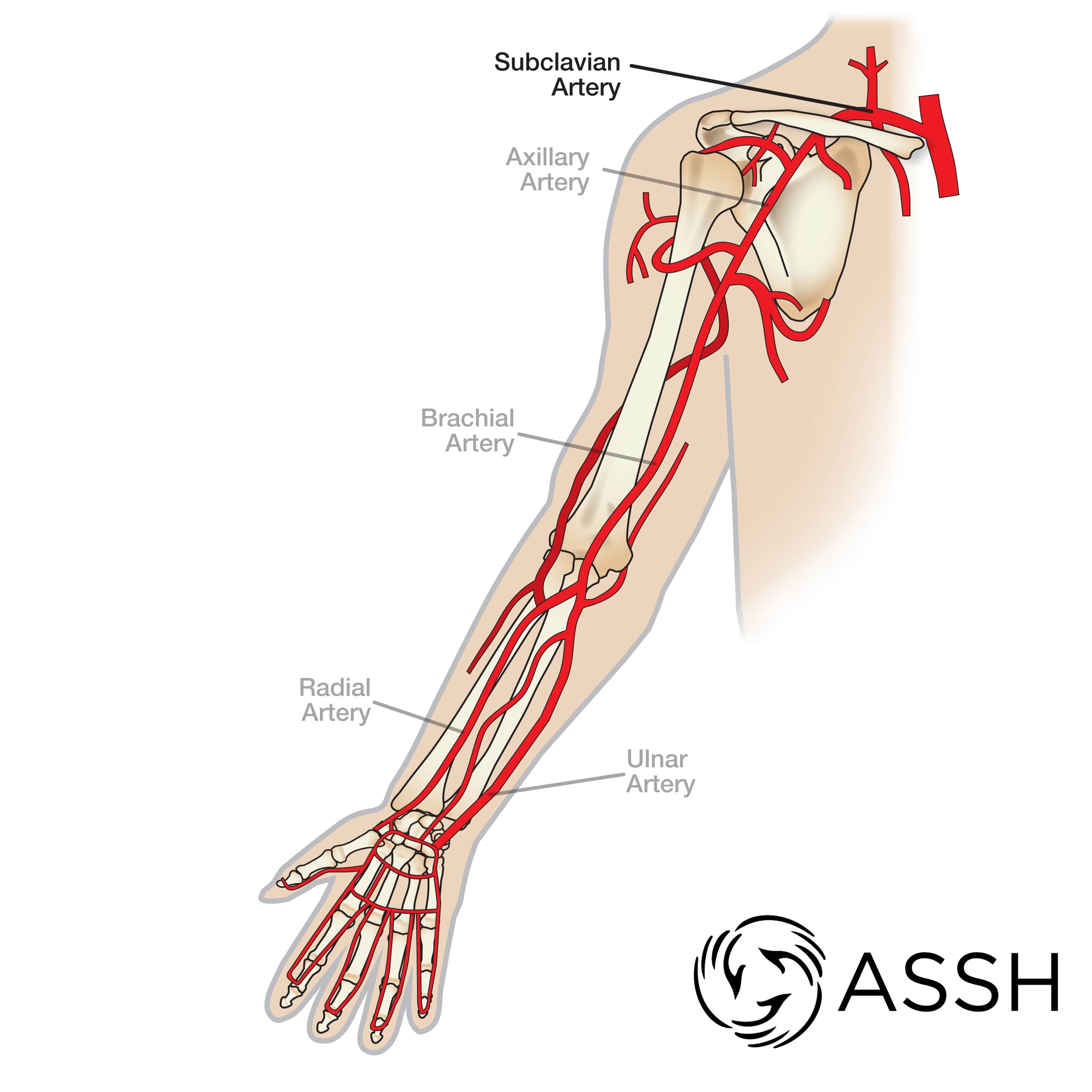
forearm blood supply
brachial artery
radial artery and vein
ulnar artery and vein
compartments of forearm & how
interosseous membrane divides forearm into posterior and anterior compartments
interosseous membrane
hold ulnar and radial bones together
has a free upper margin, distal to radial tuberosity and a small, circular opening located distally
allows for passage of vessels between anterior and posterior compartments
anterior compartment
flexor compartment
common features:
assist in movements of the wrist
flexion of digits
pronation
innervated by median or ulnar nerve
divided into three layers:
superficial (4)
intermediate (1)
deep (3)
anterior compartment: superficial layer muscles
pronator teres
flexor carpi radialis
palmaris longus
flexor carpi ulnaris
share common origin from medial epicondyle
extend distally into wrist/hand (expect pronator teres)
innervated by median nerve → expect flexor carpi ulnaris = ulnar nerve
pronator teres
O:
humeral head - medial epicondyle of humerus
ulnar head - coronoid process of ulna
I:
middle of convexity of lateral shaft of radius
N:
median nerve
A:
flexes and pronates the forearm at elbow
Flexor carpi radialis
O:
medial epicondyle
I:
base of 2nd metacarpal
N:
median nerve
A:
wrist flexion
radial deviation/abduction
palmaris longus
O:
medial epicondyle
I:
apex of palmar aponeurosis
distal ½ of flexor retinaculum
N:
median nerve
A:
assists in wrist flexion
helps tighten tissue of palmar paoneurosis
sometimes absent in one or both forearms
flexor carpi ulnaris
O:
humeral head - medial epicondyle
ulnar head - olecranon and posterior border of ulna
I:
pisiform
hook of hamate
5th metacarpal
N:
ulnar nerve
A:
wrist flexoin
ulnar deviation/adduction
flexor digitorum superficialis
only muscle in the intermediate layer
O:
humeroulnar head - medial epicondyle
radial head - superior ½ anterior border of radius
I:
shaft of middle phalanx of digits 2-5
N:
median nerve
A:
MCP and PIP flexion
splits into the 4 tendons in distal forearm
4 tendons pass through the carpal tunnel under the flexor retinaculum to the medial 4 fingers
at level of carpal tunnel the tendons to digits 3/4 are superficial to the tendons digits 2/5
flexor retinaculum
aka transverse carpal ligament
strong fibrous band crossing the volar/palmar surface of the wrist
roof of carpal tunnel
theoretically prevents bowstringing of flexor tendons (maintains correct alignment of tendons passing across carpals
anterior compartment: deep layer muscles
flexor digitorum profundus
flexor pollicis longus
pronator quadratus
innervated by median nerve → except FDP to digits 4-5
flexor digitorum profundus
O:
proximal ¾ of medial and anterior ulna
interosseous membrane
I:
base of distal phalanges of medial four fingers
N:
digits 2-3 = median nerve
digits 4-5 = ulnar nerve
A:
flex DIP
splits into 4 tendons in distal forearm
passes through carpal tunnel deep to FDS
FDS/FDP distal relationship
insertion: FDS tendon splits and travels around FDP tendon to insert onto the base of the shaft of the middle phalanges of medial 4 fingers
Camper’s Chiasm
aka tendinous chiasm
the area where FDS splits and FDP travels through
FDP lies deep to FDS
FDP travels beyond FDS insertion onto the base of the distal phalanx where it flexes DIP joint
Flexor Pollicis Longus
O:
anterior surface of radius
interosseous membrane
I:
base of distal phalanx of thumb
N:
median (anterior interosseous)
A:
flexes all joints of thumb
one tendon in the distal forearm that passes through carpal tunnel, lateral to tendons of FDS/FDP
Pronator Quadratus
flat square muscle in distal forearm
O:
distal ¼ of anterior ulna
I:
distal ¼ of anterior radius
N:
median (anterior interosseous)
A:
pronation
holds radius and ulna together
posterior compartment of the forearm - both layers
aka extensor compartment
common features for muscles:
assist in movements of wrist
extend digits
supination
innervated by radial nerve
all pass through tunnels created by extensor retinaculum which help guide, position, and align tendons as they pass from f/a into hand
divided into 2 layers - superficial (5) and deep (6)
posterior compartment: superficial layer
muscles:
brachioradialis
extensor carpi radialis longus
extensor carpi radialis brevis
extensor digitorum communis
extensor digiti minimi
extensor carpi ulnaris
commonalities:
originate from supracondylar ridge and lateral epicondyle of humerus
innervated by radial nerve
all have tendons extending into the hand other than brachioradialis
brachioradialis
O:
proximal ⅔ of supracondylar ridge of humerus
I:
lateral distal end of radius proximal to styloid
N:
radial nerve
A:
flex elbow when forearm is in neutral
extensor carpi radialis longus
O:
lateral supraepicondylar ridge of humerus
I:
dorsal aspect of base of 2nd metacarpal
N:
radial nerve
A:
strong wrist extensor
abducts at wrist
extensor capri radialis brevis
O:
lateral epicondyle (common extensor tendon)
I:
dorsal aspect of base of 3rd metacarpal
N:
radial nerve
A:
strong wrist extensor
abducts at wrist
lies deep to ECRL
extensor digitorum communis
O:
lateral epicondyle (common extensor tendon)
I:
into extensor hood (triangular shaped CT)
into base of dorsal surface of middle and distal phalanges of medial 4 fingers
N:
radial (posterior interosseous) nerve
A:
MCP extension
forms 4 tendons in distal forearm
tendons pass under extensor retinaculum
extensor retinaculum
a strong fibrous band stretching obliquely across the back of the wrist and holds down the extensor tendons of the fingers and thumb
helps guide, position, and align tendons as they travel from the forearm into the hand (synovial sheaths for lubrication and protection)
extensor digiti minimi
O:
lateral epicondyle
I:
extensor expansion of 5th digit
N:
radial (posterior interosseous) nerve
A:
5th digit MCP and IP extension
extensor carpi ulnaris
O:
lateral epicondyle
posterior border of ulna
I:
dorsal aspect of base of 5th metacarpal
N:
radial (posterior interosseous) nerve
A:
wrist extension
ulnar deviation
posterior compartment: deep layer
muscles:
abductor pollicis longus
extensor pollicis brevis
extensor indicis
extensor pollicis longus
supinator
commonalities:
originate from posterior surface of radius and ulna, and interosseous membrane - expect supinator
innervated by radial nerve
under extensor retinaculum - except supinator
abductor pollicis longus
O:
posterior proximal ½of radius
ulna
interosseous membrane
I:
base of 1st metacarpal
N:
radial (posterior interosseous) nerve
A:
thumb abduction
MCP extension
extensor pollicis brevis
O:
posterior distal ⅓ of radius
interosseous membrane
I:
dorsal aspect of base of proximal phalanx of thumb
N:
radial (posterior interosseous) nerve
A:
extension of MCP and CMC thumb
extensor pollicis longus
O:
posterior middle ⅓ ulna
interosseous membrane
I:
dorsal aspect of base of distal phalanx of thumb
N:
radial (posterior interosseous) nerve
A:
extension of all joints of the thumb
CMC joint
Extensor Indicis
O:
posterior distal ⅓ of ulna
interosseous membrane
I:
extensor expansion of index (2nd) finger
N:
radial (posterior interosseous) nerve
A:
extension of 2nd digit
extend hand at wrist
supinaotr
O:
lateral epicondyle
annular and radial collateral ligaments
proximal ulna at supinator crest
I:
lateral posterior and anterior surface of proximal ⅔ of radius
N:
radial nerve
A:
supination
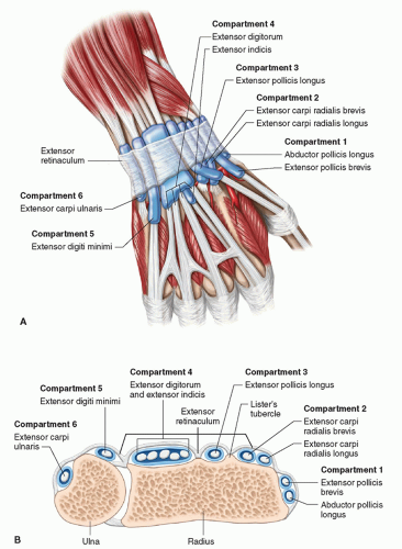
Dorsal extensor compartments
6 tunnels:
1: APL and EPB
2: ECRL and ECRM
3: EPL
4: EDC and EI
5: EDM
6: ECU
pronation muscles
pronator teres
pronator quadratus
wrist extension muscles
ECRL and ECRB = strongest
ECU
Long finger extensors assist slightly
wrist flexion muscles
FCR and FCU - strongest
long finger flexors assist (FDP, FDS)
radial deviation of wrist muscles
FCR
ECRL, ECRB
APL
ulnar deviation of wrist muscles
FCU
ECU
finger flexion muscles
FDS - PIP joint
FDP - DIP joint
digit extension muscles
EDC
EI
EDM