Seasons Structure Wk3 LO's

0.0(0)
Studied by 0 people
call kaiCall Kai
Locked
learnLearn
examPractice Test
spaced repetitionSpaced Repetition
heart puzzleMatch
flashcardsFlashcards
GameKnowt Play
Card Sorting

1/58

flashcard set

Earn XP

Description and Tags

Blood Flow Through the Heart, Blood Supply to the Heart & Pathology of Cellular Injury to the Heart

Last updated 8:36 PM on 8/20/26
Name
Mastery
Learn
Test
Matching
Spaced
Call with Kai
Chat

No analytics yet

Send a link to your students to track their progress

59 Terms

1
New cards

LO 1: where do coronary arteries arise from & what do they do

The heart’s "self-service" fuel system consists of coronary arteries arising from the aorta to deliver oxygen and cardiac veins that return deoxygenated blood to the right atrium via the coronary sinus.
<p><span style="background-color: transparent; font-family: &quot;Times New Roman&quot;, serif;">The heart’s "self-service" fuel system consists of coronary arteries arising from the aorta to deliver oxygen and cardiac veins that return deoxygenated blood to the right atrium via the coronary sinus.</span></p>
2
New cards

LO 1: arterial origin

  • left & right coronary arteries


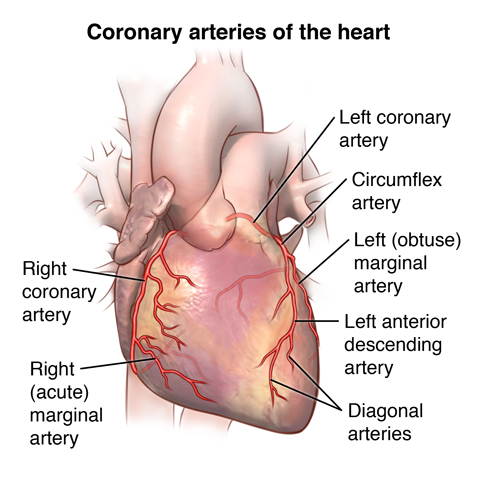
Arterial Origin: The left and right coronary arteries (LCA, RCA) are the very first branches of the ascending aorta, emerging just superior to the aortic valve cusps.
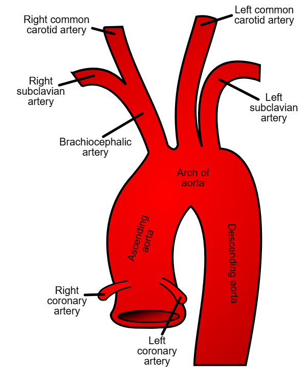
<p><span style="background-color: transparent; font-family: &quot;Times New Roman&quot;, serif;"><strong>Arterial Origin:</strong> The left and right coronary arteries (LCA, RCA) are the very first branches of the ascending aorta, emerging just superior to the aortic valve cusps.</span></p>
3
New cards

LO 1: right coronary artery branches (RCA)

  • what are the branches


  • sinoatrial (SA) nodal branch

  • right marginal branch

  • posterior interventricular branch (in posterior interventricular groove)
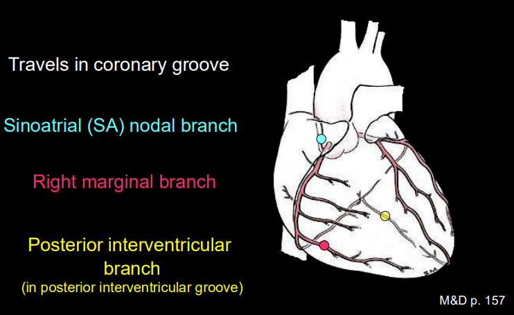

<ul><li><p>sinoatrial (SA) nodal branch</p></li><li><p>right marginal branch </p></li><li><p>posterior interventricular branch (in posterior interventricular groove)</p></li></ul><p></p>
4
New cards

LO 1: right coronary artery branches (RCA)

  • locations


Right Coronary Artery (RCA) Branches:

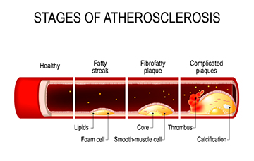
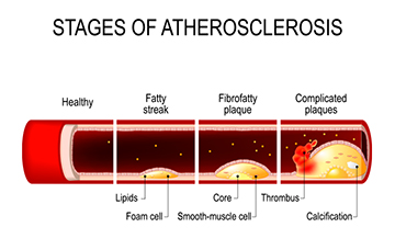
  • SA Nodal Branch: Usually passes posterior to the superior vena cava (SVC) to the pacemaker.

  • Right Marginal Branch: Travels along the acute (right) margin toward the apex.

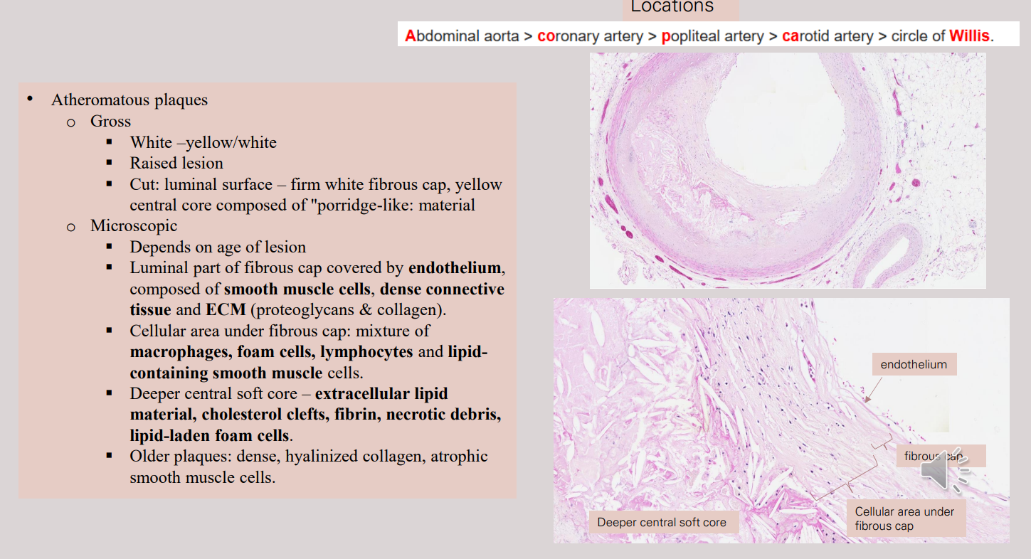
  • Posterior Interventricular Branch (PDA): Runs in the posterior interventricular groove.
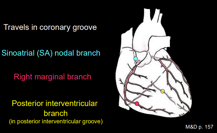

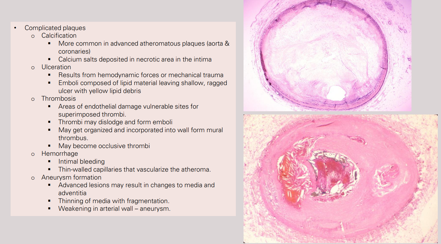
<p><span style="background-color: transparent; font-family: &quot;Times New Roman&quot;, serif;"><strong>Right Coronary Artery (RCA) Branches:</strong></span></p><ul><li><p><span style="background-color: transparent; font-family: &quot;Times New Roman&quot;, serif;"><strong>SA Nodal Branch:</strong> Usually passes posterior to the superior vena cava (SVC) to the pacemaker.</span></p></li><li><p><span style="background-color: transparent; font-family: &quot;Times New Roman&quot;, serif;"><strong>Right Marginal Branch:</strong> Travels along the acute (right) margin toward the apex.</span></p></li><li><p><span style="background-color: transparent; font-family: &quot;Times New Roman&quot;, serif;"><strong>Posterior Interventricular Branch (PDA):</strong> Runs in the posterior interventricular groove.</span></p></li></ul><p></p>
5
New cards

LO 1: left coronary artery branches

  • what are the branches


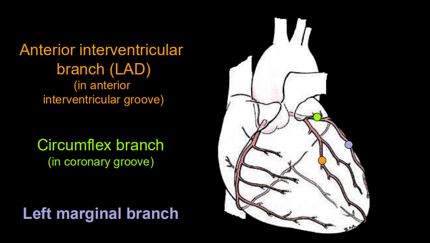
  • anterior interventricular branch (left anterior descending aka LAD) in the anterior interventricular groove

  • circumflex branch (in coronary groove)

  • left marginal branch


<ul><li><p>anterior interventricular branch (left anterior descending aka LAD) in the anterior interventricular groove</p></li><li><p>circumflex branch (in coronary groove)</p></li><li><p>left marginal branch </p></li></ul><p></p>
6
New cards

LO 1: left coronary artery branches

  • locations

  • LAD what are branches called

  • LCX what are branches called

  • what is a possible variation


  1. Left Coronary Artery (LCA) Branches:

    • Left Anterior Descending (LAD): Passes along the anterior interventricular groove; it gives off diagonal branches.

    • Circumflex Artery (LCX): Follows the coronary groove to the left; it gives off left marginal branches.

    • Variation: A ramus intermedius may arise as a third branch directly from the left main coronary.
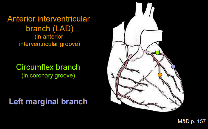

<ol><li><p><span style="background-color: transparent; font-family: &quot;Times New Roman&quot;, serif;"><strong>Left Coronary Artery (LCA) Branches:</strong></span></p><ul><li><p><span style="background-color: transparent; font-family: &quot;Times New Roman&quot;, serif;"><strong>Left Anterior Descending (LAD):</strong> Passes along the anterior interventricular groove; it gives off <strong>diagonal branches</strong>.</span></p></li><li><p><span style="background-color: transparent; font-family: &quot;Times New Roman&quot;, serif;"><strong>Circumflex Artery (LCX):</strong> Follows the coronary groove to the left; it gives off <strong>left marginal branches</strong>.</span></p></li><li><p><span style="background-color: transparent; font-family: &quot;Times New Roman&quot;, serif;"><em>Variation:</em> A <strong>ramus intermedius</strong> may arise as a third branch directly from the left main coronary.</span></p></li></ul></li></ol><p></p>
7
New cards

LO 1: what does the great cardiac vein run with

left anterior descending artery (LAD)
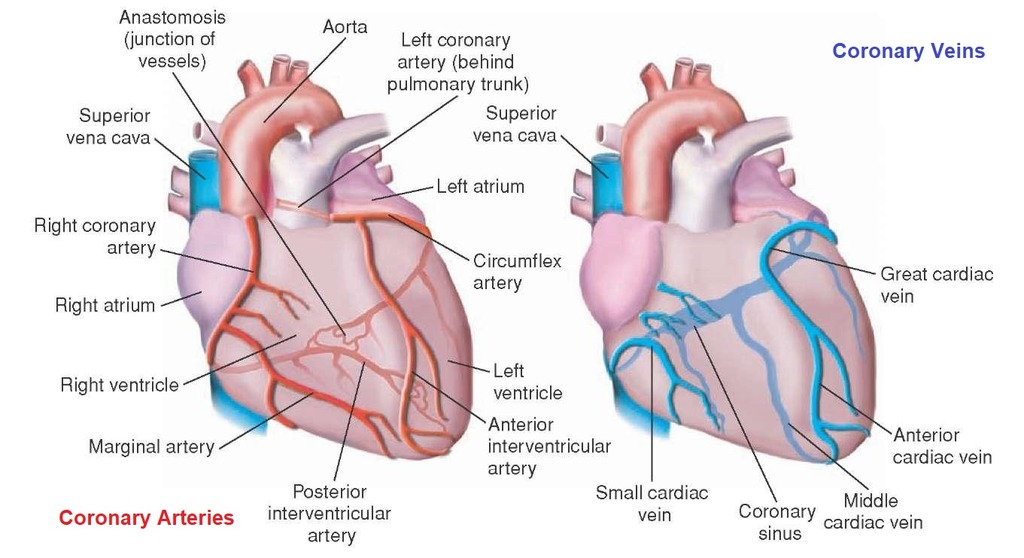
<p>left anterior descending artery <span style="background-color: transparent; font-family: &quot;Times New Roman&quot;, serif;">(LAD)</span></p>
8
New cards

LO 1: what does the middle cardiac vein run with

The Middle Cardiac Vein runs with the posterior interventricular branch (PDA) in posterior interventricular groove.
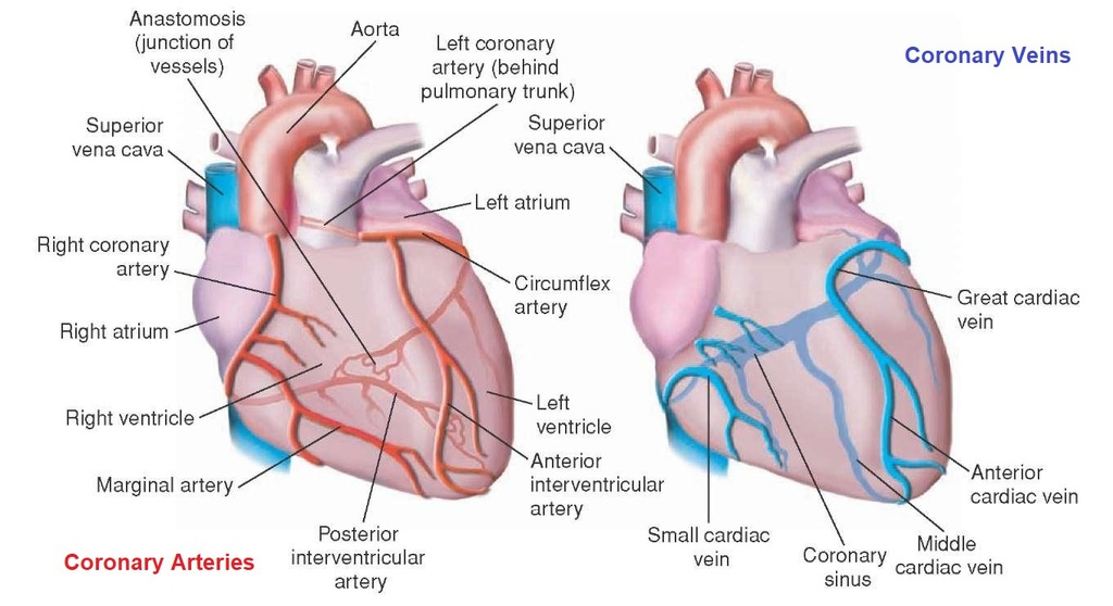
<p><span style="background-color: transparent; font-family: &quot;Times New Roman&quot;, serif;">The <strong>Middle Cardiac Vein</strong> runs with the posterior interventricular branch (PDA) in posterior interventricular groove.</span></p>
9
New cards

LO 1: what does the small cardiac vein run with

The Small Cardiac Vein runs with the right coronary (RCA) marginal branch.
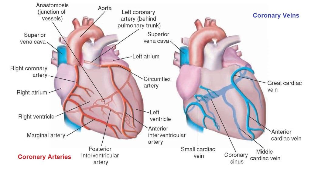
<p><span style="background-color: transparent; font-family: &quot;Times New Roman&quot;, serif;">The <strong>Small Cardiac Vein</strong> runs with the  right coronary (RCA) marginal branch.</span></p>
10
New cards

LO 1: what do the great cardiac vein, middle cardiac vein, & small cardiac vein drain into

All these drain into the Coronary Sinus, a large vein on the posterior heart that empties into the Internal Opening (Os) in the right atrium, located between the IVC and the tricuspid valve.
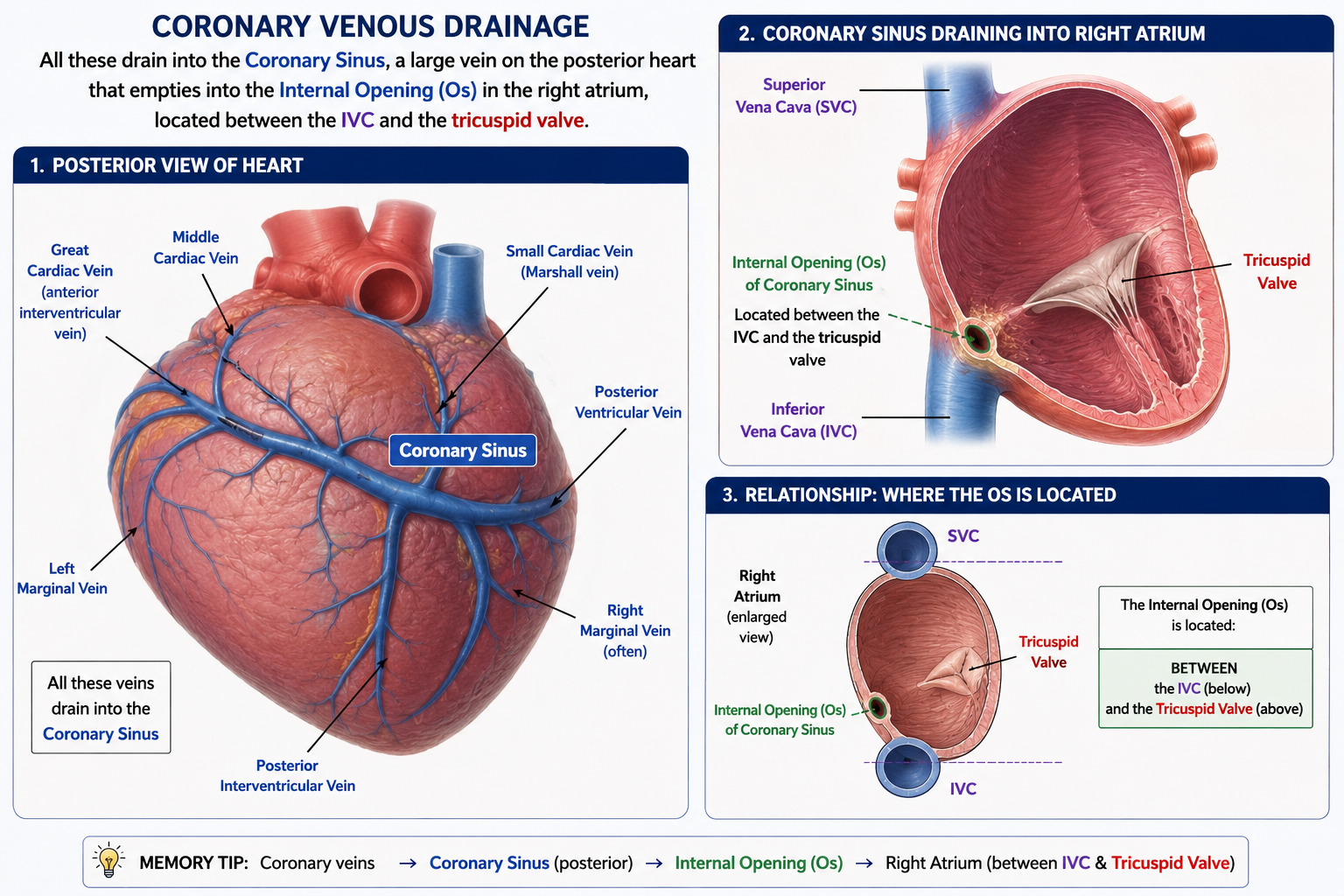
<p><span style="background-color: transparent; font-family: &quot;Times New Roman&quot;, serif;">All these drain into the <strong>Coronary Sinus</strong>, a large vein on the posterior heart that empties into the <strong>Internal Opening (Os)</strong> in the right atrium, located between the IVC and the tricuspid valve.</span></p>
11
New cards

LO 2: what is dominance determined by

Defining Dominance: Dominance is determined by which coronary artery gives rise to the Posterior Descending Artery (PDA) (posterior interventricular branch).

12
New cards

LO 2: right dominance

  • what it is

  • % of ppl


Right Dominance (80–90% of people): The PDA arises from the RCA.

13
New cards

LO 2: left dominance

  • what it is

  • % of ppl


Left Dominance (10–20% of people): The PDA arises from the LCX.

14
New cards

LO 2: in a right-dominant person what kind of MI does a RCA occlusion cause & what blood supply is at risk

Infarct Territory: In a right-dominant person, an RCA occlusion causes an inferior wall MI and risks the blood supply to the AV node.

<p><span style="background-color: transparent; font-family: &quot;Times New Roman&quot;, serif;"><strong>Infarct Territory:</strong> In a right-dominant person, an RCA occlusion causes an inferior wall MI and risks the blood supply to the AV node.</span></p>
15
New cards

LO 2: which vessels collateralize at the apex

Collateral Circulation: The PDA and LAD often collateralize at the apex; the extent of this determines how well a patient survives a sudden blockage in one of those vessels.

16
New cards

LO 2: what dominance is a patient with an inferior wall MI and they also develop a heart block

Clinical Correlation A patient with an inferior MI (ST elevation in leads II, III, aVF) is likely right-dominant. If they also develop a heart block, it confirms the RCA is the culprit because it is likely supplying the AV node.

17
New cards

LO 3: SA node

  • where is it located

  • what is it’s blood supply from


Sinuatrial (SA) Node:

  • Location: Superior end of the crista terminalis, near the SVC/Right Atrium junction.

  • Supply: SA nodal artery, arising from the RCA (60%) or LCA/LCX (40%).
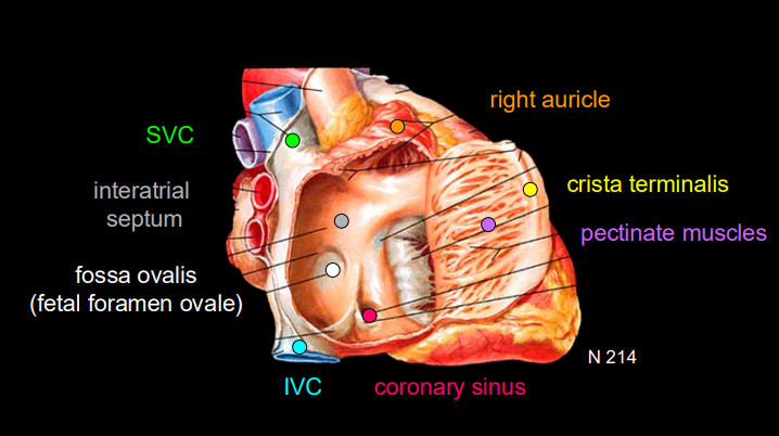

<p><span style="background-color: transparent; font-family: &quot;Times New Roman&quot;, serif;"><strong>Sinuatrial (SA) Node:</strong></span></p><ul><li><p><span style="background-color: transparent; font-family: &quot;Times New Roman&quot;, serif;"><strong>Location:</strong> Superior end of the crista terminalis, near the SVC/Right Atrium junction.</span></p></li><li><p><span style="background-color: transparent; font-family: &quot;Times New Roman&quot;, serif;"><strong>Supply:</strong> SA nodal artery, arising from the <strong>RCA (60%)</strong> or LCA/LCX (40%).</span></p></li></ul><p></p>
18
New cards

LO 3: AV node

  • location

  • what is it’s blood supply


Atrioventricular (AV) Node:

  • Location: Floor of the right atrium near the opening of the coronary sinus.

  • Supply: AV nodal artery, arising from the RCA (85–90%) or LCX (10–15%).
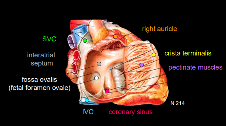

<p><span style="background-color: transparent; font-family: &quot;Times New Roman&quot;, serif;"><strong>Atrioventricular (AV) Node:</strong></span></p><ul><li><p><span style="background-color: transparent; font-family: &quot;Times New Roman&quot;, serif;"><strong>Location:</strong> Floor of the right atrium near the opening of the coronary sinus.</span></p></li><li><p><span style="background-color: transparent; font-family: &quot;Times New Roman&quot;, serif;"><strong>Supply:</strong> AV nodal artery, arising from the <strong>RCA (85–90%)</strong> or LCX (10–15%).</span></p></li></ul><p></p>
19
New cards

LO 3: Bundle of His

  • location

  • blood supply


Bundle of His (AV Bundle):

  • Location: Passes from the AV node through the fibrous skeleton into the interventricular septum.

  • Supply: Primarily the LAD (anterior interventricular artery).
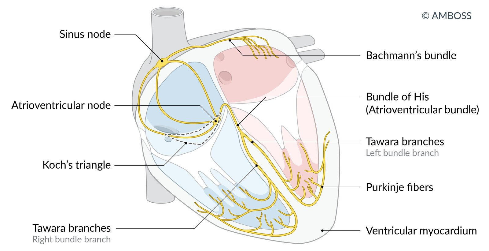

<p><span style="background-color: transparent; font-family: &quot;Times New Roman&quot;, serif;"><strong>Bundle of His (AV Bundle):</strong></span></p><ul><li><p><span style="background-color: transparent; font-family: &quot;Times New Roman&quot;, serif;"><strong>Location:</strong> Passes from the AV node through the fibrous skeleton into the interventricular septum.</span></p></li><li><p><span style="background-color: transparent; font-family: &quot;Times New Roman&quot;, serif;"><strong>Supply:</strong> Primarily the <strong>LAD</strong> (anterior interventricular artery).</span></p></li></ul><p></p>
20
New cards

LO 3: moderator band (septomarginal trabecula)

  • what does it contain

  • where does it carry the signal to


Moderator Band (Septomarginal Trabecula):

  • Contains the Right Bundle Branch; carries the signal to the anterior papillary muscle of the right ventricle.
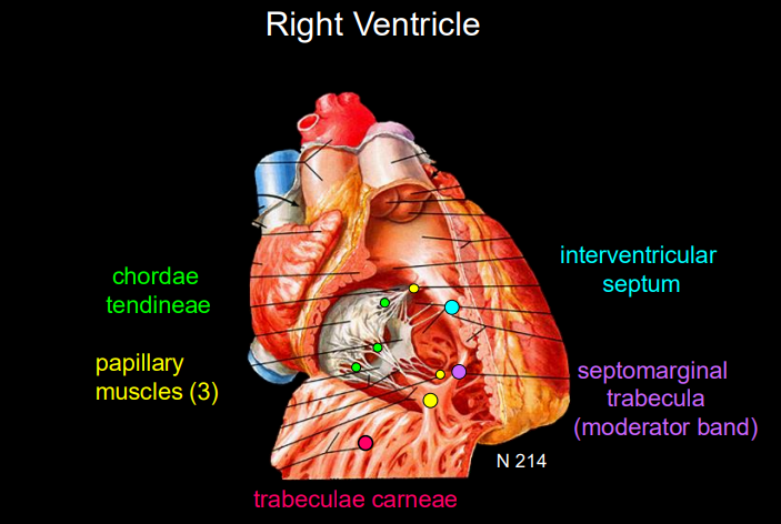

<p><span style="background-color: transparent; font-family: &quot;Times New Roman&quot;, serif;"><strong>Moderator Band (Septomarginal Trabecula):</strong></span></p><ul><li><p><span style="background-color: transparent; font-family: &quot;Times New Roman&quot;, serif;">Contains the <strong>Right Bundle Branch</strong>; carries the signal to the anterior papillary muscle of the right ventricle.</span></p></li></ul><p></p>
21
New cards

LO 4: what is the mediastinum divided into & by what

Division: Divided into Superior and Inferior by a plane through the sternal angle (Angle of Louis) to T4/T5.
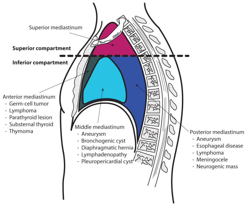
<p><span style="background-color: transparent; font-family: &quot;Times New Roman&quot;, serif;"><strong>Division:</strong> Divided into <strong>Superior</strong> and <strong>Inferior</strong> by a plane through the sternal angle (Angle of Louis) to T4/T5.</span></p>
22
New cards

LO 4: inferior mediastinum

  • what are the 3 divisions & what do they contain


Inferior Mediastinum Subdivisions:

  • Anterior: Fat and thymus remnants.

  • Middle: Contains the Heart and pericardium.

  • Posterior: Contains the esophagus, thoracic aorta, and azygous system.
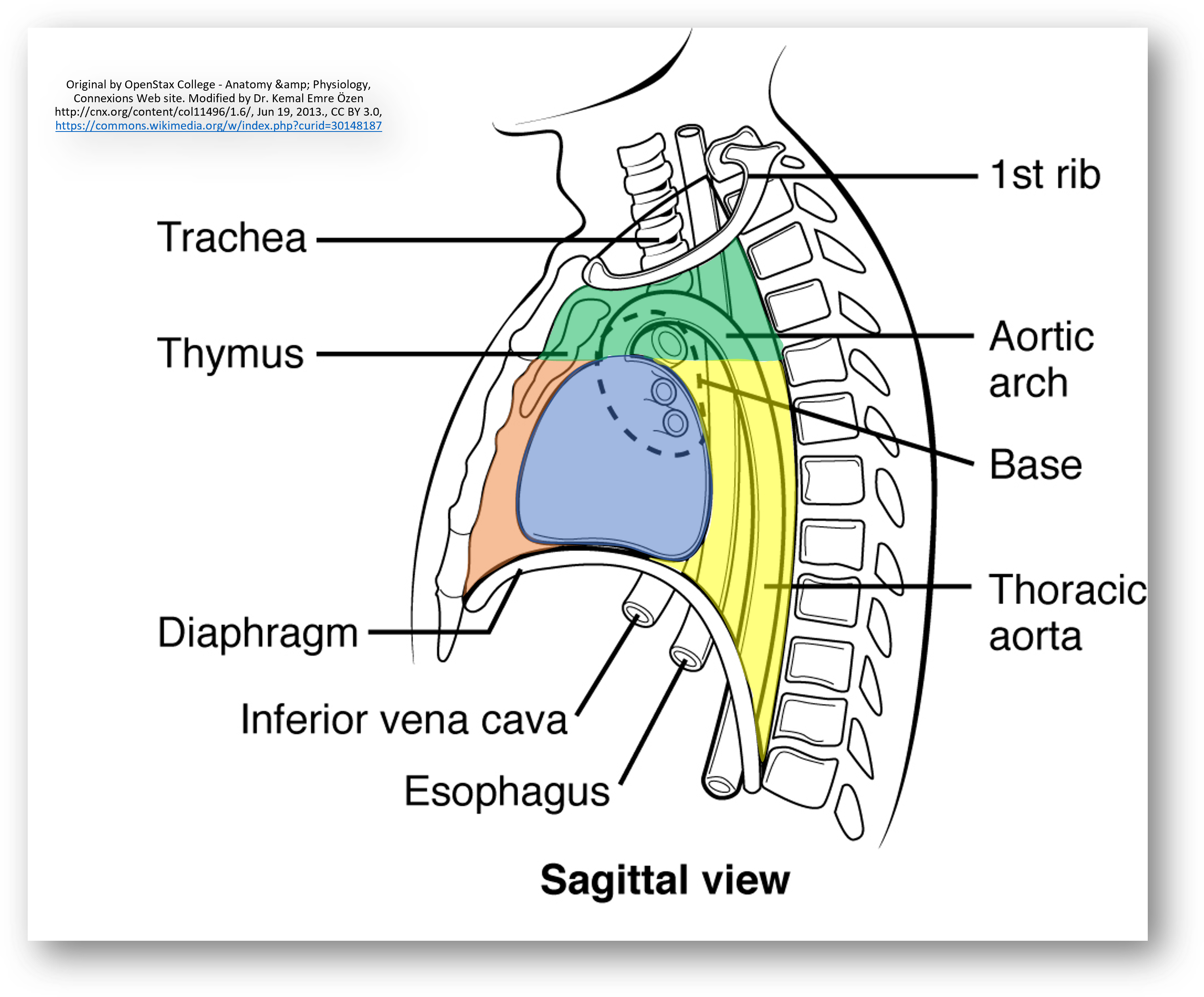

<p><span style="background-color: transparent; font-family: &quot;Times New Roman&quot;, serif;"><strong>Inferior Mediastinum Subdivisions:</strong></span></p><ul><li><p><span style="background-color: transparent; font-family: &quot;Times New Roman&quot;, serif;"><strong>Anterior:</strong> Fat and thymus remnants.</span></p></li><li><p><span style="background-color: transparent; font-family: &quot;Times New Roman&quot;, serif;"><strong>Middle:</strong> Contains the <strong>Heart</strong> and pericardium.</span></p></li><li><p><span style="background-color: transparent; font-family: &quot;Times New Roman&quot;, serif;"><strong>Posterior:</strong> Contains the esophagus, thoracic aorta, and azygous system.</span></p></li></ul><p></p>
23
New cards

LO 4: position of the heart

  • what forms the apex

  • what is the base formed by

  • what is the anterior surface formed by

  • where are the esophagus & descending aorta in relation to the heart

  • where do the phrenic nerves run


Position of the Heart:

  • Apex: Formed by the left ventricle; points inferolaterally at the 5th left intercostal space midclavicular line.

  • Base: Posterior aspect formed mostly by the Left Atrium.

  • Anterior surface: Mostly Right Ventricle.

  • Relations: Esophagus and descending aorta are posterior to the heart. Phrenic nerves (C3-C5) run anterior to the lung roots along the pericardium.
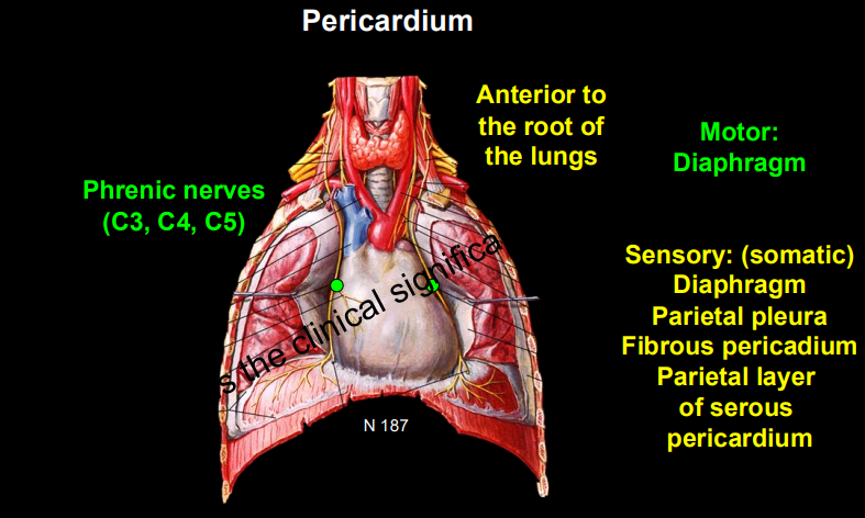

<p><span style="background-color: transparent; font-family: &quot;Times New Roman&quot;, serif;"><strong>Position of the Heart:</strong></span></p><ul><li><p><span style="background-color: transparent; font-family: &quot;Times New Roman&quot;, serif;"><strong>Apex:</strong> Formed by the left ventricle; points inferolaterally at the <strong>5th left intercostal space</strong> midclavicular line.</span></p></li><li><p><span style="background-color: transparent; font-family: &quot;Times New Roman&quot;, serif;"><strong>Base:</strong> Posterior aspect formed mostly by the <strong>Left Atrium</strong>.</span></p></li><li><p><span style="background-color: transparent; font-family: &quot;Times New Roman&quot;, serif;"><strong>Anterior surface:</strong> Mostly <strong>Right Ventricle</strong>.</span></p></li><li><p><span style="background-color: transparent; font-family: &quot;Times New Roman&quot;, serif;"><strong>Relations:</strong> Esophagus and descending aorta are <strong>posterior</strong> to the heart. Phrenic nerves (C3-C5) run <strong>anterior</strong> to the lung roots along the pericardium.</span></p></li></ul><p></p>
24
New cards

LO 4: what portion of the mediastinum is the heart in & what intercostal space is the apex at

  • Heart is in the middle inferior mediastinum.

  • apex at the left 5th intercostal space and its base formed by the left atrium.
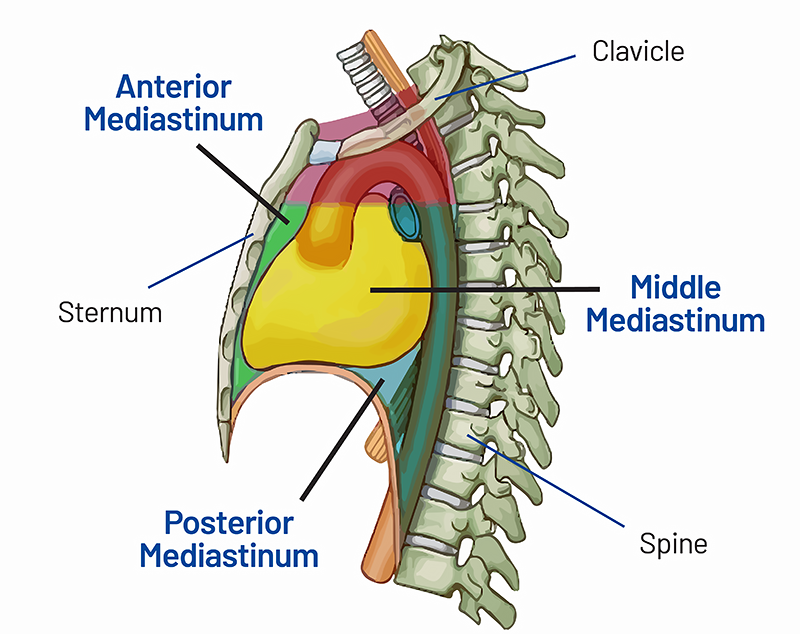

<ul><li><p><span style="background-color: transparent; font-family: &quot;Times New Roman&quot;, serif;">Heart is in the <strong>middle</strong> inferior mediastinum.</span></p></li><li><p><span style="background-color: transparent; font-family: &quot;Times New Roman&quot;, serif;">apex at the left 5th intercostal space and its base formed by the left atrium.</span></p></li></ul><p></p>
25
New cards

LO 5:

  • where are the coronary arteries located in terms of heart muscle layers

  • which layer is most vulnerable to ischemia & why


Anatomical Vulnerability:

  • Coronary arteries sit on the epicardial surface and dive down.

  • The subendocardium (inner layer) is the last to get blood and faces the highest pressure from the ventricle, making it the most vulnerable to ischemia.


26
New cards

LO 5: Subendocardial Ischemia (NSTEMI / Stable Angina)

  • what is the cause

  • what occurs physiologically


Subendocardial Ischemia (NSTEMI / Stable Angina):

  • Cause: Partial occlusion or increased demand (e.g., exercise with 70% stenosis).

  • Physiology: Blood reaches the surface but can't reach the deepest "end of the line" subendocardium


27
New cards

LO 5: Transmural Ischemia (STEMI / Prinzmetal)

  • what is the cause

  • what occurs physiologically


  • Prinzmetal angina, also known as vasospastic or variant angina, is a type of chest pain caused by a sudden, severe spasm of the coronary arteries that temporarily reduces blood flow to the heart

Transmural Ischemia (STEMI / Prinzmetal):

  • Cause: Complete occlusion by a thrombus or severe, prolonged vasospasm.

  • Physiology: The entire thickness of the wall is starved of oxygen.


28
New cards

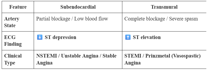
LO 5: subendocardial MI

  • artery state

  • ECG finding

  • clinical type name


knowt flashcard image
29
New cards

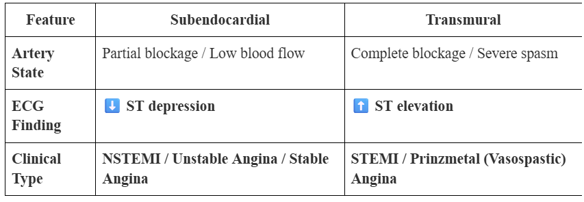
LO 5: transmural MI

  • artery state

  • ECG finding

  • clinical type name


knowt flashcard image
30
New cards

LO 6: what leaks out when myocyte membranes are damaged by necrosis

Mechanism: When myocyte membranes are damaged by necrosis, Troponin I and T leak into the blood.

31
New cards

LO 6: unstable angina

  • what it is

  • troponin status


Unstable Angina: Ischemia without necrosis. Troponins remain negative.

32
New cards

LO 6: NSTEMI & STEMI

  • what do they both involve

  • troponin status


NSTEMI & STEMI: Both involve necrosis. Troponins are positive.

33
New cards

LO 6: troponin time course

  • rise

  • peak

  • duration/how long it remains elevated


Time Course:

  • Rise: 2–4 hours after onset.

  • Peak: ~24–48 hours.

  • Duration: Remains elevated for 7–10 days.


34
New cards

LO 6: A patient has chest pain at rest and ST depression on ECG. If Troponin is negative at 6 hours, it is __________. If it is positive, it is an ___________

A patient has chest pain at rest and ST depression on ECG. If Troponin is negative at 6 hours, it is Unstable Angina. If it is positive, it is an NSTEMI.

35
New cards

LO 7: what do these represent on ECG

  • p-wave

  • PR interval

  • QRS complex

  • T wave

  • ST segment


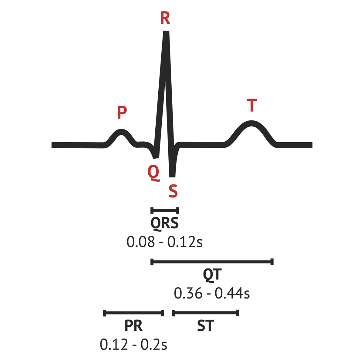
  1. P Wave: Atrial depolarization (squeeze).

  2. PR Interval: Time for the signal to travel from the SA node through the AV node (the "gatekeeper").

  3. QRS Complex: Ventricular depolarization (the big squeeze). Should be narrow.

  4. T Wave: Ventricular repolarization (the reset).

  5. ST Segment: The pause between depolarization and repolarization; it should be flat on the "isoelectric line".

<ol><li><p><span style="background-color: transparent; font-family: &quot;Times New Roman&quot;, serif;"><strong>P Wave:</strong> Atrial depolarization (squeeze).</span></p></li><li><p><span style="background-color: transparent; font-family: &quot;Times New Roman&quot;, serif;"><strong>PR Interval:</strong> Time for the signal to travel from the SA node through the AV node (the "gatekeeper").</span></p></li><li><p><span style="background-color: transparent; font-family: &quot;Times New Roman&quot;, serif;"><strong>QRS Complex:</strong> Ventricular depolarization (the big squeeze). Should be narrow.</span></p></li><li><p><span style="background-color: transparent; font-family: &quot;Times New Roman&quot;, serif;"><strong>T Wave:</strong> Ventricular repolarization (the reset).</span></p></li><li><p><span style="background-color: transparent; font-family: &quot;Times New Roman&quot;, serif;"><strong>ST Segment:</strong> The pause between depolarization and repolarization; it should be flat on the "isoelectric line".</span></p></li></ol><p></p>
36
New cards

Lo 8: STEMI

  • ECG

  • what does this mean about heart muscle


STEMI: ST-segment Elevation (the "Fireman's Helmet"). Indicates full-thickness (transmural) damage.

37
New cards

LO 8: NSTEMI

  • ECG

  • what does this mean about heart muscle


NSTEMI: ST-segment Depression or T-wave inversion (no elevation). Indicates partial-thickness (subendocardial) damage.

38
New cards

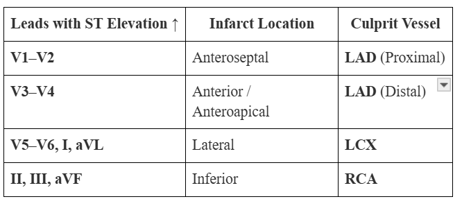
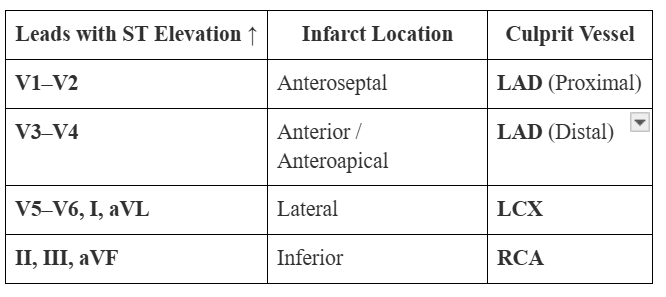
LO 9: leads V1-V2

  • infarct location

  • vessel involved


knowt flashcard image
39
New cards

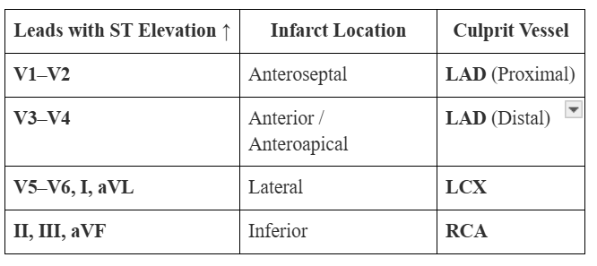
LO 9: leads V3-V4

  • infarct location

  • vessel involved


knowt flashcard image
40
New cards

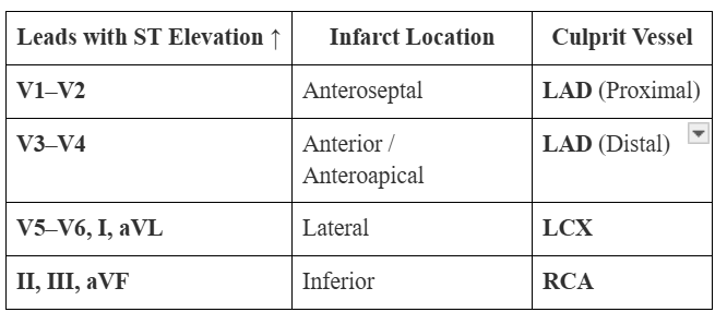
LO 9: leads V5-V6, I, aVL

  • infarct location

  • vessel involved


knowt flashcard image
41
New cards

LO 9: leads II, III, aVF

  • infarct location

  • vessel involved


knowt flashcard image
42
New cards

LO 9: vessel

  • V1–V4 =

  • II, III, aVF =

  • I, aVL, V5–V6 =


  • V1–V4 = LAD.

  • II, III, aVF = RCA.

  • I, aVL, V5–V6 = LCX.


43
New cards

LO 10: myocardium response to ischemia/injury

  • 0-30 min

  • gross

  • micro

  • is this reversible


0–30 min: Reversible injury. Gross/Micro: None.

<p><span style="background-color: transparent; font-family: &quot;Times New Roman&quot;, serif;"><strong>0–30 min:</strong> Reversible injury. <strong>Gross/Micro:</strong> None.</span></p>
44
New cards

LO 10: myocardium response to ischemia/injury

  • 30min-4hrs

  • gross

  • micro

  • is this reversible


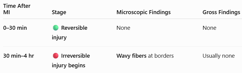
30 min – 4 hours: Irreversible injury begins. Micro: Wavy fibers at borders.
<p><span style="background-color: transparent; font-family: &quot;Times New Roman&quot;, serif;"><strong>30 min – 4 hours:</strong> Irreversible injury begins. <strong>Micro:</strong> Wavy fibers at borders.</span></p>
45
New cards

LO 10: myocardium response to ischemia/injury

  • 4-24hrs

  • gross

  • micro


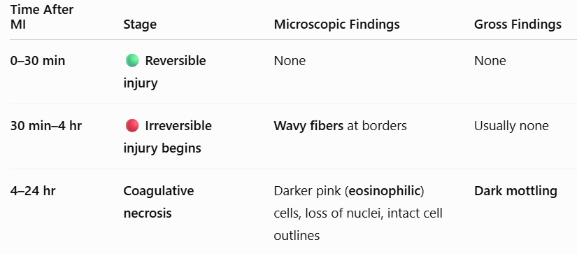
4–24 hours: Micro: Coagulative necrosis (darker pink cells, loss of nuclei, intact outlines). Gross: Dark mottling.
<p><span style="background-color: transparent; font-family: &quot;Times New Roman&quot;, serif;"><strong>4–24 hours:</strong> <strong>Micro:</strong> Coagulative necrosis (darker pink cells, loss of nuclei, intact outlines). <strong>Gross:</strong> Dark mottling.</span></p>
46
New cards

LO 10: myocardium response to ischemia/injury

  • 1-3 days

  • gross

  • micro


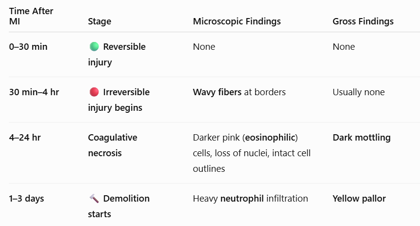
1–3 days: Demolition starts. Micro: Heavy neutrophil infiltrate. Gross: Yellow pallor.
<p><span style="background-color: transparent; font-family: &quot;Times New Roman&quot;, serif;"><strong>1–3 days:</strong> <strong>Demolition starts.</strong> <strong>Micro:</strong> Heavy neutrophil infiltrate. <strong>Gross:</strong> Yellow pallor.</span></p>
47
New cards

LO 10: myocardium response to ischemia/injury

  • 3-7 days

  • gross

  • micro


3–7 days: Peak danger. Micro: Macrophages cleaning up debris. Gross: Central yellow softening (risk of rupture!).
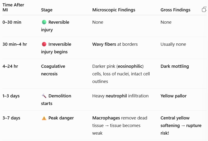
<p><span style="background-color: transparent; font-family: &quot;Times New Roman&quot;, serif;"><strong>3–7 days:</strong> <strong>Peak danger.</strong> <strong>Micro:</strong> Macrophages cleaning up debris. <strong>Gross:</strong> Central yellow softening (risk of rupture!).</span></p>
48
New cards

LO 10: myocardium response to ischemia/injury

  • 1-3 weeks

  • gross

  • micro


1–3 weeks: Reconstruction. Micro: Granulation tissue (new capillaries, fibroblasts). Gross: Red, hyperemic border.
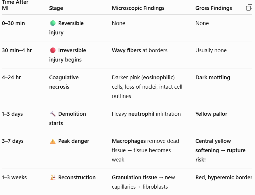
<p><span style="background-color: transparent; font-family: &quot;Times New Roman&quot;, serif;"><strong>1–3 weeks:</strong> <strong>Reconstruction.</strong> <strong>Micro:</strong> Granulation tissue (new capillaries, fibroblasts). <strong>Gross:</strong> Red, hyperemic border.</span></p>
49
New cards

LO 10: myocardium response to ischemia/injury

  • >2 months

  • gross

  • micro


>2 months: Finished. Micro: Dense collagenous scar. Gross: White scar.
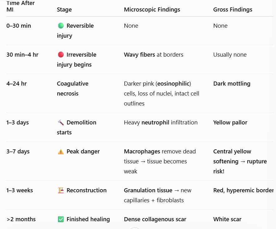
<p><span style="background-color: transparent; font-family: &quot;Times New Roman&quot;, serif;"><strong>&gt;2 months:</strong> <strong>Finished.</strong> <strong>Micro:</strong> Dense collagenous scar. <strong>Gross:</strong> White scar.</span></p>
50
New cards

LO 10: myocardium response to ischemia/injury

  • Wavy fibers = earliest sign (______).

  • Neutrophils =_______ days; Macrophages = _______ days.

  • Day 3-7 is the _________ _____ ____.


  • Wavy fibers = earliest sign (30m-4h).

  • Neutrophils = 1-3 days; Macrophages = 3-7 days.

  • Day 3-7 is the highest rupture risk.


51
New cards

LO 11: what is arthrosclerosis

Atherosclerosis is a chronic inflammatory response to vessel wall injury, resulting in a "pimple" of fat and scarring inside an artery.
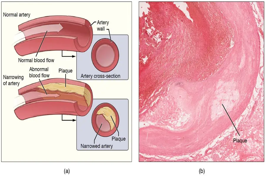
<p><span style="background-color: transparent; font-family: &quot;Times New Roman&quot;, serif;">Atherosclerosis is a chronic inflammatory response to vessel wall injury, resulting in a "pimple" of fat and scarring inside an artery.</span></p>
52
New cards

LO 11: arthrosclerosis

  • endothelial dysfunction: what are triggers


Endothelial Dysfunction: Triggers (smoking, HTN) injure the lining.

53
New cards

LO 11: arthrosclerosis

  • fatty streak: what does this look like & what are they made from


Fatty Streak: The earliest visible sign; flat yellow dots made of Foam Cells (macrophages that ate LDL).
<p><span style="background-color: transparent; font-family: &quot;Times New Roman&quot;, serif;"><strong>Fatty Streak:</strong> The earliest visible sign; flat yellow dots made of <strong>Foam Cells</strong> (macrophages that ate LDL).</span></p>
54
New cards

LO 11: what are foam cells

Fatty Streak: The earliest visible sign; flat yellow dots made of Foam Cells (macrophages that ate LDL).
<p><span style="background-color: transparent; font-family: &quot;Times New Roman&quot;, serif;"><strong>Fatty Streak:</strong> The earliest visible sign; flat yellow dots made of <strong>Foam Cells</strong> (macrophages that ate LDL).</span></p>
55
New cards

LO 11: mature plaque (atheroma)

  • fibrous cap: what it’s composed of

  • necrotic core: what it’s composed of


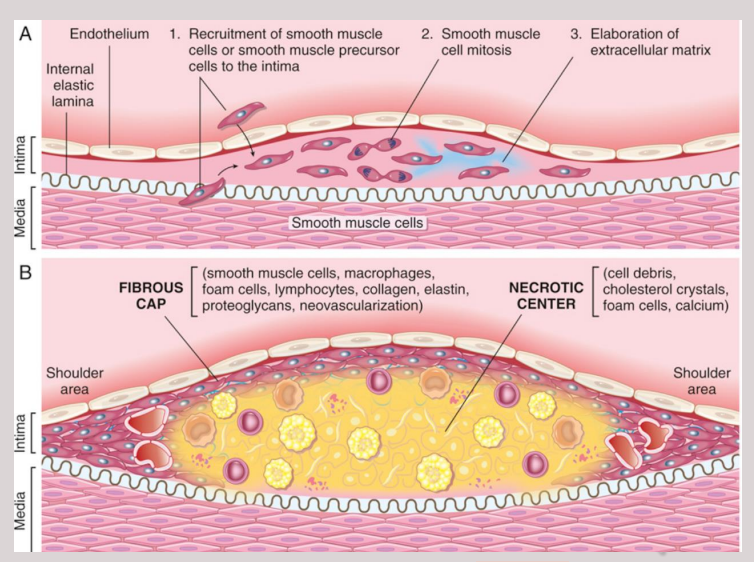
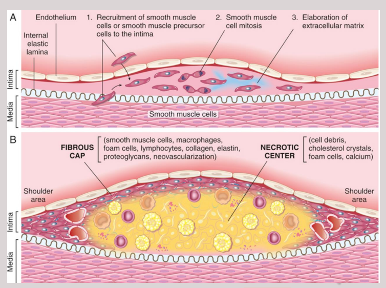
The Mature Plaque (Atheroma):

  • Fibrous Cap: A hard "roof" composed of smooth muscle cells, dense collagen, and proteoglycans.

  • Necrotic Core: A "cheesy" center under the cap containing extracellular lipids, cell debris, and Cholesterol Clefts (needle-shaped spaces where crystals once were).

<p><span style="background-color: transparent; font-family: &quot;Times New Roman&quot;, serif;"><strong>The Mature Plaque (Atheroma):</strong></span></p><ul><li><p><span style="background-color: transparent; font-family: &quot;Times New Roman&quot;, serif;"><strong>Fibrous Cap:</strong> A hard "roof" composed of smooth muscle cells, dense collagen, and proteoglycans.</span></p></li><li><p><span style="background-color: transparent; font-family: &quot;Times New Roman&quot;, serif;"><strong>Necrotic Core:</strong> A "cheesy" center under the cap containing extracellular lipids, cell debris, and <strong>Cholesterol Clefts</strong> (needle-shaped spaces where crystals once were).</span></p></li></ul><p></p>
56
New cards

LO 11: complicated plaque

  • what does it show


Complicated Plaque: Can show calcification, ulceration, or thrombosis (the trigger for an MI).
<p><span style="background-color: transparent; font-family: &quot;Times New Roman&quot;, serif;"><strong>Complicated Plaque:</strong> Can show <strong>calcification</strong>, <strong>ulceration</strong>, or <strong>thrombosis</strong> (the trigger for an MI).</span></p>
57
New cards

LO 11: what is the risk with plaques that have a thin fibrous cap

Plaques with a thin fibrous cap and many macrophages are "vulnerable" plaques; they are more likely to rupture and cause a sudden heart attack than large, stable, thick-capped plaques.

58
New cards

LO 11: Atherosclerosis is characterized by an intimal plaque containing a fibrous cap and a necrotic lipid core with_______ and ________

Atherosclerosis is characterized by an intimal plaque containing a fibrous cap and a necrotic lipid core with foam cells and cholesterol clefts
<p><span style="background-color: transparent; font-family: &quot;Times New Roman&quot;, serif;">Atherosclerosis is characterized by an intimal plaque containing a fibrous cap and a necrotic lipid core with foam cells and cholesterol clefts</span></p>
59
New cards

LO 11: Atherosclerosis is characterized by an intimal plaque containing a _________ and a _______________with foam cells and cholesterol clefts

Atherosclerosis is characterized by an intimal plaque containing a fibrous cap and a necrotic lipid core with foam cells and cholesterol clefts
<p><span style="background-color: transparent; font-family: &quot;Times New Roman&quot;, serif;">Atherosclerosis is characterized by an intimal plaque containing a fibrous cap and a necrotic lipid core with foam cells and cholesterol clefts</span></p>