GI - Histology I - Salivary Gland, Esophagus & Stomach
1/72
There's no tags or description
Looks like no tags are added yet.
Name | Mastery | Learn | Test | Matching | Spaced |
|---|
No study sessions yet.
73 Terms
What glands account for 70% of mucous secretion?
minor salivary glands
the parenchyma of the submandibular gland is derived from what germ layer?
oral ectoderm
the stroma of the submandibular gland is derived from what germ layer?
ectomesenchyme
salivary gland epithelium consists of what two cells types?
- secretory
- myoepithelial
at the end of the ducts are...
acini
singular = acinus (grape)
describe the morphology of a serous acinus
- pyramid-shape cell
- perfectly round nuclei located near the base of the cell
What explains the flattened appearance of nuclei in a mucous acinus?
when H&E staining is done, the organic solvent causes the mucinogen to expand, pushing the nuclei basally, or near the basement membrane
compare morphology of serous and mucous acini
serous
- round nuclei found near the base of the cell
- pyramid - shaped cells
mucous
- flattened nuclei found near the base of the cell
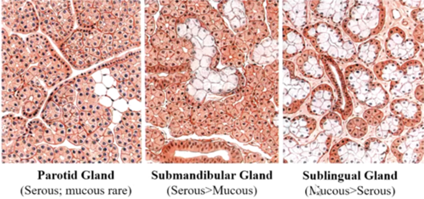
compare the histological appearance of the parotid, submandibular and sublingual glands
since parotid gland secretions are almost always serous (mucous in rare cases), you wouldn't expect to see many unstained areas (mucous units) whereas in submandibular, you would see some unstained areas but not as many as you would see in the sublingual gland, where there is more mucous units than serous units
what type of epithelium is found in the intercalated duct?
simple cuboidal
what type of epithelium is found in the striated duct?
simple columnar
what type of epithelium is found in the excretory duct?
pseudostratified
how do you expect the cytoplasm of striated ducts to appear in H&E staining? Why?
striated ducts have eosinophilic staining due to large numbers of mitochondria
what happens in the intercalated ducts?
reabsorbs Cl- and secretes HCO3-
- antimicrobials: lysozyme and lactoferrin..
the striations of the striated ducts are due to...
infoldings of the basement membrane into the cytoplasm
describe the histological appearance of nuclei in the striated ducts
they are pushed toward the lumen due to the striations or infoldings of the basement membrane into the cytoplasm
between the striations of the striated ducts, what do you expect to see?
mitochondria
usually, cell nuclei are located within the basal 1/3 but in striated ducts, where are they?
more apical
What is the purpose of striations in striated ducts?
to increase surface area to hold more ion pumps and do more ion exchange
what is the main function of the excretory duct?
to transport saliva to the oral cavity
does saliva modicifcation happen everyhwere?
NO, there is no salivary modification in the excretory duct
what cell helps to expel secretions?
myoepithelial cells
the myoepithelial cells are found between...
secretory cells and basement membrane
most glands secrete mucus either exclusively or if mixed in a greater proportion. what is the exception to this?
the gustatory (Ebner) glands in the tongue
- vallate and circumvallate papillae
what is the purpose of the gustatory (Ebner) glands in the tongue
- vallate and circumvallate papillae
to flush the vallate and circumvallate taste buds
What is Sjogren's syndrome?
chronic autoimmune disorder affecting salivary & lacrimal glands leading to xerostomia and dry eyes
what is the 3rd most common autoimmune disease next to RA and lupus
Sjogren's syndrome
What is sialolithiasis?
formation of calculi or mineralized concentrations that may block a salivary duct
significance of sialolithiasis
may lead to periodic pain in ducts or gland as well as swelling of the gland due to obstruction of duct by these concretions
is xerostomia more common in males or females?
females
significance of xerostomia
places patients at risk for tooth decay, opportunistic oral infections, gum disease
What is mumps?
acute infectious disease causing inflammation and swelling of the parotid gland with occasional involvement of other salivary glands
mumps typically involves which gland?
parotid
- occasionally includes others
what is the cause of mumps?
contagious infection by mumps virus
the following is a description of the significance of what disease?
typically a childhood disease, although adults may be affected; metastatic disease may involve other organs (e.g. testes, ovaries, breasts, meninges)
mumps
can mumps be metastatic?
YES
presentation: 13 y/o male w/ 2d history of facial swelling on It side; pain w/out fever or ear discharge; recently began tuba
What is the diagnosis?
pneumoparotid
how would you treat a pneumoparotid?
parotid massages = foamy secretions from duct
a pneumoparotid is commonly seen in which population?
seen in wind instrument players, glassblowers, etc.
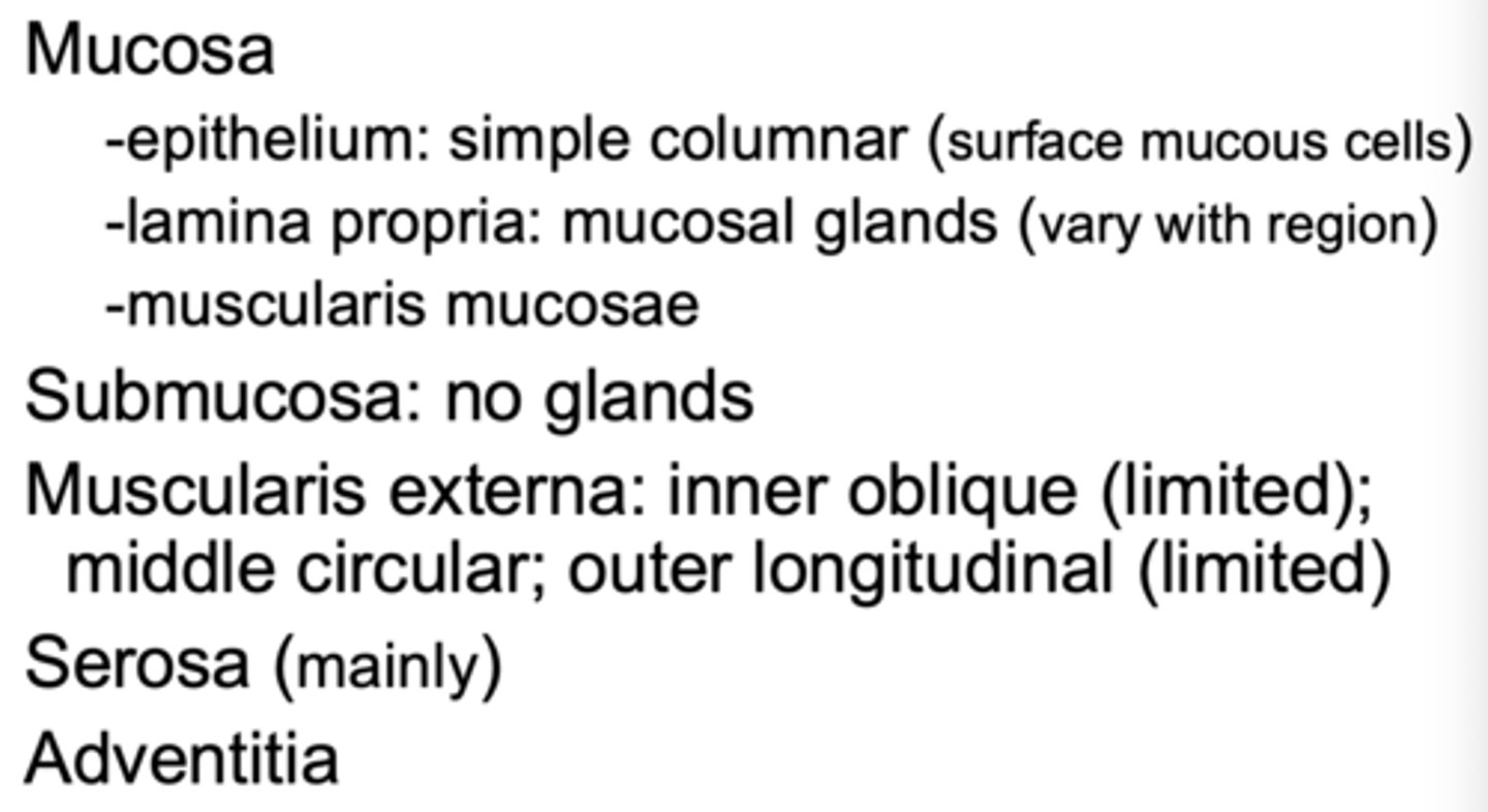
what forms the mucosa layer of the esophagus? (3)
- epithelium: SSNKE
- lamina propria
- muscularis mucosa
What are the 4 layers of the esophagus?
mucosa, submucosa, externa, adventitia
the adventitia of the esophagus is located where? what is the significance of this?
located in the thorax and provides easy passage for the spread of cancer
where is the serosa of the esophagus located?
in the abdominal cavity
the inner muscularis externa of the esophagus has muscle fibers running in what direction?
circular
the outer muscularis externa of the esophagus has muscle fibers running in what direction?
longitudinal
what type of muscle is found in the upper 1/3 muscularis externa of the esophagus?
skeletal muscle
what type of muscle is found in the middle 1/3 muscularis externa of the esophagus?
a mix of skeletal and smooth muscle
what type of muscle is found in the lower 1/3 muscularis externa of the esophagus?
smooth muscle
what is contained in the submucosa of the esophagus? (2)
- loose CT
- esophageal glands
what is found in the mucosal lining of the esophagus? (3)
- SSNKE
- lamina propria
- muscularis mucosae
what two structures have submucosal glands?
- esophagus
- duodenum of small intestine
the ducts of the esophagus have what type of epithelium?
stratified cuboidal
the connection in epithelium of the stomach and the esophagus is known as the...
squamocolumnar junction
- a zig-zag line
What is Barrett's esophagus?
abnormal squamocolumnar junction
- metaplasia to gastric or intestinal mucosa
Barrett's esophagus is metaplasia from the usual squamocolumnar junction to?
gastric or intestinal mucosa
Barrett's esophagus (metaplasia) involves which of the following?
a) pyloric sphincter
b) squamocolumnar junction
c) enteroendocrine cells
d) pyloric glands
B
are there glands in the submucosa of the stomach?
NO
- glands are only found in the submucosa of the esophagus and duodenum
what are the layers of the stomach?
mucosa
submucosa
muscularis
serosa (mainly)
adventitia
where are the mucosal glands of the stomach located?
the lamina propria
What are the 3 types of stomach mucosal glands?
- cardial gastric glands
- gastric glands proper (parietal, zygomatic chief)
- pyloric glands
in which set of glands are the parietal and zygomatic chief cells?
in the gastric (fundic) glands proper
Parietal cells secrete what?
How many layers of muscles are there in the muscularis externa layer of the stomach?
3
What are the 3 muscle layers of the muscularis externa layer of the stomach?
inner oblique (limited)
middle circular
outer longitudinal (limited)
what covers the inner surface of the stomach and the gastric pits?
surface mucous cells
what do mucous neck cells secrete?
a thin, acidic mucus that is different from the mucus secreted elsewhere in the body
- they are PAS positive
which cells are PAS positive?
mucous neck cells
parietal cells are similar in appearance to what?
a fried egg
what cell discussed contains intracellular canaliculi seen histologically?
parietal cells
parietal cells are abundant in mitochondria, how would you expect them to present histologically?
pink cytoplasm/eosinophilic staining
the inner oblique muscles of the stomach are used for what motion?
churning
a cluster of chief cells will show up how, histologically?
basophilic cytoplasm due to RER
Which one of the following pairs is IMPROPERLY matched?
a). gastric chief cell - pepsinogen
b) gastric glands - submucosa
c) gastric parietal cell - HCl and intrinsic factor
d) gastric pits - surface mucosa cells
e) enteroendocrine cell - gastrin
b) gastric glands - submucosa
- the only structures with glands in the submucosa are the esophagus and duodenum