Adult 2 Unit 2
 Call Kai
 Learn
Learn Practice Test
 Spaced Repetition
 Match
Match Flashcards
 Knowt Play
1/67

There's no tags or description
Looks like no tags are added yet.
Name | Mastery | Learn | Test | Matching | Spaced | Call with Kai | Chat |
|---|
No analytics yet
Send a link to your students to track their progress
68 Terms
Kidney and Urinary Systems
Provides hormones involved in:
red blood cell production
bone metabolism
Control of blood pressure
Regulates fluid and electrolytes
Removes wastes
Structures
Kidneys
Ureters
Bladder
Urethra
Diagnostic Studies
Urinalysis and urine culture
Renal function tests (refer to Table 48-5)
Ultrasonography
CT and MRI
Nuclear scans
Endoscopic procedures
Biopsies
IV urography
Retrograde pyelography
Cystography
Renal angiography
The KDIGO Clinical Practice Guideline for AKI (2024) defines AKI (Acute Kidney Injury) as:
Increase in serum creatinine by 0.3 mg/dL or more within 48 hours
OR
Increase in serum creatinine to 1.5 or more times (50% or more) the patient’s baseline, which is known or presumed to have occurred within the prior 7 days
OR
Urine output less than 0.5 mL/kg/h for greater than 6 hours
AKI / ARF
1368-1373
Acute renal failure can have many etiologies
Characterized by a sudden onset and rapid decline of renal function
Almost a complete loss of function in a period of hours to days
Kidneys unable to remove metabolic wastes
Cannot perform regulatory functions
Substances accumulate
Disruptions in both endocrine and metabolic functions
Fluid, electrolyte and acid-base problems develop
Acute vs Chronic
Acute –
lethargic, ill, dry skin & mucous membranes, central nervous symptom s/s, decreased GFR of sudden onset (GFR- 125mL/min/1.73m2)
Chronic –
progressive, irreversible loss of function, eventually affects other organ systems leading to end-stage renal disease (ESRD)
Acute Kidney Injury
Characterized by a rapid loss of kidney function.
Rise in serum creatinine level and/or a reduction in urine output.
Serum creatinine: 0.6-1.2 mg/dL (male) 0.4-1.0 mg/dL (female)
Can develop over hours or days
Acute Renal Failure
Risk Factors
Hx of HTN, DM
Recent use/exposure to nephrotoxic agents, heavy metals or organic solvents
Recent severe hypotensive episode
Presence of tumor or obstruction
Recent infections
Idiopathic / unknown
Wide range of possibly life threatening metabolic issues
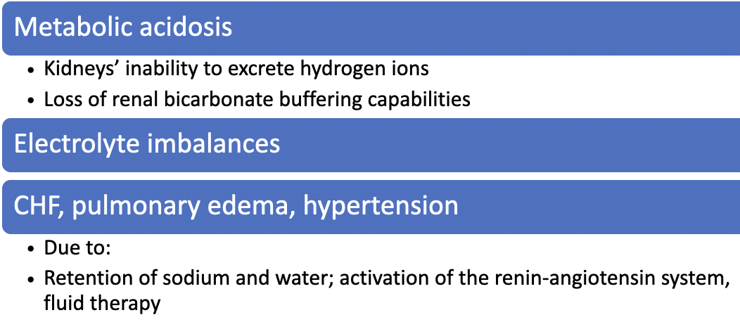
Metabolic acidosis
Fluid / electrolyte imbalances
Effects of Aging on Urinary System/Gerontologic Considerations
Between ages 30 and 90
Size and weight of kidneys decrease 20% to 30%
By seventh decade
Loss of 30% to 50% of glomerular function
Atherosclerosis accelerates decrease of renal size with age
Physiologic changes
Loss Between ages 30 and 90
Size and weight of kidneys decrease 20% to 30%
By seventh decade
Loss of 30% to 50% of glomerular function
Atherosclerosis accelerates decrease of renal size with ageof elasticity and muscle support
Decreased renal blood flow
Results in decreased GFR
Altered hormonal levels result in
Decreased ability to concentrate urine
Altered excretion of water, sodium, potassium, and acid
Prostate enlargement
Gerontologic Considerations
More susceptible to AKI
Dehydration due to polypharmacy- diuretics & laxatives
Illness and immobility
Hypotension
Aminoglycoside therapy
Obstructive disorders
Surgery
Infection
Under normal conditions, the aging kidney is able to maintain homeostasis.
However, after abrupt changes in blood volume, acid load, or other insults, the kidney may not be able to function effectively because much of its reserve has been lost.
Categories of ARF
Chart 49-4
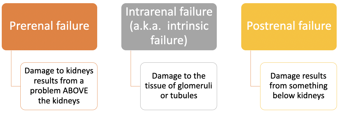
Causes: Prerenal, Intrarenal, Postrenal
Image
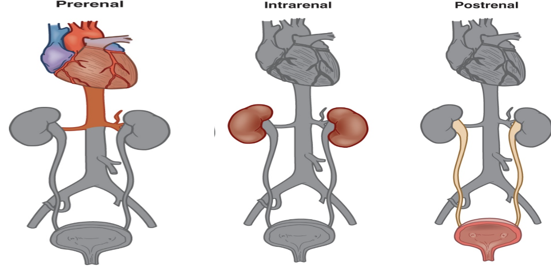
Prerenal Causes
Volume depletion
Hemorrhage, overuse of diuretics, excessive GI losses (vomiting, diarrhea), severe dehydration
Impaired cardiac function
Heart failure, MI, cardiogenic shock, valve disease, damage to renal artery
Vasodilation
Medications, anaphylaxis, sepsis
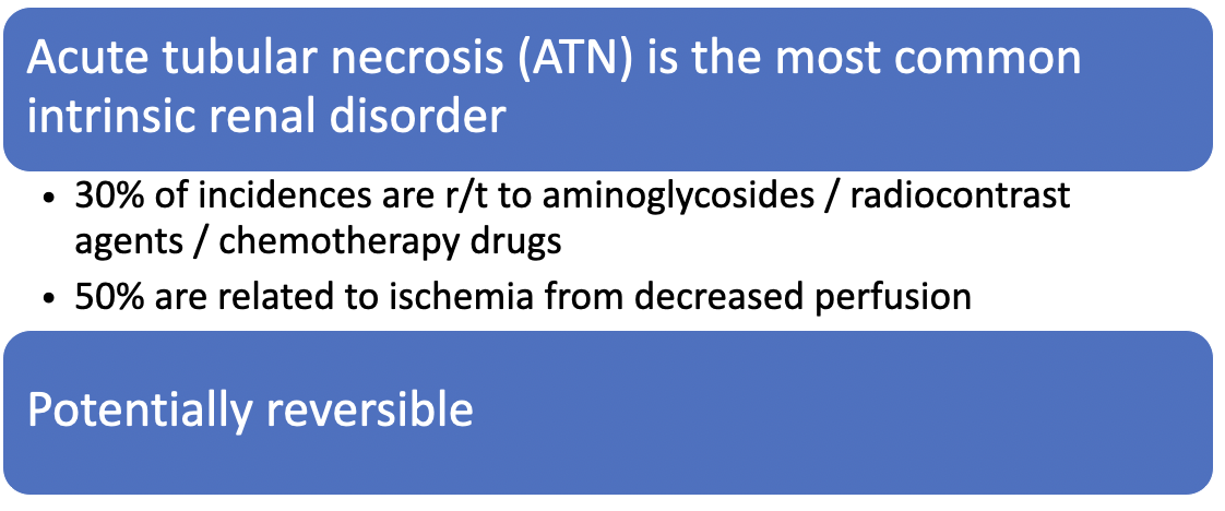
Intrarenal Causes
Prolonged renal ischemia
Myoglobinuria (burns, massive trauma, injuries involving a large amount of muscle damage); hemoglobinuria
Nephrotoxic agents
Aminoglycosides antibiotics , NSAIDS, radiopaque contrast agents
Infection
Pyelonephritis, glomerulonephritis, etc. (e. coli, various bacteria, fungi, protozoan or viral causes)
Intrarenal Failure
Image
Postrenal Causes
Obstruction in flow of urine from ureters to external urethral opening
Urolithiasis (stones), blood clots or stricture
Tumors (prostate, ovarian or cervical or colon cancer)
Bladder dysfunction
Obstruction (benign prostatic hyperplasia, obstructed indwelling urinary catheter, ureteral obstructions)
Trauma
Obstructions to urine outflow
Image
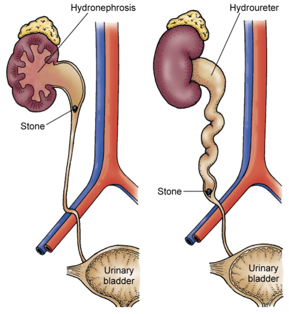
Obstructions to urine outflow
Obstruction
Increased pressure in kidney structures
Pressures interfere with function
Can’t filter or regulate wastes / fluids
Tissue injury occurs
GFR decreases
Hydronephrosis
Kidney Failure
Acute Kidney Injury Progression
AKI may progress through phases: oliguric, diuretic, and recovery.
When a patient does not recover from AKI, then CKD may develop.
Oliguria
UOP < 400 mL/day
Minimum amount of urine required to rid the body of waste products is 400 mL/day
Anuria:
UOP < 50 mL/day
Uremia:
Excess of urea and other nitrogenous wastes in blood
Azotemia
Abnormal concentration of nitrogenous wastes in blood
Diagnostics
Image
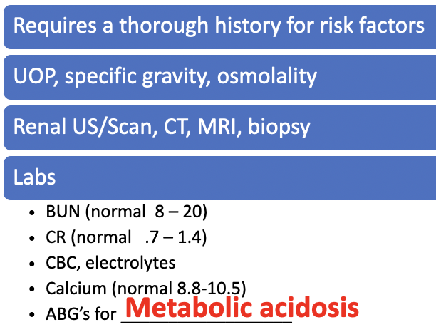
Urine Studies
Image
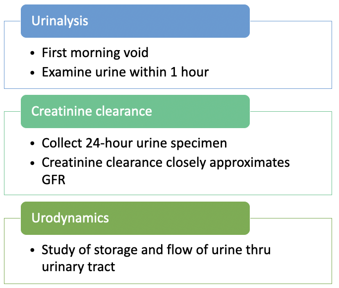
Radio-contrast induced nephropathy
Contrast-induced nephropathy
Some contrast agents can result in nephrotoxic injury
Major cause of hospital acquired ARF
Identify patients at risk before procedures
Baseline Cr > 2.0 mg/Dl = high risk
Recommendations
pre-procedure hydration may be considered
Use of other diagnostic procedures - ultrasound
Client receiving metformin
Should be held 48 hours prior and post procedure to decrease risk of lactic acidosis*
Follow your institution’s policy. Guidelines and opinions vary
Phases of AKI / ARF
Image

Oliguric Phase
Image
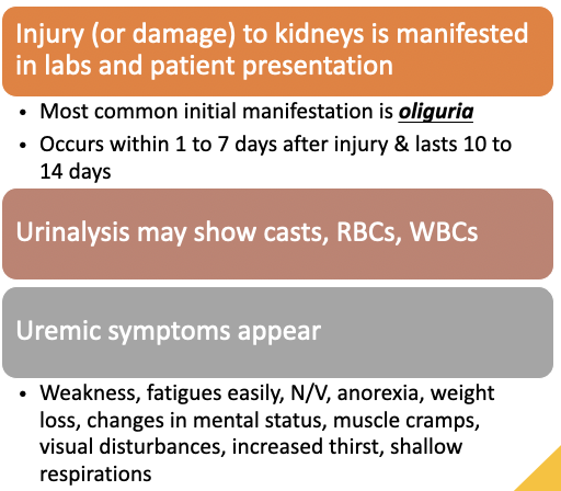
Oliguric Phase: Clinical Manifestations
Fluid overload
Contributes to heart failure, pulmonary edema, pericardial or pleural effusions
Elevated BUN, CR
Neuro symptoms
Fatigue, difficulty concentrating, seizures, stupor, coma
Hypermagnesemia
Kidneys cannot excrete magnesium
Hypocalcemia:
Kidneys cannot produce the active component of vit D in order to absorb calcium through GI tract
Hyperphosphatemia
Low calcium = high phosphorus
Metabolic acidosis
Impaired kidney cannot excrete hydrogen ions
Serum bicarbonate decreases
Existing levels depleted attempting to buffer acids
Also production is affected
Severe acidosis develops
Kussmaul respirations
Diuretic Phase: Clinical Manifestations
Begin to see increases in UOP
1 to 3 L (can get up to 5 L / a day!)
Glomerular filtration is recovering
Improvement in ability to excrete wastes
BUT – can’t concentrate urine
Lab values begin to normalize
Uremic symptoms may still be present
Volume may reach normal or elevated levels
At risk for excess fluid loss
Monitor for:
Hyponatremia
Hypokalemia
Dehydration
Recovery Phase
Image
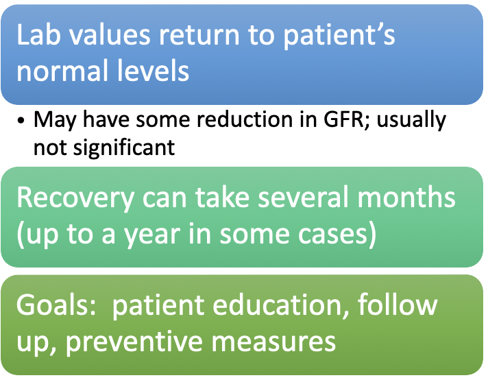
AKI Complications
Image
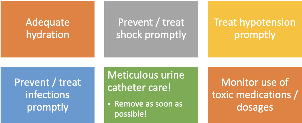
Prevention of AKI/ARF
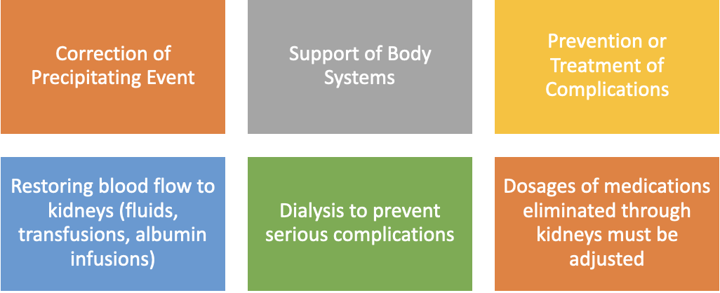
AKI Medical Management
Pharmacologic Therapy
Loop Diuretics
Furosemide or bumetanide may be prescribed to promote diuresis.
Help remove excess fluid and manage fluid overload.
May increase urine output but do not repair kidney damage.
IV Fluids and Volume Replacement
IV fluids may restore kidney perfusion in patients with prerenal AKI caused by hypovolemia.
Blood products may be administered when volume or blood replacement is indicated.
Albumin may be prescribed when hypovolemia is caused by low protein levels.
Medication Safety
Discontinue medications that are toxic to the kidneys.
Adjust medication dosages according to kidney function or creatinine clearance.
Avoid radiographic contrast when possible.
Nutritional Therapy
AKI increases metabolism and tissue breakdown, placing patients at high risk for malnutrition. Nausea and vomiting may further reduce nutritional intake.
Monitor weight daily.
Weight loss may indicate that calorie intake is below the patient’s needs.
Weight gain, failure to lose weight, or hypertension may indicate fluid retention.
Provide a high-protein, high-calorie diet.
Individualize nutrition based on the cause of AKI, degree of tissue breakdown, kidney replacement therapy, other health conditions, and nutritional status.
Include a renal dietitian as an essential member of the healthcare team.
Nutritional Therapy
Image

Nutritional imbalances in AKI
Caused by nausea, vomiting, inadequate dietary intake
Impaired glucose use and protein synthesis
Increased tissue catabolism
Weight monitoring
Daily weight checks are essential
Loss of 0.2–0.5 kg (0.5–1 lb) per day may indicate negative nitrogen balance (caloric intake < requirements)
Weight gain, no weight loss, or development of hypertension may indicate fluid retention
Nutritional support considerations
Based on: underlying cause of AKI, catabolic response, type/frequency of renal replacement therapy (RRT), comorbidities, and nutritional status
Protein replacement is individualized to maximize benefit and minimize uremic symptoms
Dietary approach
High-carbohydrate meals to spare protein for growth/tissue healing
Restrict sodium, potassium, and phosphorus-containing foods (e.g., bananas, citrus fruits/juices, dairy products)
Phases of AKI
How long they last
Oliguric phase: lasts 10–14 days
Diuretic phase: urine output increases, signals beginning of recovery phase
Fluid and electrolyte management
Guided by blood chemistry results and daily weights
Replace sodium, potassium, and water as needed, avoiding over- or underhydration
Post-diuretic phase
High-protein, high-calorie diet
Gradual return to normal activities
Focused Renal Assessment
Physical (cardiopulmonary, renal and overall hemodynamic status)
Previous renal problem history
Recent use of nephrotoxic substances
Recent exposure to heavy metals or organic solvents
Hypotensive episode of > 25 minutes
Tumors or clots in renal area
Presence of infection
Nursing Assessment
Image
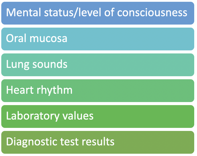
Nursing Assessment
Pulmonary
Assess for signs of circulatory overload.
Cardiac
Assess for signs of decreased cardiac output
Auscultate heart sounds for presence of third heart sound (could indicate heart failure)
Nursing Diagnoses and Collaborative Problem
Image
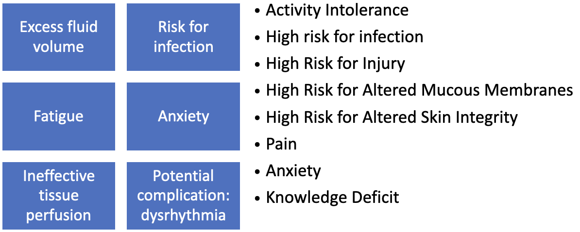
Nursing Interventions - AKI
Monitor fluid / electrolytes
I’s and O’s (include all emesis, drainage, stools)
Lab values
S/sx of hyperkalemia (cardiac* and musculoskeletal systems)
Weigh daily
Monitor heart rhythms, serum electrolytes
Monitor s/sx metabolic acidosis
If peripheral edema is present, move the patient gently and reposition often.
Give oral and IV fluids as prescribed BUT administer IV medications in least amount of fluid possible.
Administer medications (e.g., diuretics) as prescribed.
Diuretic therapy requires close supervision because reduced blood volume can result in inadequate renal perfusion.
Client may require renal replacement therapy if indicated.
Therapy clears the body of excess fluid and waste products. Renal replacement therapy may be needed to clear solutes, and dehydration should be treated.
Nursing Interventions: Hyperkalemia
Image

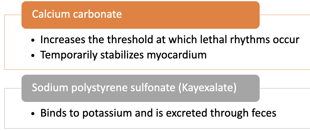
Treatment Options for Renal Failure
Hemodialysis
Peritoneal dialysis
Continuous renal replacement therapies (CCRT)
Renal Replacement Therapy (RRT)
Alternative or adjunctive method for treating ARF
Means by which uremic toxins and fluids are removed
Acid–base status/electrolytes adjusted slowly and continuously
Renal Replacement Therapy (RRT)
Indications
Volume overload
Elevated serum potassium level
Metabolic acidosis
BUN level > 120 mg/dL (43 mmol/L)
Significant change in mental status
Pericarditis, pericardial effusion, or cardiac tamponade
Evaluation - AKI
The expected outcomes are that the patient with AKI will
Regain and maintain normal fluid and electrolyte balance
Adhere to the treatment regimen
Experience no complications
Have complete recovery
Health Promotion - AKI
Image
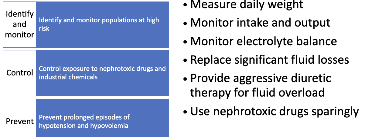
Urolithiasis and Nephrolithiasis
Image

Urolithiasis refers to stones anywhere in the urinary tract.
Nephrolithiasis refers specifically to kidney stones.
Stones may develop in one or both kidneys and vary greatly in size.
Kidney stones are common and frequently recur.
Urolithiasis and Nephrolithiasis
How Stones Form
Stones develop when substances in the urine become highly concentrated and form crystals. Common substances include:
Calcium oxalate
Calcium phosphate
Uric acid
Calcium-based stones are the most common.
Urolithiasis and Nephrolithiasis
Major Risk Factors
Dehydration
Urinary tract infections
Urinary stasis
Immobility
High blood or urine calcium
Abnormal urinary tract anatomy
Gout or metabolic disorders
Inflammatory bowel disease, ileostomy, or bowel resection
Certain medications and supplements
Types of Stones
Image

Urolithiasis and Nephrolithiasis
Clinical Manifestations
Deep aching pain in the costovertebral or flank area
Hematuria and sometimes pyuria
Pain radiating toward the bladder, genitalia, or thigh
Urinary frequency or urgency with only a small amount of urine passed
Nausea, vomiting, diarrhea, or abdominal discomfort
Fever and chills if infection is present
Renal colic causes sudden, severe flank pain with costovertebral tenderness, nausea, and vomiting.
Ureteral colic causes severe, wave-like pain that radiates to the thigh or genital area. Blood may appear in the urine as the stone irritates the urinary tract.
Bladder stones may cause irritation, UTI, hematuria, or urinary retention.
Urolithiasis and Nephrolithiasis
Assessment and Diagnostic Findings
Urinalysis: Checks for blood, infection, crystals, and other abnormalities.
Imaging: May include kidney, ureter, and bladder x-rays; noncontrast CT; or abdominal ultrasound.
Blood tests: Evaluate substances associated with stone formation.
24-hour urine collection: Measures calcium, uric acid, creatinine, sodium, urine pH, and total urine volume.
Health history: Reviews diet, medications, and family history of kidney stones.
Stone analysis: Determines the stone’s chemical composition and may identify the underlying metabolic disorder.
Urolithiasis and Nephrolithiasis
Medical Management
Treatment goals are to:
Remove or help the stone pass
Relieve pain and obstruction
Treat infection
Prevent kidney damage
Identify the stone type and prevent recurrence
General Management
Administer opioids or NSAIDs for renal colic.
NSAIDs also decrease prostaglandins, swelling, and ureteral pressure.
Encourage fluids unless contraindicated by vomiting, heart failure, or fluid restrictions.
Recommend 8–10 glasses of water daily and a urine output greater than 2 L/day.
Nutritional and Medications by Stone Type
Calcium stones: Liberal fluids; calcium restriction may be recommended for certain patients. Thiazide diuretics may reduce urinary calcium.
Uric acid stones: Follow a low-purine diet and avoid foods such as organ meats, shellfish, anchovies, asparagus, and mushrooms. Allopurinol may lower uric acid and reduce stone size.
Cystine stones: Increase fluids, follow a low-protein diet, and alkalinize the urine with potassium alkali salts.
Oxalate stones: Increase fluids and limit high-oxalate foods, including spinach, Swiss chard, chocolate, peanuts, and pecans.
Urolithiasis and Nephrolithiasis
Interventional Procedures
Ureteroscopy
Extracorporeal shock wave lithotripsy
Percutaneous stone removal
Urolithiasis and Nephrolithiasis
Surgical Management
Nephrolithotomy
Urterolithotomy
Cystotomy
Pyelolithotomy
Nephrectomy
Ureteroscopy
Image
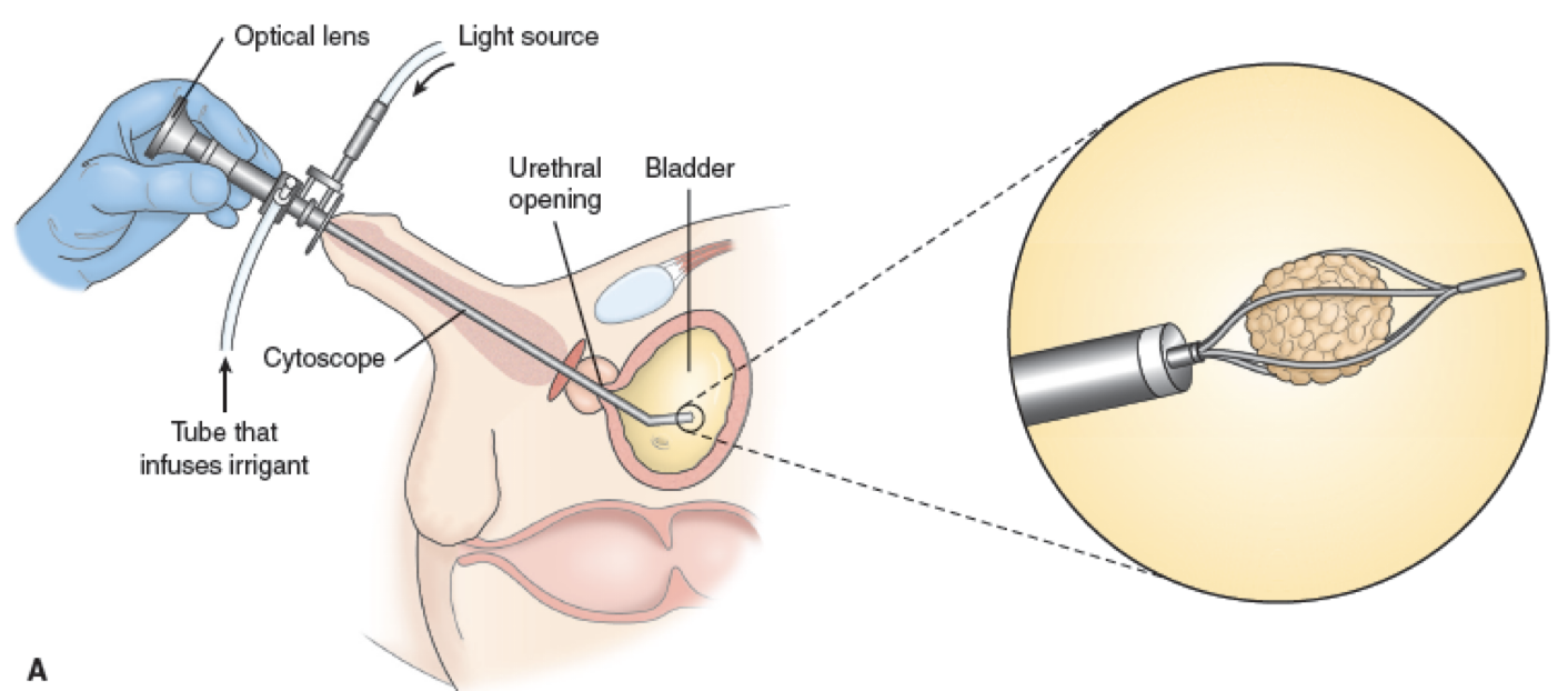
Percutaneous Nephrolithotomy
Image
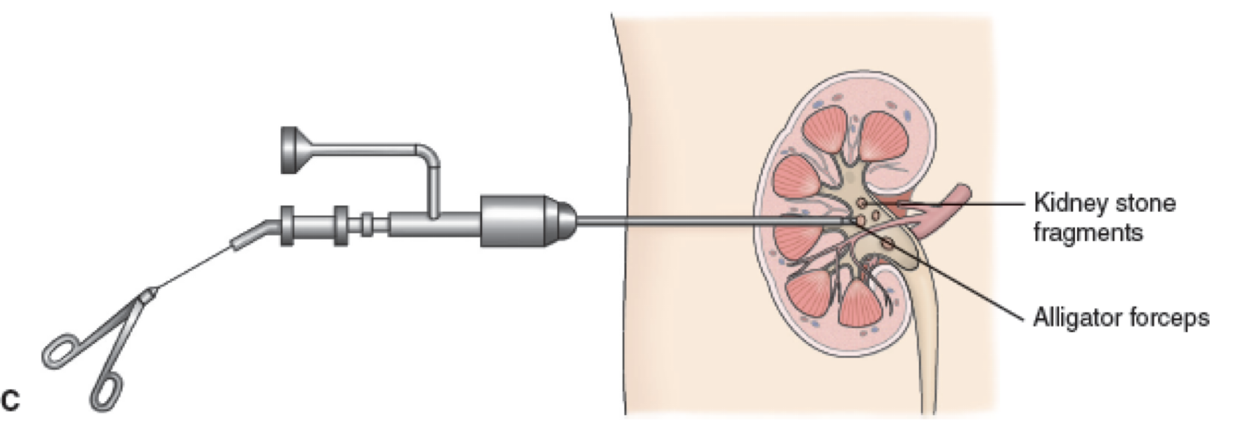
Patient Education for Renal Calculi

Nursing Interventions for Renal Calculi
Assess
Pain
n/v/d
Abd Distention
UTI
Obstruction
Strain urine
Increase fluid intake
Monitor I/O
Monitor for infection
Sudden increase in pain
VS
Patient education