Human Morph Exam 2
 Call Kai
 Learn
Learn Practice Test
 Spaced Repetition
 Match
Match Flashcards
 Knowt Play
1/70

Earn XP
Description and Tags
Skeletal system
Name | Mastery | Learn | Test | Matching | Spaced | Call with Kai | Chat |
|---|
No analytics yet
Send a link to your students to track their progress
71 Terms
What are the components of the skeletal system?
Bones of skeleton
Cartilage
Ligaments
Other connective tissue
Bones Overview
-primary organ of the skeletal system
-Rigid framework of body
-Bone types: Compact & spongy
Compact bone
Dense/cortical bone
80% of bone mass
Spongy bone
cancellous/trabecular bone
located internal to compact bone
looks porous
20% of bone mass
Cartilage
-semirigid CT that is more flexible than bone
Types: Hyalone cartilage & Fibrocartilage
Hyaline Cartilage
attaches ribs to sternum
covers ends of some bonds
present within growth plates
used as a model for bone formation
Fibrocartilage
weight0bearing cartilage that can withstand compression
composes : intervertebral discs, pubic symphsis (joint where the fibrocatilage connects to allow for a small amount of movement) & menisci of knee
Structures composed of dense regular CT
-Ligaments (connect bones to bone)
-tendons (connect muscle to bone)
Function of skeletal system
-support & protrecting
-levers for movement
-Hematopoiesis (blood cell production that ocurs in red bone marrow CT)
-stores mineral & energy reserves (Calcium & phosphate)
Tissue types in Bone
-Connective (comoact & spoongy bone, dense irregular connective tissue, reticular connective tissued, blood)
-Nervous tissue (detects & monitors sensory stimuli about status of the bone)
Classification of bones
bones are determined by shape
-Long bones
-Short bones
-Flat bones
-Irregular bones
Long Bones
greater in length than width (femur, humerus)
Short Bones
length is almost = to width (carpals & tarsals)
-outside is composed of compact bone that is covered by periosteum; interior is spongy bone & has no medullary cavity
FLat Bones
-flat, thin surfaces that may be curved (cranial bones)
outside is composed of compact bone that is covered by periosteum; interior is spongy bone & has no medullary cavity
Diploe: spong bone in skull
Irregular bones
elaborate, sometimes complex shapes (vertebrae)
outside is composed of compact bone that is covered by periosteum; interior is spongy bone & has no medullary cavity
Anatomy of the long bone
Diaphysis
Medullary Cavity
Epiphysis
Metaphysis
Epiphyseal plate
periosteum
Endosteum

Diaphysis (long bone)
-elongated, cylindrical shaft that provides leverage & weight support
-compact bone w/ thin spicules of spongy bone extending inward
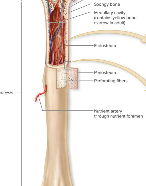
Medullary (marrow) cavity- long bone
-hollow, cylindrical space inside the diaphysis
-contains red bone marrow in kids & yellow bone marrow in adults
Epiphysis
-knob region at each end of the long bone
Proximal epiphysis: end of the bone closest to trunk
Distal epiphysis: end fartherst from trunk
composed of compact bone on the outer thin layer & spongy bone on the inner region
Articular cartilage: covers the joint surface reducing friction and absorbing shock in moving joints.
It is a thin layer of hyaline cartilage

Metaphysis
Region where the bone widens & transfers weight between the diaphysis & epiphysis

Epiphyseal plate
-located w/in metaphysis
-this is the growth plate; it provides lengthwise bone growth
-has a thin layer of hyaline cartilaage
-for adults the epiphyseal line is the remnant of the epiphyseal plate

Periosteum
-covering & linning of bone
-Tough sheath covering outer surface of bone
-Outer fibrous layer (dense irregular CT that protects bone from surrounding structures, it anchors blood vessels & nerves to bone surface & is an attachment site for ligaments & tendons)
-Inner cellular layer (includes osteoprogenitor cells, osteoblasts)
-Attached to bone by numerous collagen fibers (perforating fibers)

Endosteum
-covers internal surface of bone w/in medullary cavity
-thin later of CT containing osteoprogenitor cells & osteoblasts & osteoclast

Blood supply of the blood
-bone is highly vascularized (spongy bone)
-Nutrient foramen is a small hole in bone that allows artery entrance & vein to exit
Nerve supply of the bone
-blood vessels also go through foramen
-innervate bone, periosteum, endosteum, marrow cavity
mostly sensory nerves
Bone Marrow
this is the soft ct of the bone
consists of red & yellow bone marrow
Red Bone Marrow
(myeloid tissue)
Hematopoietic (forms blood cell)
Consists of reticular ct, develops blood cells & adipocytes
Children: this is found in spongy bone & medullary cavity of long bones
Adults: found only in specific areas of axial skeleton (skull, vertebrae, ribs, sternum, ossa coxae, proximal epiphyses of humerus & femur)
Yellow bone marrow
-red bone marrow degeneration as you mature
-fatty substance
-may convert back to red bone marrow during severe anemia or any condition w/ reduced eythrocytes (red blood cells)
-faciliates production of more erythrocytes
Bone CT (Osseous CT)
-primary component of bone
-composed of cells & extraceulluar matrix
Cells of bone
-these 4 types are found in bone CT:
Osteoprogenitor cells
Osteoblasts
Osteocytes
Osteoclasts
Osteoprogenitor cells
-stem cells come from mesenchyme
-cell division yield stem cell & a committed cell (matures to become an osteoblast)
cells are located in the periosteum & endosteum
Osteoblasts
-forms from osteoprogenitor stem cells
-they synthesize & secrete osteoid
-is entrapped w/in the matrix
-differentiate into osteocyes
Osteoid
initial semsisolid organic form of the bone matrix from the osteoblasts that calcifies later
Osteocytes
-mature bone cells that come from osteblasts
-detects stress on bone; triggering new bone formation
Osteoclasts
-large, multinuclear, phagocytic cells
-derived from fused hematopoietic cells
-ruffled border increases surface area exposed to bone
-involved in bone resoprtion
-w/in/ adjacent to a depression/pit on bone surface (resoprtion lacuna)
Inorganic componenets of the bone matrix
-Salt cyrstals, calcium phosphate, Ca3(PO4)2
-interacts w/ calcium hydroxide to form crystals, hydroxyapatite, a10(PO4)6(OH)2
-calcium carbonate, sodium, magnesium, sulfate, flouride is incorporate into crystals
-Crystals deposite aorund collagen fibers
-They harden the natrix & account for rigidity of bones
Organic components of bone matrix
-osteoblasts produce osteoid that contains collagen proteins & semisolid ground substance of preteoglycans & glycoproteins
give bone tensile strength by resisting stretching
-contributes to bone flexibility
Bone formation
-starts w/ secretion of osteoid
Calcification occurs bcuz hydroxyapatite crystals (calcium & phosphate ions precipates out) & deposit
Bone formation requires:
Vit D (to enhance Ca2+ absorption from GI tract)
Vit C: for collagen formation
Calcium & phosphate for calcification
Bone resoprtion
occurs when blood calcium levels are low
Bone matrix becomes destroyed by substances released from osteoclasts
Proteolytic enzymes are released from lysosomes w/in osteoclasts to chemically digest organic matrix components
Calcium & phosphate is dissolved by HCL acid into ions & enter the blood
Process of Calcification
Osteoblasts produce osteoid
Osteoid forms collagen matrix
Hydroxyapatite crystals form
Crystals depost on collagen fibers
Mineralized bone matrix is produced
What is the calcification of bone
deposition of hydroxyapitite crystals w/in the osteoid organic matrix
Osteons (compact bone)
small cylindrial strucutes (Haversian systems)
-basic functional & structural unit of mature compact bone
-oritented parallel to bone diaphysis that looks like bull’s eye target

Osteon Components
-Central (Haversian) canal:
-Concentric lamellae
-Osteocytes
-Canaliculi
Central (Haversian) canal
Cylindrical channel at center of osteon & parallel to it
Blood vessels & nerves extend through channel
Concentric lamellae
rings of bone CT
surrounds central canal
collagen fibers that give bone strength & resilience; 90 degrees from previous & next lamellae
Osteocytes
-Mature bone cells
-found in small spaces between concentric lamellae (lacunae)
-purpose is to maintain bone matrix
Canaliculi
-Tiny connecting channels w/in bone CT
-extends from lacuna—-lamella & connects to lacunae & central canal
-house osteocyte projections that allow cell contact
-allows exchange of nutrients, minerals, gases & wastes betwween blood vessels & osteocytes

Difference between lacuna & lacunae
-Lacuna: 1 single space/pocked w/in the bone matrix that houses 1 osteocyte
-Lacunae: plural- multiple spaces/pockets throughout the bone tissue that has osteocytes
Structures in long bone (not part of osteon)
-Perforatiing canals
-Circumferential lamellae
-Interstitial lamellae
-Trabeculae
-Parallel Lamella
Parallel lamellae
-bone matrix
-osteocytes are found between these lamellae
-canaliculi radiate from lacunae

Trabeculae
-open lattice of narrow rods & plates of bones
-the bone marrow fills the spaces and theres a meshwork of crisscrossing bars
-purpose: resisting stress
Perforating (Volkmann) canals
-perpendicular to central canals
-connects central canals w/in different osteons

Circumferential lamellae
-External: rings of bun run internal to periosteum
-Internal: rings of bone that go internal to the endosteum
-They run the entire circumference of the bone
Interstitial lamellae
-components of compact bone between osteons
Properties of hyaline cartilage connective tissue
-these cartilage cells are scattered through matrix of protein fibers & embededd in gel-like ground substance (proteoglycans but not Ca2+)
-very resilent & flexible
-high % of H2O
-Avascular & no innervation
-compressible & a good shocks absorber
Hyaline cartilage CT
-Chondroblasts: produce cartilage matrix
-Chondrocytes: chondroblasts encased in the matrix, found in lacunae & they maintain the matrix
-Perichondrium: dense irregular CT that covers cartilage & helps maintain its shape
Ossification (osteogenesis)
is the formation & development of bone CT
-starts in the embryo & continues through childhood & adolescence
~8-12wks of embryonic development: Skeleton starts forming (from intramembranous ossification/endochondral ossification)
What is intramembranous ossification?
bone growth w/in a membrane that produces flat bones of skull, some facial bones, nadible & central part of the clavicle
aka dermal ossification
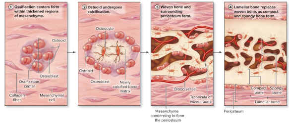
What are the (4) steps of intramembranous ossification?
-Ossification centers form w/in thickened regions of mesenchyme starting @~8th wk of development
Some cells become osteoprogenitor cells, or osteoblasts secreting osteoid
-Osteoid Undergoes calcification
(Ca2+ salts deposit onto osteoid & crystalize & the entrapped cells become osteocytes)
-Woven bone (primary) & surrounding periosteum form
Bone is immature & poorly organized until the mesenchyme around the woven forms periosteum
-Lamellar bone replaces woven bone, as compact & spongy bone forms
compact & spongy bone form from trabeculae
this is the typical structure of a flat cranial bone (2 external layers of compact bone & a layer of spongy bone in between)
Endochondral ossification
starts w/ hyaline cartilage
-produces most bones of skeleton (upper & lower limbs, pelvis, vertebrae, ends of clavicle)
What are the steps of endochondral ossification
-Hyaline cartilage model of bone forms
-Bone 1st replaces hyaline cartilage in the diaphysis
-Bone replaces hyaline cartilage everything except epiphyseal plates & articular cartilage
-By a person’s late 20’s, all epiphyseal plates have ossified & lengthwise bone growth is complete