TEST 1 - CH 4,5,6,8
 Call Kai
 Learn
Learn Practice Test
 Spaced Repetition
 Match
Match Flashcards
 Knowt Play
1/94

There's no tags or description
Looks like no tags are added yet.
Name | Mastery | Learn | Test | Matching | Spaced | Call with Kai | Chat |
|---|
No analytics yet
Send a link to your students to track their progress
95 Terms
What lies in the retroperitoneum?
Suprarenal glands, AO + IVC, most of duodenum, pancreas, ureters, ascending/descending colon, kidneys, esophagus, rectum
Layers or the artery
Tunica intima (inner) - Endothelium that lines the lumen of all vessels
Tunica media (middle) - Smooth muscle cells and elastic fibers
Tunica adventitia (outer) - Collagen fibers
Proximal
When a structure is closer to the body midline or point of attachment to the trunk. For example, the knee is proximal to the ankle.
Distal
Farther from the midline or point of attachment to the trunk. For example, the ankle is distal to the knee
Cephalic/Superior/Cranial
Towards the head
Caudal/Inferior/caudad
Towards to the feet
Anterior/Ventral
The front (belly) surface of the body
Posterior/Dorsal
The back surface of the body
Medial
The superoinferior body axis that goes right through the midline of the body; Medial structures are those that are closer to the midline of the body than they are to another structure.
Ex) The aorta is medial to the kidneys
Lateral
Toward the side of the body; Ex) The ovaries are lateral to the uterus
Sagittal plane
Lengthwise plane running from front to back; Divides the body or any of its parts into right and left sides
Midsagittal plane
Divides the body into equal halves
Transverse plane
Divides the body or any of its parts into upper and lower portions
Coronal plane
Lengthwise plane that runs from side to side and divides the body into anterior and posterior portions
What are the abdominal regions?
From top to bottom:
Right Hypochondrium, epigastrium, Left hypochondrium
Right lumbar region, umbilical region, left lumbar region
Right iliac fossa, hypo-gastrium, left iliac fossa
What region of the abdomen is the liver in?
Right lobe - Majority is in the right hypochondrium and epigastrium
Left lobe - Left hypochondrium and epigastrium
Gallbladder would also be in the right hypochondrium
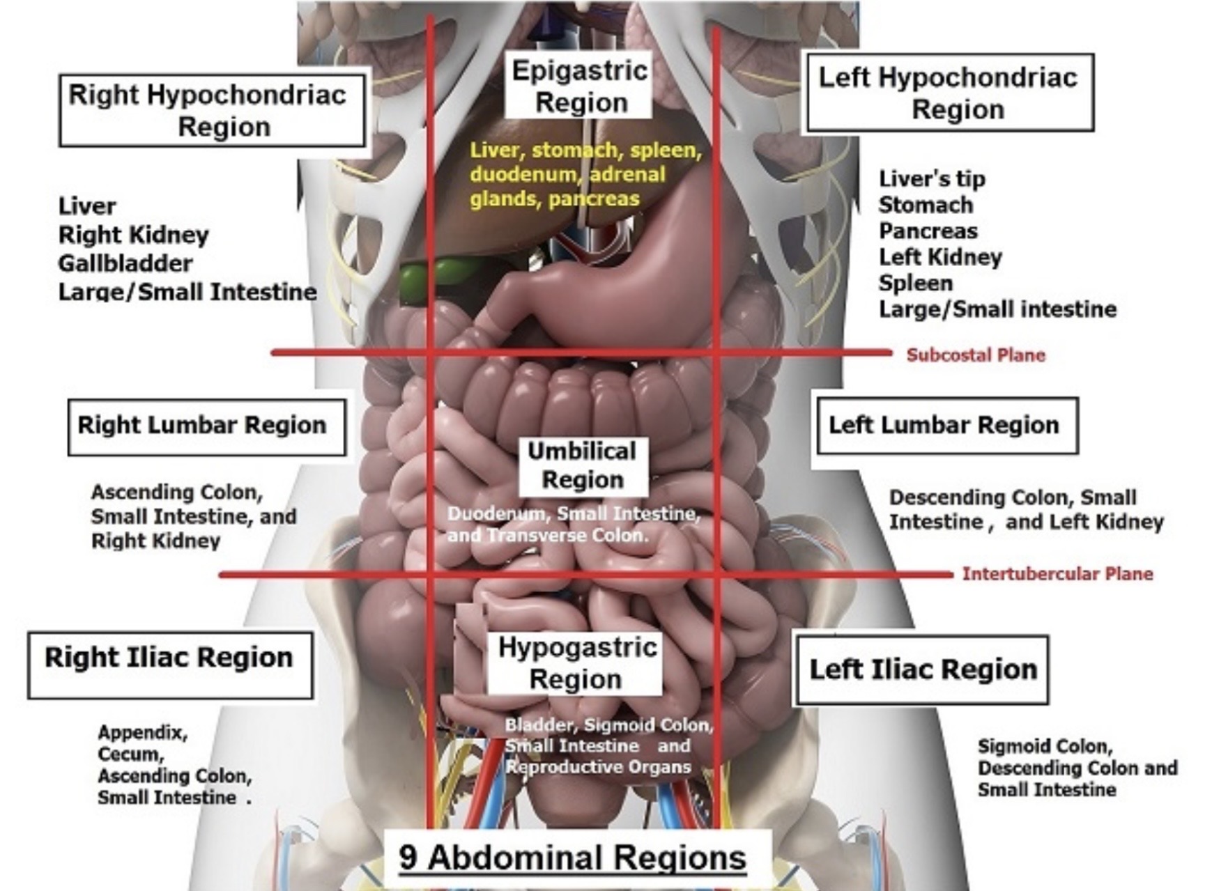
Major organs in the epigastrium
Stomach and Pancreas

Major organs in the left hypochondrium
Spleen

Major organs in the right hypochondrium
Majority of right lobe of the liver and gallbladder

Major organs of the right lumbar region
Ascending colon, small intestine, and right kidney

Major organs of the umbilical region
Duodenum, small intestine, and transverse colon

Major organs of the left lumbar region
Descending colon, small intestine, and left kidney

Major organs of the right iliac fossa
Appendix, cecum, ascending colon, small intestine

Major organs of the hypo-gastrium
Bladder, sigmoid colon, small intestine and reproductive organs

Major organs or the left iliac fossa
Sigmoid colon, descending colon and small intestine
What is anatomic position?
Assumes that the body is standing erect, the eyers are looking forward, and the arms are at the sides with the palms and toes directed forward

The patient positions
What is the typical patient position for abdominal examination?
Supine
Parietal peritoneum
Outer layer that lines the abdominal wall
Visceral peritoneum
Inner layer that covers the abdominal organs
Artifacts
Anything that does not properly present the structures or motion imaged; Caused by some problematic aspect of the imaging technique; Some artifacts are helpful and should be used to the sonographer’s advantage but some hinder the diagnostic process.
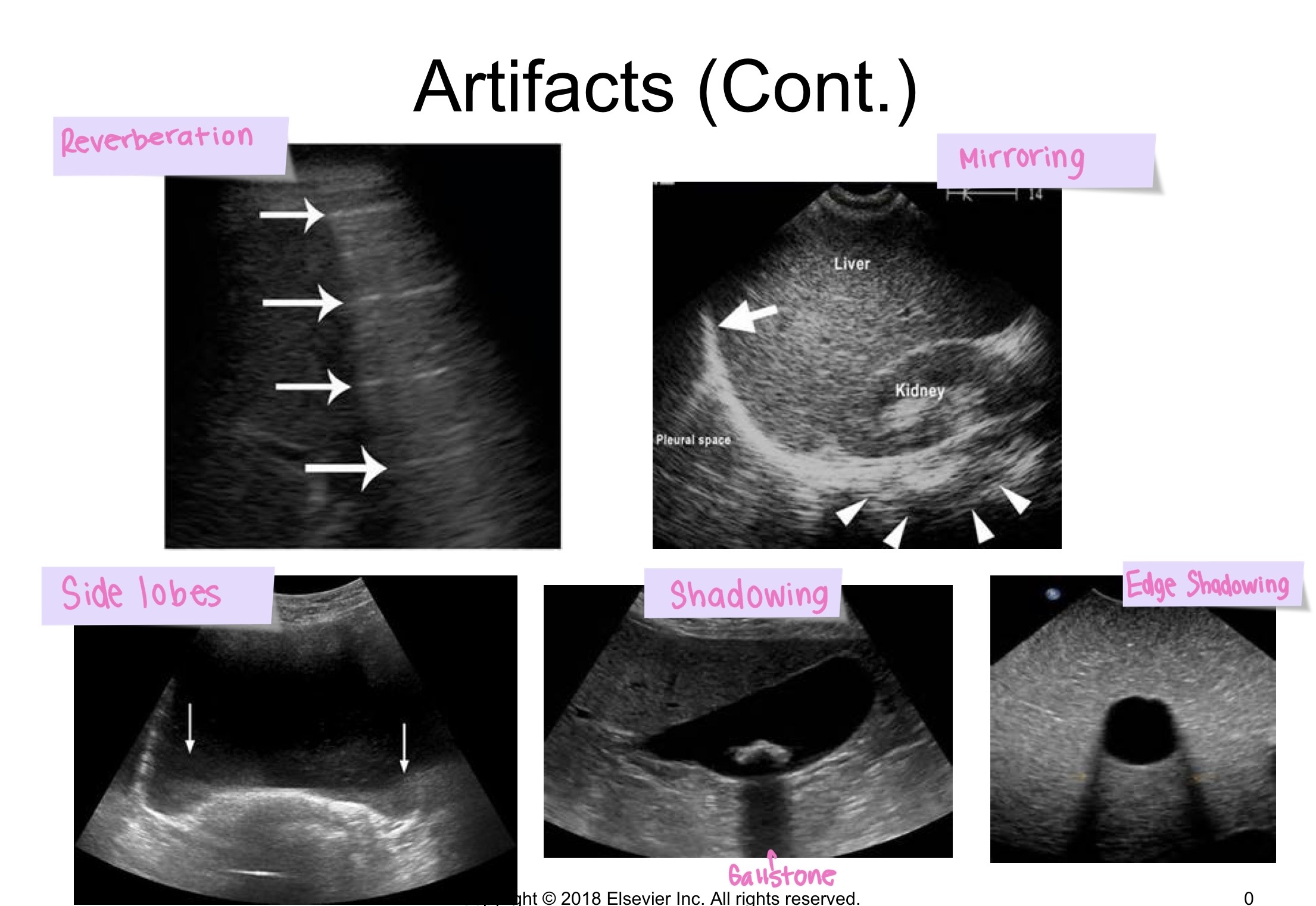
Examples of artifacts are
Reverberation, mirror, side lobe, shadowing, and edge shadowing
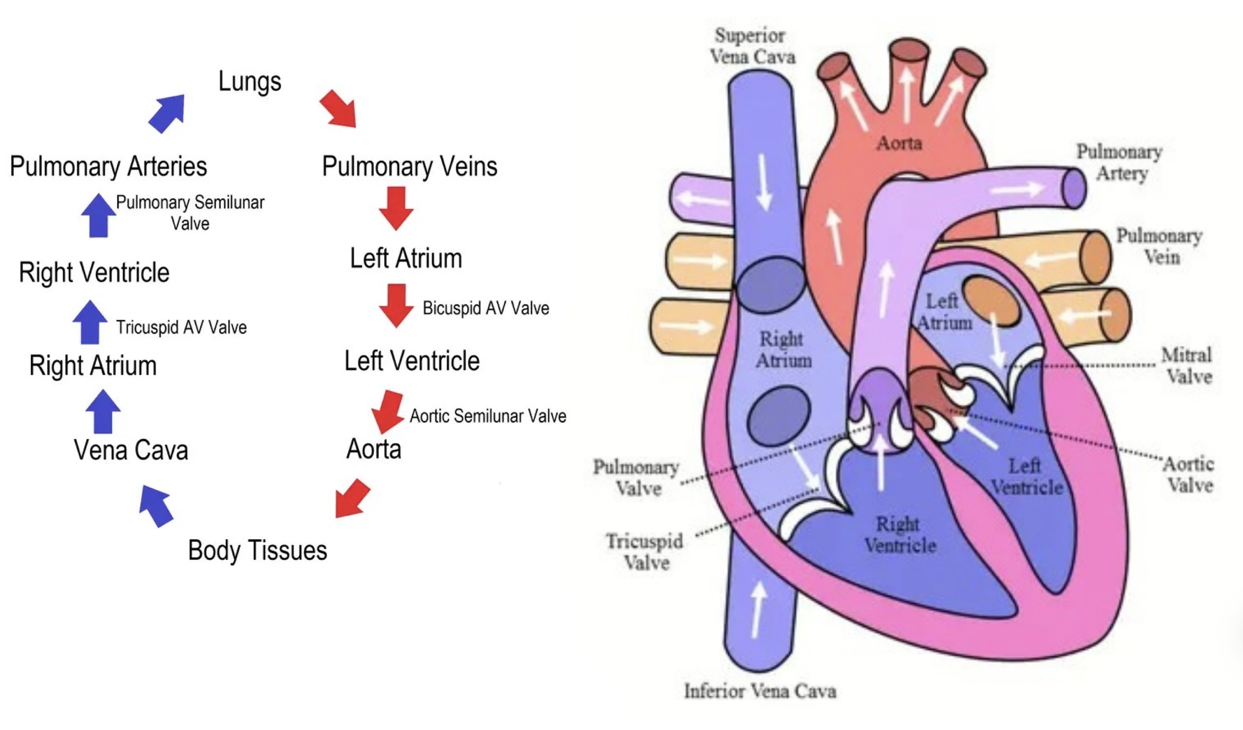
Blood flow through the heart
Arteries carry blood away from the heart and veins carry blood to the heart and back from the tissues.
Red = Oxygen
Blue = No oxygen

What branches off of the aortic arch?
Brachiocephalic trunk (only on the right side of the aortic arch)
Off the Brachiocephalic trunk are the Rt Subclavian and Rt Common Carotid Artery
Left common carotid artery
Left subclavian artery
What is another name for the brachiocephalic trunk?
Innominate
Anterior branches of the aorta
Celiac trunk, Common Hepatic Artery, SMA, IMA
Lateral branches of the aorta
Phrenic arteries, renal arteries, and gonadal arteries
What is the seagull sign
It is basically how the hepatic artery and splenic artery look in a TRV view
What is an aneurysm
A permanent localized dilation of an artery, with an increase in diameter greater than 1.5 times its normal diameter
What is a true aneurysm
A type of aneurysm that involves all three layers (intima, media, adventitia)
What percentage of aneurysms are infrarenal
95%

Types of aneurysms
Saccular - Bulges out on one side and the most common type of cerebral aneurysm
Fusiform - Bulges or balloons out on all sides of the blood vessel; Overall, most common type
What are risk factors of Abdominal Aortic Aneurysms (AAA)?
Tobacco
Hypertension
Vascular disease
Chronic obstructive pulmonary disease (COPD)
Genetics - Family history of AAA
Iliac and popliteal (behind the knee) aneurysm association
What are causes of AAA?
Atherosclerosis (plaque buildup) #1 cause
Trauma (after transection)
Congenital defect
Syphilis (involving the ascending aorta and arch)
Mycosis (fungal dissection)
Symptoms of AAA
Palpable abdominal mass, back pain, drop in hematocrit (rupture), may also be asymptomatic
Treatment for an aneurysm <4cm in diameter
Follow it every 6 months with intervention if the patient becomes symptomatic
Treatment for an aneurysm 4-5cm in diameter
Surgical intervention may be suggested if the patient is in good health
Treatment for an aneurysm >5-6cm in diameter
May benefit from a surgical repair, especially if the patient has other factors for rupture like hypertension, smoking, COPD
Treatment for aneurysms >6-7cm in diameter
Pose the greatest risk; Risk increases with age and other medical problems
Surgical intervention and AAA
Suggested with associated rneal and iliac involvment
Length of the infrarenal aortic neck is important to help determine the surgical approach
Endovascular Stent Grafts
Treatment for aneurysms that is less invasive; usually for infrarenal aneurysm
Basically, a large fabric-covered stent that is compressed and positioned through a catheter through the patient’s groin. Once in place, the graft is expanded to create a new path for blood flow.
What is the mortality rate if an aortic aneurysm ruptures?
50%
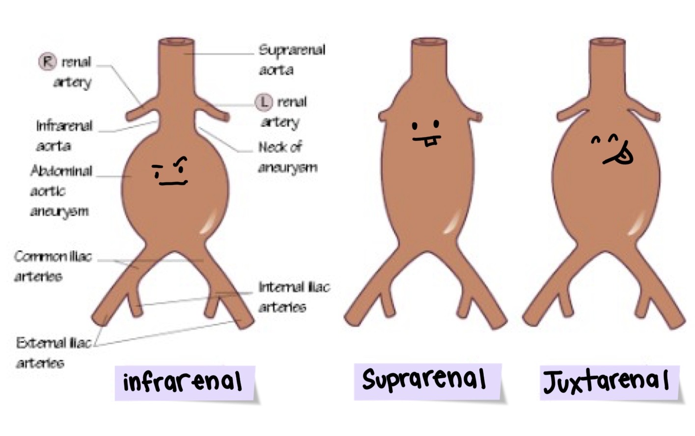
What are the most common locations for aneurysms
Infrarenal - Below the renal arteries
Suprarenal - Above the renal arteries
Juxtarenal - Just below the renal arteries

Types of Endoleaks
Type I - Gap between graft and vessel
Type II - most common, flow through branch vessels into aneurysm sac
Type III - Defect/misalignment of endograft materials
Type IV - Porous graft material
Type V (“endotension”) - No evidence of leak but continued enlargement of aneurysm

Pseudoaneurysm
A pulsative hematoma that results from the leakage of blood into the soft tissue abutting the punctured artery, with subsequent fibrous encapsulation and failure of the vessel wall to heal
Blood escapes through a hole in the intima of the vessel wall bit is contained by the deeper layers of the aorta or by the adjacent tissue
When are pseudoaneurysms seen
After invasive procedures (often cardiac cath)
Why are pseudoaneurysms false?
They don’t involve all three layers of the artery

What is aortic dissection
A separation (tear) of the intima from the media of the aortic wall; Increased pressure on outer wall can eventually rupture; They can extend into the carotid arteries or down the aorta to the femoral arteries
High mortality risk
Where do aortic dissections usually originate?
Aortic arch in the thorax
What are the causes of aortic dissection
Marfan syndrome - connective tissue disorder
Chronic hypertension
AAA
Iatrogenic cardiac intervention - caused by medical treatment
Congenital heart anomalies
Coarctation of aorta
Trauma
What are symptoms of aortic dissection
Severe abdominal pain
Shortness of breath
Dizziness or fainting
Rapid, weak pulse
Heavy sweating
A significant difference in blood pressure on right and left side
Stroke-like symptoms can also occur
Example of aortic dissection on ultrasound (looks like cracks)

What is the origin of the IVC
Formed by the union of the common iliac veins posterior to the right common iliac artery at the level of the fifth lumbar vertebra; Ascends vertically through the retroperitoneal space on the right side of the AO, posterior to the liver and piercing the central tendon of the diaphragm at the level of the eighth thoracic vertebra to enter the right atrium of the heart
What is the most common origin of pulmonary emboli?
Venous thrombosis from the lower extremities
What are IVC filters?
AKA Greenfield Filter; Surgical and angiographic placement of transvenous filters into the IVC have been used to prevent recurrent embolization patients who cannot tolerate anticoagulants
Appearance of greenfield filter (always important to check medical records because they can look like clots)

What forms the portal vein
Formed posterior to the pancreas by the union of the SMV and splenic veins
How long is the portal vein trunk?
5-7cm in length
Where does the portal vein carry blood to
Carries blood from the intestinal tract to the liver by means of its two main branches: the right and left portal veins
Where does the portal vein drain blood from
Drains blood from the gastrointestinal tract; from the lower end of the esophagus to the upper end of the anal canal; and from the pancreas, gallbladder, bile ducts, and spleen
How do the Lt and Rt portal veins course through the liver
Transversely, so scans display their longest extent
SMV spatial relationship to the pancreas?
Anterior to the uncinate of the pancreas, and posterior to the neck
Characteristics of the splenic vein
A tributary of the portal circulation that begins at the hilum of the spleen, where it is formed by the union of several veins; Runs along the posteromedial border of the pancreas and anterior to the SMA to form the portal vein
What is the portal triad? Mickey Mouse Sign
Part of the portal system that contains a branch of the portal vein, hepatic artery, and bile duct contained within a connective tissue sheath. This gives the portal vein an echogenic wall
SMV location
Passes anterior to the third part of the duodenum and posterior to the neck of the pancreas where it joins the splenic vein to form the main portal vein
What small veins does the SMV also drain
the middle colic vein (transverse colon), right colic vein (ascending colon), and pancreatic duodenal vein
Where does the SMV receive tributaries from?
Receives tributaries that correspond to the branches of the SMA where it is joined by the inferior pancreaticoduodenal vein to the right gastroepiploic vein from the right aspect of the greater curvature of the stomach

(Low) non-resistive vessels
Have a high diastolic component and supply organs that need constant perfusion
Ex) Internal carotid artery, hepatic artery, renal artery

(High) Resistive vessels
Have very little or even a reversed flow in diastole and supply organs that do not need a constant blood supply
Ex) External carotid artery and Iliac and brachial arteries
Low Resistance waveforms
More of a rounded systolic peak with significant flow during diastole and a noticeable end diastolic velocity
High Resistance waveforms
Sharp systolic peak and has a reversed diastolic flow due to resistance peripheral vascular bed.
Often triphasic
Rapid systolic peak followed by a brief reversal of flow and then a smaller forward flow in diastole
Plug Flow
Typically seen in large arteries, in which most cells are moving at the same velocity across the entire diameter of the vessel; A clear window under systole is typical of plug flow
When plug flow is present, the volume of blood flow can be calculated
Laminar flow
Smaller vessels and is bullet shaped
Turbulent flow
Basically, the blood is just going crazy and going in all types of directions. Happens with stenosis or in curved vessels.
Budd-Chiari Syndrome
Thrombosis of the haptic veins is present; Hepatic veins are small with echogenic material
Presence of normal flow excludes Budd-Chiari syndrome

Triphasic
Waveform that touches the baselines three times

Biphasic
Waveform that touches the baseline twice

Monophasic
Waveform that doesn’t touch the baseline

Nutcracker syndrome
Occurs when the left renal vein is compressed between the SMA and the aorta.
Can cause:
Venous hypertension (collaterals)
Retrograde flow into gonadal veins (pelvic congestion)

May Thunder Syndrome (Iliac Vein Compression)
Compression of the left common iliac vein against the lumbar vertebrae by the overlying right common iliac artery. This increases the risk for deep vein thrombosis

Celiac Band Syndrome (Median arcuate ligament syndrome)
Compression of celiac axis by the median arcuate ligament of the diaphragm; Clinically an abdominal bruit is noted on expiration, but disappears with inspiration
Expiration - The MAL compresses celiac axis causing a “stenosis”
Inspiration - Celiac axis straightens and the “stenosis” disappears and velocities return to normal
What is the aortas waveform like
Triphasic, high resistance
What is the renal arteries waveform like
low resistance
What is the hepatic veins waveform like
Bidirectional, pulsatile due to tricuspid regurgitation
What is the portal veins waveform like
Hepatopedal (pedal toward the liver) and undulatory