Unit 1 Practical Exam - Part 2
 Call Kai
 Learn
Learn Practice Test
 Spaced Repetition
 Match
Match Flashcards
 Knowt Play
1/32

Earn XP
Description and Tags
Anatomy of the microscope, cell cycle and mitosis stages, and cell anatomy
Name | Mastery | Learn | Test | Matching | Spaced | Call with Kai | Chat |
|---|
No analytics yet
Send a link to your students to track their progress
33 Terms
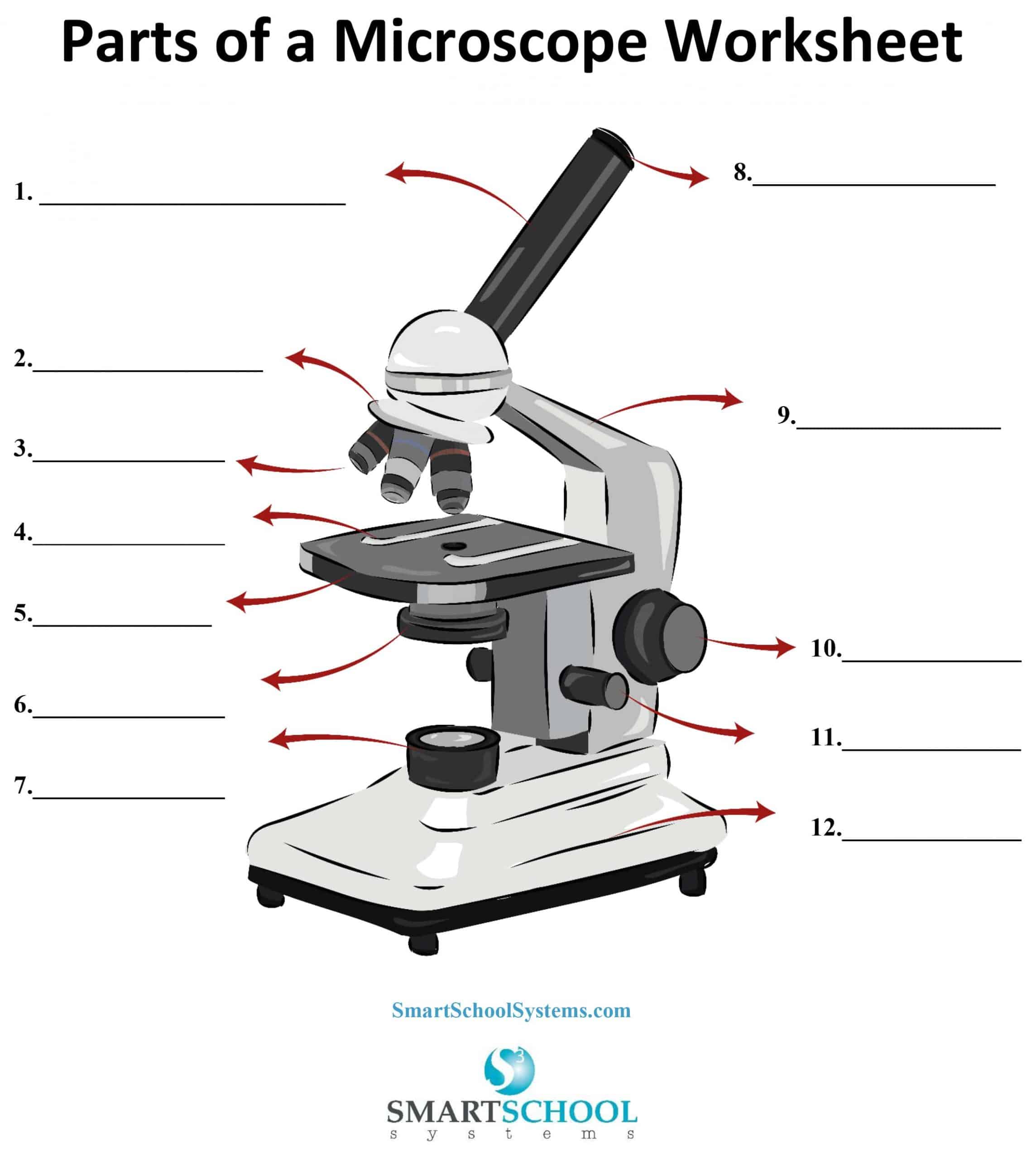
1 - 10x magnification; adjustable; one contains pointer
Eyepeice

2 - circular strucuture that supports the four objective lenses
Nosepeice

3 - the primary lenses located on the rotating nosepeicethat provide different msnification levels snd form the initial enlarged specimine 4x, 10x, 40× 100x
Objective Lens

4 - Holds slides in place
Stageclip

5 - pltaform that holds the stage
Stage

6 - focuses and concentrates light on specimen
condenser

7 - light source
Illuminator

9 - frame extending upward from the base
Arm
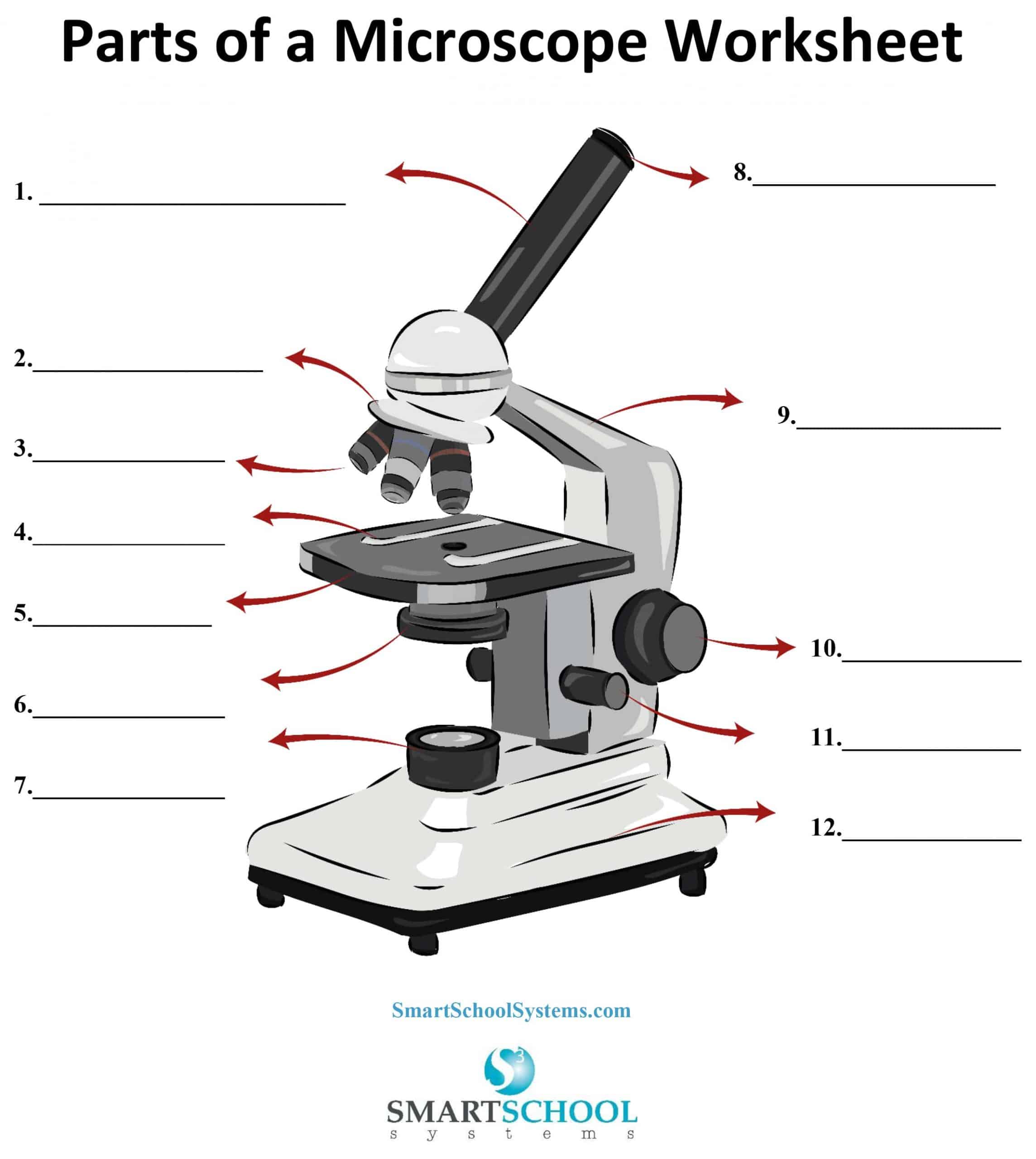
10 - Large movements; scanning & low power only (4x, 10x)
Course adjustment nob

11 - Small movements; high power & oil immersion
Fine adjustment nob
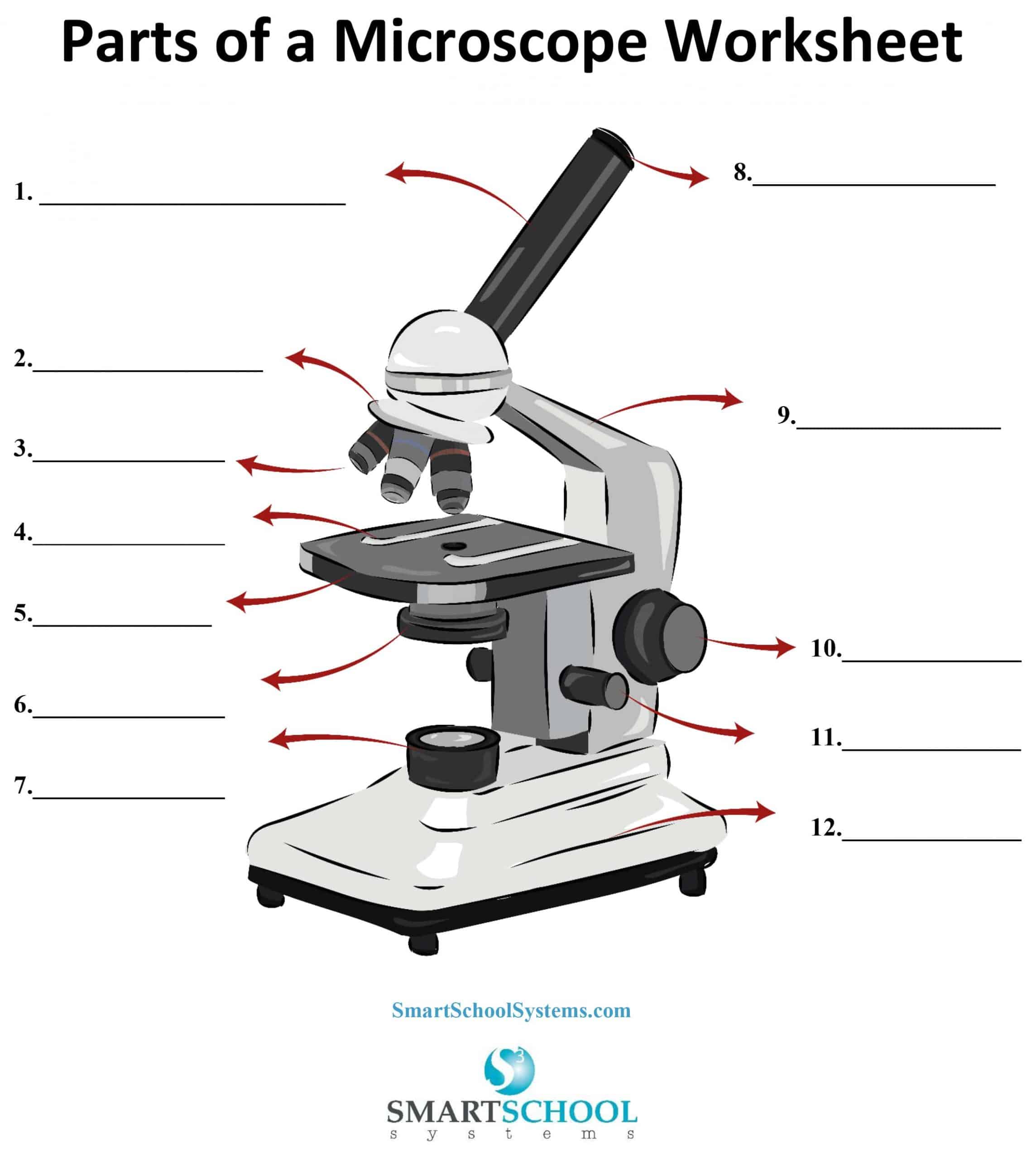
12 - Solid foundation that rests on the table
Base

Not labeled - opening in the stage for light passage
Aperture

Not labeled - moves slide on the stage
Stage controller

Not labeled - controls light intensity
Brigtness adjustment

During _____ a cell doesn't look like it is actively dividing, but critical changes are happening under the microscope
Interphase

The loose, DNA "spaghetti" bunches up into tight, visible, X-shaped chromosomes.
Prophase
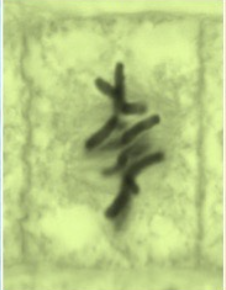
During _____, the cell's chromosomes align perfectly along the center of the cell.
Metaphase
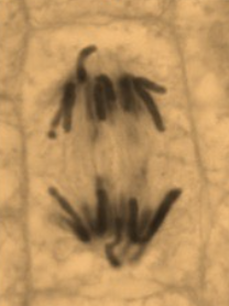
During ________, the main visual event is the separation and movement of chromosomes to opposite sides of the cell.
Anaphase
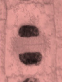
The cell looks like it is undoing the steps of prophase to return to a normal state
Telaphase
Cell fully separates into 2
Cytokinesis
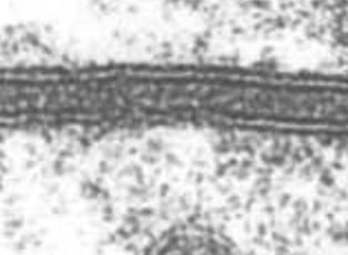
It reveals a distinct "railroad track" or trilaminar (three-layered) appearance. OUTER layer
Plasma Membrane

A small, hollow barrel or cylinder
Centrioles
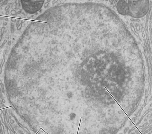
Appears as a dense, membrane-bound spherical structure filled with dark patches of inactive heterochromatin and lighter regions of active euchromatin
Nucleous
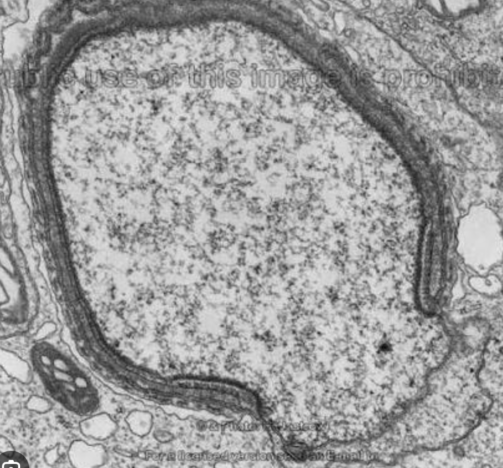
Appears as a double-membrane boundary that surrounds the cell nucleus
Nuclear envelope

Look like donut-shaped or flower-like ring structures studding the surface of the nuclear envelope
Nuclear pore
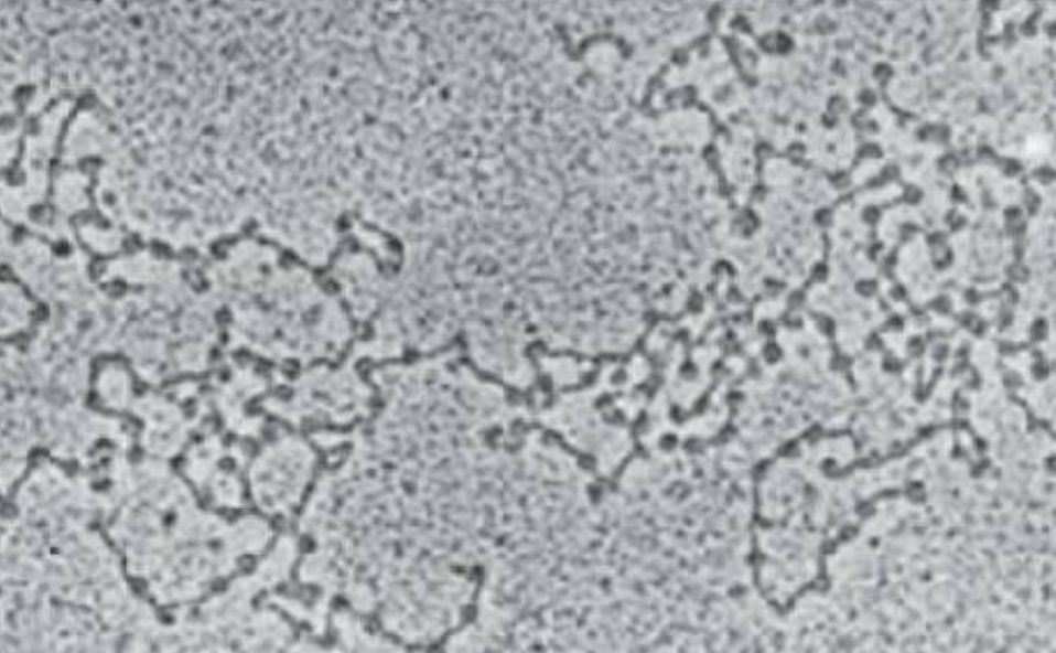
Under a microscope, _______ looks like a diffuse, grainy, or thread-like mass in a standard light microscope, but high-powered electron microscopes reveal a detailed "beads on a string" structure
Chromatoin
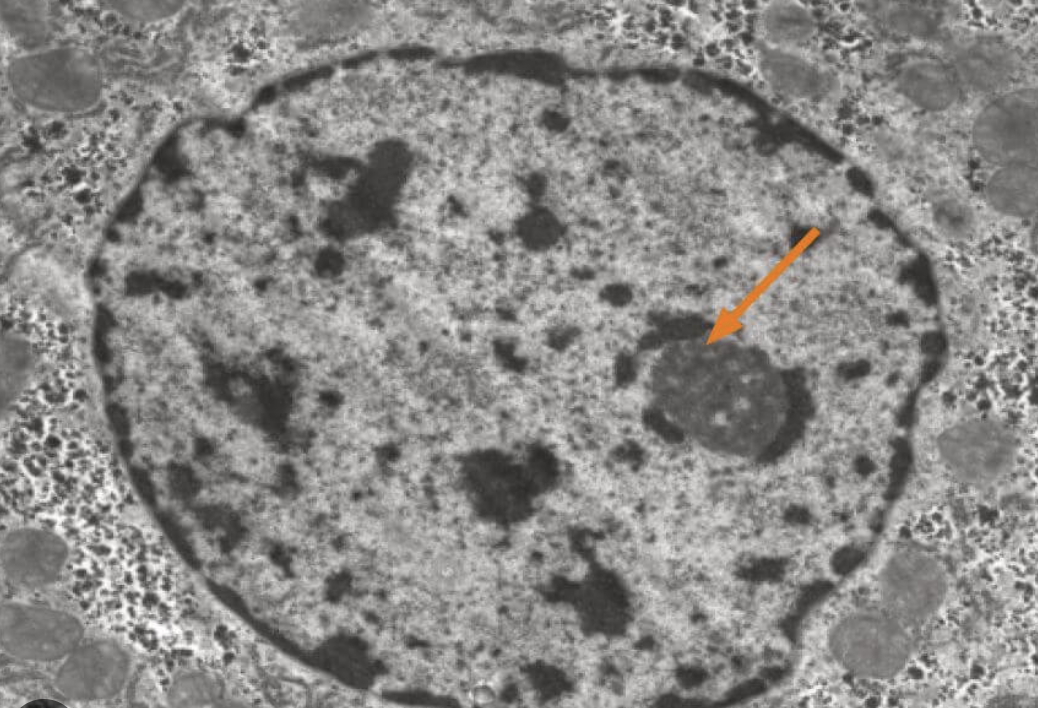
Appears as a large, dense, dark, round or oval spot inside the cell's nucleus
Nucleolus
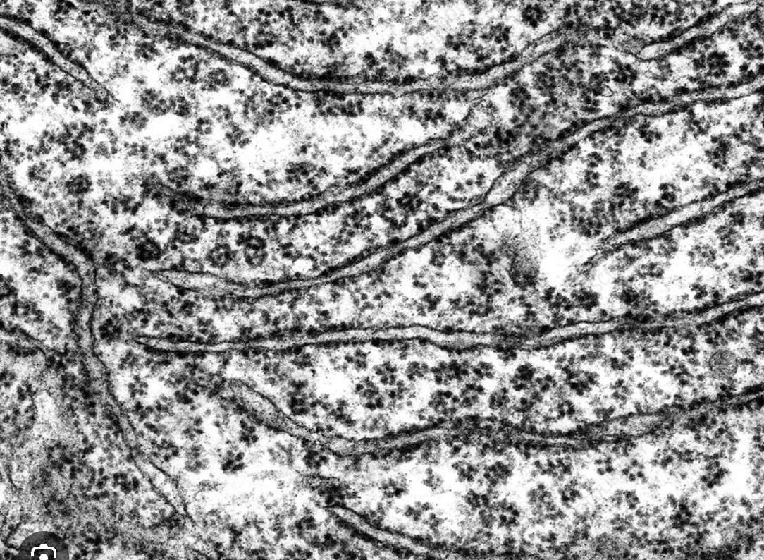
Looks like a series of flattened, interconnected, sheet-like sacs called cisternae that are studded with tiny, dark, granular dots representing ribosomes
Rough ER

Under a transmission electron microscope, _____ look like tiny, dark, bead-like dots
Ribosome

Looks like a network of delicate, interconnected tubes and flat, curved sacs (cisternae) with a clean, unstudded surface
Smooth ER

Reveals individual vesicles as tiny, round, membrane-bound sacs.
Secretory Vesticle

Appears as a stack of curved, flattened, membrane-bound sacs called cisternae surrounded by small transport vesicles
Golgi Apparatus
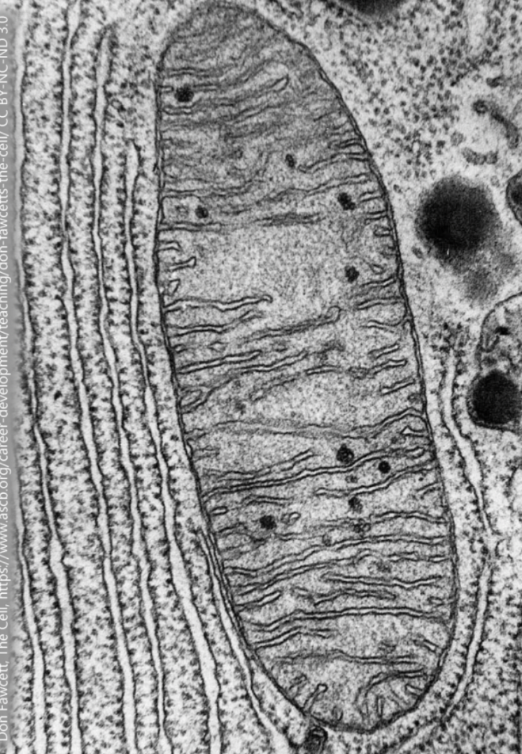
look like tiny, oval, sausage-shaped, or bean-like structures, similar in size and shape to bacteria
Mitochondria