L11: Ankle Sprains & Ligamentous Instability
 Call Kai
 Learn
Learn Practice Test
 Spaced Repetition
 Match
Match Flashcards
 Knowt Play
1/96

There's no tags or description
Looks like no tags are added yet.
Name | Mastery | Learn | Test | Matching | Spaced | Call with Kai | Chat |
|---|
No analytics yet
Send a link to your students to track their progress
97 Terms
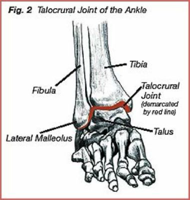
What is the talocrural joint composed of?
Relationship between the tibia, fiibula, and the talus
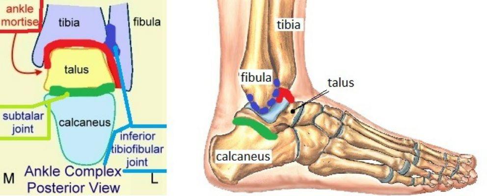
What is the subtalar joint composed of?
Relationship between the talus and calcaneus
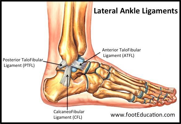
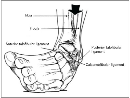
What are the static stabilizers of the lateral ankle?
Anterior Talofibular Ligament (ATFL)
Posterior Talofibular Ligament (PTFL)
Calcaneofibular Ligament (CFL)
Of the static stabilizers which is the most likely to be injured / torn?
The Anterior Talofibular Ligament is most often injured with an ________ ankle sprain
Inversion
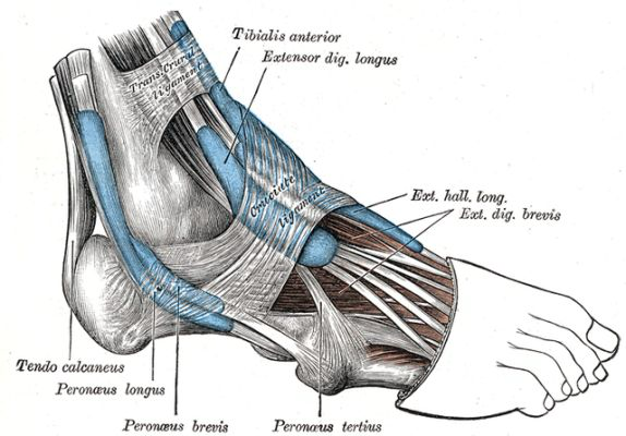
What are the dynamic stabilizers of the lateral ankle?
Fibularis Longus m.
Fibularis Brevis m.
Fibularis Tertius m.
What is the "purpose" of the lateral dynamic stabilizers?
Bring the medial side of the foot "back down"
What is the action of each lateral dynamic stabilizer?
Fibularis Longus = Plantarflexion + Eversion
Fibularis Brevis = Plantarflexion + Eversion
Fibularis Tertius = Dorsiflexion + Eversion
What are the static stabilizers of the medial ankle?
Deltoid Ligament Complex
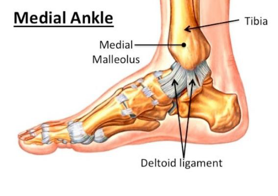
What are the dynamic stabilizers of the medial ankle?
Tibialis Posterior m.
Flexor Digitorum Longus m.
Flexor Hallucis Longus m.
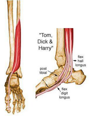
What is the action of each of the medial dynamic stabilizers?
Tibialis Posterior = Plantarflexion + Inversion
Flexor Digitorum Longus = Plantarflexion + Inversion
Flexor Hallucis Longus = Plantarflexion + Inversion
What is the "purpose" of the medial dynamic stabilizers?
Stabilizes for eversion movement to get the lateral side of the foot "back down"
The ________ consists of the gastroc and soleus and is considered to be a ________ dynamic stabilizer for the medial ankle
Triceps surae, Secondary
A lateral ankle sprain is also known as an ________ ankle sprain
Inversion
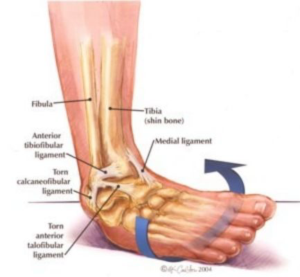
What is the typical MOI for a lateral ankle sprain?
Plantarflexion + Inversion
Examples -- stepping in a hole, jumping and landing ""weird""
Common in sports like basketball and volleyball
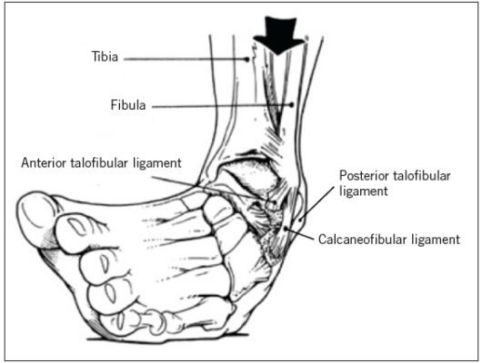
For patients with a lateral ankle sprain, a lot of them have pain up the lateral side of the shin. Why?
Since the ligaments "gave out" now the dynamic stabilizers are required to "work harder" in that instance to cause regular positioning
When you injure the ligaments, the dynamic stabilizers contract so hard that you also can sprain these (hence why the pain can radiate superiorly along these tendons / muscles)
What impact does swelling have following an ankle sprain?
Arthrogenic Inhibition
In a Grade 1 Lateral Ankle Sprain what ligament(s) are typically injured?
Usually the Anterior Talofibular Ligament
In a Grade 2 Ankle Sprain what ligament(s) are typically injured?
Usually the Anterior Talofibular Ligament (ATFL) and Calcaneofibular Ligament (CFL)
In a Grade 3 Ankle Sprain what ligament(s) are typically injured?
What are the clinical signs/symptoms of a Grade 1 Lateral Ankle Sprain?
Stretching of the Ligaments (usually ATFL)
Point Tenderness / tender to palpation over ATFL
Limited Dysfunction
No Laxity
Able to Bear Full Weight
Little/No Edema
No Bruising
What are the clinical signs/symptoms of a Grade 2 Lateral Ankle Sprain?
Partial tearing of ligaments (usually ATFL and CFL)
Point and Diffuse Tenderness
Point tenderness = along ATFL
Can also be generalized
Slight to Moderate Dysfunction
Antalgic Gait
Pain with FWB (may need AD to ambulate)
Mild to Moderate Edema
Often see Bruising
What are the clinical signs/symptoms of a Grade 3 Lateral Ankle Sprain?
Substantial Tearing of the ligaments (ATFL, CFL, & PTFL)
Point and Diffuse Tenderness
Moderate to Severe Dysfunction
Moderate to Severe Laxity
Limited / Inable to Weight Bear without AD
Severe Edema
Bruising
What special test(s) of the ankle would be positive with a Grade 1 Lateral Ankle Sprain?
What special test(s) of the ankle would be positive with a Grade 2 Lateral Ankle Sprain?
What special test(s) of the ankle would be positive with a Grade 3 Lateral Ankle Sprain?
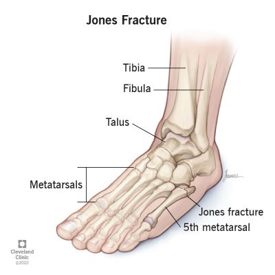
What is a Jones Fracture?
Avulsion of the Peroneus Brevis / Fracture of the 5th Metatarsal
Caused by Inversion / Lateral Ankle Sprain
Should be suspicious of this if your patient complains of pain distally
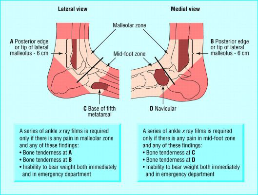
What is the purpose of the Ottawa Foot & Ankle Rules?
Helps us to determine if someone needs imaging for a possible fracture
According to the Ottawa Ankle Rules, imaging is required if there is bone tenderness in what locations of the malleolar zone?
If there is bone tenderness at the posterior/tip of the lateral or medial malleolus
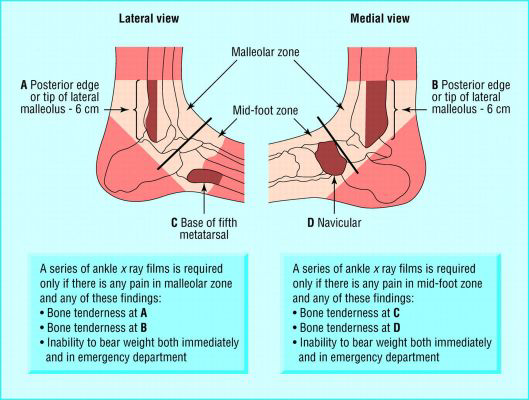
According to the Ottawa Ankle Rules, imaging is required if there is bone tenderness in what locations of the midfoot zone?
If there is bone tenderness at the base of the 5th metatarsal or on the navicular
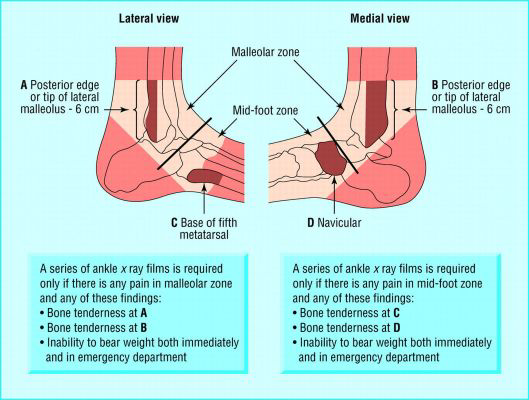
According to the Ottawa Ankle Rules, imaging is required if the patient cannot ________ (both immediately & in ED)
Bear weight
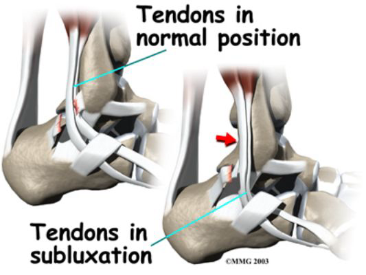
What tendon injury can result from an inversion ankle sprain?
Peroneal Tendon Subluxation
A tear of the peroneal tendon retinaculum can occur with an inversion ankle sprain
This causes the peroneal tendon to sublux / dislocate and move anteriorly
What is the Anterior Positional Fault of the Fibula?
After injury, the fibula is shifted more anteriorly due to the talus moving anteriorly
When should you NOT test for the Anterior Positional Fault of the Fibula?
If a patient has a high ankle sprain / syndesmotic ankle sprain
How is a peroneal subluxation treated?
Retinaculum that is torn will be repaired
The groove will be made deeper
What are signs/symptoms of Osteochondritis Dissecans of the Talar Dome?
Will either see no swelling or delayed swelling
This is indicative of an articular cartilage injury
Shear Forces and Compression Forces are painful
Patient may report a "catching" or something "stopping" their ROM
Pain with palpation over areas of articular cartilage
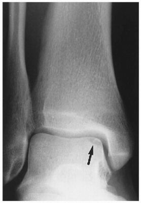
What are potential surgical interventions for Osteochondritis Dissecans of the Talar Dome?
Scope and Debridement
Microfracture
OATs
** NWB for 6 weeks (then tx like lateral ankle sprain)
What are the Lateral Ankle Sprain Tx/Rehab Goals during the Acute Phase of Healing?
Protect injured structures or prevent re-injury
Treat pain, inflammation, and effusion (if present)
Re-Establish ROM
Re-Establish Neuromuscular Control and Strength
What are the Lateral Ankle Sprain Tx/Rehab Goals during the Sub-Acute Phase of Healing?
Protect injured structures or prevent re-injury
Treat pain, inflammation, and effusion (if present)
Re-Establish ROM
Re-Establish Neuromuscular Control and Strength
Re-Esablish Proprioception and Agility
What are the Lateral Ankle Sprain Tx/Rehab Goals during the Maturation Phase of Healing?
Re-Establish Neuromuscular Control and Strength
Re-Esablish Proprioception and Agility
Re-Establish Functional Skills
What is typical ROM for great toe extension?
60 degrees of extension
Osteochondritis Dissecans of the Talar Dome is commonly seen in patient with ________
Chronic ankle instability
Osteochondritis Dissecans of the Talar Dome is commonly located at the ________ portion
Anterolateral
What are the two limitations that are seen in patients that have transitioned out of a walking boot?
Limited Ankle DF
Limited Great Toe Extension
What are four strengthening options for ankle sprain treatment?
BAPS Board
Theraband
CKC Weight Bearing
PNF Patterns
What are two treatment options to improve mobility afted transitioning from a walking boot?
Ankle Mobilizations (Posterior Gliding)
Talocrural Traction
A high ankle sprain is also known as a ________ ankle sprain
Syndesmotic
What is the typical MOI for a high ankle sprain?
Planted foot with forceful ER and DF
What structures can be involved in a high ankle sprain?
Tibial-Femoral Syndesmosis
Anterior-Inferior Tibiofibular Ligament (AITFL)
Posterior-Inferior Tibiofibular Ligament (PITFL)
Transverse Ligament
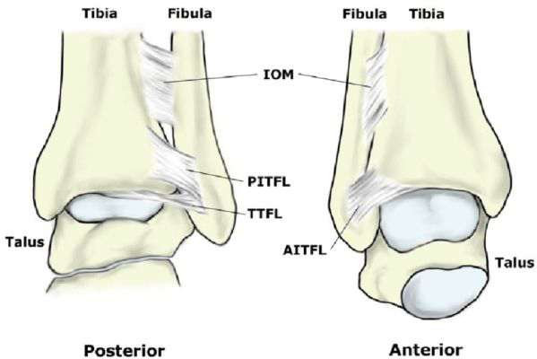
How can a high ankle sprain occur if it a contact MOI?
Space in-between the tibia and fibula should be ________; therefore, when looking at an x-ray to dx a high ankle sprain, this is something we will be looking at
< 5 mm
Discuss the healing time frame of a high ankle sprain?
Syndesmotic Injuries are slow to heal
In fact, they take 2-3x longer to heal compared to a Grade III Ankle Sprain
(bracing is not efficacious d/t rotary forces, etc. not be stopped)
Where can pain be located with a syndesmotic injury?
Pain can be on lower ankle (at distal tibiofibular joint) and/or pain can radiate up along the lenth of the tibia
Pain can also radiate below the distal tibiofibular joint (but this is less common)
A Grade 1 High Ankle Sprain can return to activity after ________
2-6 weeks
A Grade 2/3 High Ankle Sprain can return to activity after ________
3-6 months
What imaging is used to confirm a high ankle sprain?
Stress X-Ray (DF and ER)
Weight Bearing X-Ray
A Grade 1 High Ankle Sprain is indicative of a ________ to the syndesmosis
Stretch
A Grade 2 High Ankle Sprain is indicative of a ________ to the syndesmosis
Partial tear
A Grade 3 High Ankle Sprain is indicative of a ________ to the syndesmosis
Complete tear
If someone has a Grade 1 Sydesmotic Injury, what history do they report?
ER Injury
Sub-Acute Pain and Swelling
Continued Athletic Activity
If someone has a Grade 1 Sydesmotic Injury, what are the exam findings?
Mild Swelling
Mild AITFL Tenderness
Stable Ankle
(+) or (-) Squeeze Test
(+) or (-) ER Stress Test
If someone has a Grade 2 Syndesmotic Injury, what history do they report?
ER Injury
Acute Pain and Swelling
Inabiltiy to continue athletic activity
Painful gait
If someone has a Grade 2 Syndesmotic Injury, what are the exam findings?
Moderate Swelling
Moderate AITFL Tenderness
(+) or (-) Squeeze Test
(+) or (-) ER Stress Test
If someone has a Grade 3 Syndesmotic Injury, what history do they report?
ER Injury with a "pop"
Acute Severe Pain and Swellng
Inability to Walk
If someone has a Grade 3 Syndesmotic Injury, what are the exam findings?
Severe Swelling
Severe AITFL Tenderness
(+) Squeeze Test
(+) ER Test
What does Phase 1 of treatment include for a Grade 1 Syndesmotic Injury?
WB in a walking boot
ACE Wrap for compression
Pain control
Encourage motion within a pain-free ROM
Toe Yoga ("TOGA") for Circulation
What does Phase 1 of treatment include for a Grade 2 / 3 Syndesmotic Injury?
Probably not WB; this is to prevent spread of mortise
Also...
ACE Wrap for Compression
Pain Control
Encourage Motion within Pain-Free ROM
Toe Yoga for Circulation
What should be avoided within Phase 1 of treatment for a syndesmotic injury?
Closed-Kinetic Chain Exercises
Especially anything with rotation
Forced DF
This would spread the mortise (which we do not want)
What does Phase II treatment include for a syndesmotic injury?
Partial WB with an AD
Isometrics (work towards dynamic)
Weight Shifting
** really want to restore ROM and continue to avoid over-stressing tissue
What does Phase 3 treatment include for a syndesmotic injury?
Full WB
Transition out of walking boot
If still slightly unstable / there are other ligamentous injuries, patient may transition to less restrictive bracing
Improve DF
Patients have decreased ROM d/t walking boot, therefore DF is soemthing that will need to be worked on
A heel lift may need to be used during transition from walking boot to shoes to make ROM change not as vastly different
Single Leg Balance
What does Phase 4 treatment include for a syndesmotic injury?
Return to Sport
Jogging
Change of Direction (cutting / pivoting)
Able to Perform Single-Leg Heel Raise, Single-Leg Hop, and Repeated Hop
(All of these should be able to be done without pain)
Discuss the surgical repair for a syndesmotic injury.
Patient will have a ORIF to address fracture
Additionally, a syndesmotic screw (essentially, very long screw from fibula to tibia) will be placed to hold together syndesmosis
Note -- syndesmotic screw can causes issues later on after injury
Patient will then be in a boot for 4-6 weeks
After this we can treat like a lateral ankle sprain
What is a non-operative treatment option for syndesmotic injury?
Platlet-Rich Plasma (PRP) Injection
Ultrasound can be used to guide injection
Commonly seen with Grade III syndesmotic injury
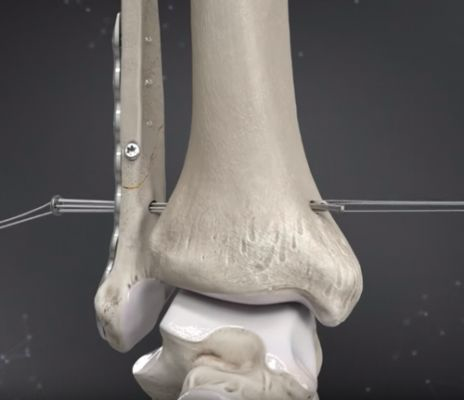
What is a Syndesmotic Tightrope?
There are two "buttons" and a "tight rope" to hold the tibia and fibula together
Allows for a better relationship between the tibia and fibula as well as the mortise
Not as "rigid" and allows for more ROM
What is a Masionneuve Fracture?
Rare & commonly missed
Comprised of 3 things:
Fracture of Proximal Fibula
Distal Tibial Fracture
Syndesmotic Injury
________ is the precursor for OA in the ankle
Chronic Ankle Instability
What are mechanical factors that can lead to chronic ankle instability?
Pathological Laxity
Arthrokinetic Restriction
Synovial Changes (thickening)
Degenerative Changes
What are functional factors that can lead to chronic ankle instability?
Impaired proprioception / joint position sense
Impaired neuromuscular control
Impaired postural control
Strength deficits
(especially for patients have had previous ankle sprain insult)
Chronic Ankle Instability (CAI) is common in individuals after they have a(n) ________
Ankle sprain
only 1 ankle sprain is needed to increase risk of chronic ankle instability
What is the problem with ankle taping?
Tape loosens over time
After approximately 45 minutes of activity -- tape has lost its structural stiffness and thus is not providing proper support
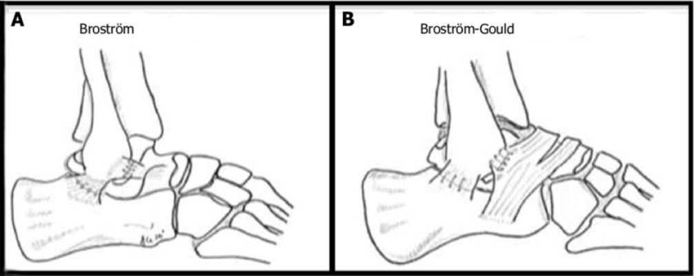
What is the Brostrum Procedure?
Stabilization Surgery for Chronic Ankle Instability
"Pants Over Vest Technique"
Remnants of the stretched ligaments are sutured back together
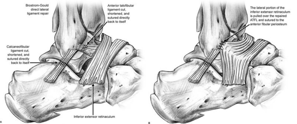
What is the Brostrum Repair Gould Modification?
Another type of Stabilization Surgery for Chronic Ankle Instability
Exact same thing as Brostrum Repair but the extensor retinaculum is flipped over to provide extra lateral instability
What is the Biotenodesis Brostrum?
Third type of Stabilization Surgery for Chronic Ankle Instability
Can be an allograft (from cadaver) or an autograft (from self)
Ligaments are made out of other structures
Examples = semitendinosus m., gracilis m., or plantaris m.
What do we need to be cautious of following all ankle repairs?
(just prevent anything that stresses the repair)
How does aggressive vs less aggressive therapy progression differ?
Aggressive:
Partial Weight-Bearing or WBAT in a Walking Boot for 2-4 weeks
Conservative / Less Aggressive:
NWB for 2-4 weeks while in a Walking Boot
Continue in Walking Boot for 6-8 Weeks w/ WBAT
Bracing
What are common interventions / exercises for patients who have undergone ankle surgery?
AROM in a pain-free range
Toe Scrunches (helps with inflammation)
Strength Quads, Hamstrings, Hip Abductors
Joint Mobilizations
If for pain -- can be performed earliy
If going for tissue extensibility / stretching -- wait for 8 weeks
Isometrics (Submax --> Max ISO --> Concentric)
Be careful of inversion
Weight Shifts
Single-Leg Balance
The ________ Classification is the most common classification system for a lateral malleolar fracture
Danis Weber
The more proximal a fibular fracture is → the increased risk for ________ and ________
Syndesmotic disruption, Ankle instability
What is the treatment for a non-displaced lateral malleolar fracture?
Patient is put in a walking boot for 6-8 weeks
What is the treatment for a displaced lateral malleolar fracture?
Patient will likely have an ORIF (d/t unstable fx or comminuted fx)
Booted for 4-6 weeks; and progress to WB once physician approval is received
After that → treat like a lateral ankle sprain!
Focus on ROM once patient out of boot
Joint mobilizations (subtalar, talocrural, and 1st MTP)
What is the treatment for a bi-malleolar fracture?
Patient will often have an ORIF
Though -- surgery may not occur right away to wait for swelling to go down (during this time, patient may be in splint)
However, if fx / dislocation could impact nerves and vascular structures they will just go ahead with surgery
Work on mobility with these patients
No joints mobs until 8-weeks post-op
What structures are involved with a trimalleolar fracture?
Medial Malleolus
Lateral Malleolus
Tibial Plafound (aka -- "3rd malleolus")
What is the typical MOI for a tri-malleolar fracture?
Often results from a significant trauma (e.g., fall)
What is the treatment for a tri-malleolar fracture?
Patient will require ORIF with hardware
Booted for 6-8 weeks
May be longer depending on the severity
Can take a long time for patients to regain functionality
Could be up to 1-2 years before back to full function