Fluid & Electrolytes 1 - Ann Hellmann
 Call Kai
 Learn
Learn Practice Test
 Spaced Repetition
 Match
Match Flashcards
 Knowt Play
1/113

There's no tags or description
Looks like no tags are added yet.
Name | Mastery | Learn | Test | Matching | Spaced | Call with Kai | Chat |
|---|
No analytics yet
Send a link to your students to track their progress
114 Terms
Extracellular fluid (ECF) aka. intravascular fluid
fluid in your bloodstream
Solvent
Water (~60%)
Solutes
Proteins and Electrolytes
Albumin
Major solute in the blood plasma that helps maintain OP.
Electrolytes (+/-)
Considered solutes.
Anions = negatively charged
Cations = positively charged
Glucose
2 Anions to Know
Phosphate PO4-
Bicarbonate HCO3-
4 Cations to Know
Sodium (Na+)
Potassium (K+)
Magnesium (Mg++)
Calcium (Ca++)
3 Compartments
ICF = 40%
ECF / intravascular fluid = 20%
Insterstitial fluid (ISF) = 40%
What does the ECF/intravascular fluid contain?
electrolytes, oxygen, glucose, other nutrients, and cellular waste products
What does the Interstitial Fluid (ISF) contain?
Na+ and water
What should the ISF NOT contain?
Proteins because they are too big.
*If proteins are present its a sign of inflammation
4 ways that fluid moves
Diffusion, Osmosis, Facilitated Transport, Active Transport
Osmosis
passive movement of water through semi-permeable membrane
water moves from LOWER → HIGHER conc. until equilibrium is reached
-use of conc. gradient
Diffusion = “Moving Down”
The passive movement of molecules from an area of higher concentration to an area of lower concentration, until equilibrium is reached.
Active Transport
is the energy-dependent process that moves ions or molecules against their concentration gradient, typically involving membrane proteins or pumps.
Facilitated Transport
Molecules pass through membrane w/ help of carrier proteins
Example of Facilitated Transport
EX) Glucose enters cell via. Insulin
-Insulin (carrier proteins) lower blood glucose by moving glucose into the cell via. facilitated transport
Active Transport
aka. the “Na+/K+ ATPase Pump”
ACTIVE process, requires ATP pump
-cell is working to move Na+/K+ against conc. gradient
3 Na+ OUT
2 K+ IN
Starling’s Law of Capillary Forces
Explains how fluid moves across capillary beds and between the blood vessels and surrounding tissues.
Controlled by 2 MAJOR OPPOSING FORCES that attempt to balance eachother out (homeostasis)
2 Opposing Forces
Hydrostatic Pressure (HP)
Osmotic Pressure (OP)
Hydrostatic Pressure (HP)
pressure exerted by water in the bloodstream created by cardiac pump
Osmotic Pressure (OP)
pressure exerted by solutes in a solution
Alterations in HP and/or OP can lead to?
Edema
Changes in HP or OP that can lead to edema
Increased preload increases HP
Low Albumin reduces OP
Ringers Lactate (Lr) Side Note
isotonic solution, maintains a normal hydrated status
*keeps fluid lvls the same
OP is the pressure needed to prevent water from moving
from a lower solute conc → a higher solute conc.
OP pulls water into where?
into the bloodstream (ECF) from the ICF AND ISF
Balance of HP vs. OP
oppose eachother, continually attempting to balance fluid movement across capillary membranes → homeostasis
Oncotic Pressure (aka. Colloidal Osmotic Pressure)
type of OP created by Albumin (protein associated w/ nutrition, can be given IV)
Oncotic Pressure Purposes
Pulls water in bloodstream
Keep water within BV
Maintains proper fluid balance between bloodstream + tissues
Oncotic Pressure Movement
ECF → (ISF) → ICF
Albumin job
stays in the bloodstream, helps pull water into capillaries to maintain OP
Osmolarity vs. Osmolality
Osmolarity = the # of osmoles of solute per 1L of solvent
Osmolality = the # of osmoles of solute per 1KG of soluvent
Osmole
unit used to measure amount of solute
ex) major solutes are Na+, K+, Mg++, Ca++, HCO3-, Glucose
Both Osmolarity and Osmolality tell us
the concentration of solutes in water
B-type Natriuretic Peptide (BNP) is linked to…
ventricle enlargement
Osmolality is used to evaluate
Hydration status
Normal Osmolality value
285-295 mOsm/L
With a high osmolality value, such as 350s, there are more _____ present.
solutes
High osmolality value = more solutes = more water attracted = ???
water moves toward the solutes by osmosis
Tonicity
amount of solutes in solution compared w/ the bloodstream
Isotonic
same tonicity as blood
-does NOT cause fluid shifts or changes in cell size
-Standard = 0.9% NaCl
Ringer’s Lactate
Similar physiological constituents as those found in blood
-Isotonic
Hypotonic
-Fewer particles (more water) than blood
-IV infusion causes shift from ECF → ICF
0.45% NaCl used to treat Dehydration
Hypertonic
-More particles (less water) than blood
-IV infusion pulls water from ICF → into ECF ; 3% NaCl
Ex) Mannitol Infusion
Mannitol Infusions (HYPERTONIC) are used to treat
cerebral edema
Hypertonic Summarized
-higher solute OUTSIDE
-more water INSIDE
= water moves OUT
= cell SHRINKS
Isotonic Summarized
-Equal solute
-Equal water
= no net movement
Normal cell
Hypotonic Summarized
-higher solute INSIDE
-more water OUTSIDE
= water moves IN
= cell SWELLING
Hypotonic fluids are used for
-Treat + Prevent dehydration
-Pull fluid out of vessels and INTO cells
Isotonic fluids are used for
Replace ECF, lytes, and expand vascular space
-Floods extravascular space → watch for s/s fluid overload
Hypertonic fluids are used for
-decrease intracellular volume
-increase vascular vol
-pulls fluid OUT of cells → INTO vessels to expand intravascular space
treat: hyponatremia, decrease cerebral edema, and high ICP

IV Fluid Tonicity Classifications (view img)
View IMG

A solution < 0.9% is considered
Hypotonic
A solution that is 0.9% is considered
Isotonic
A solution that is > 0.9% is considered
Hypertonic
IV solution quick reference
View img

Ways that out body maintains FLUID homeostasis
Kidneys
RAAS
Osmoreceptors
Thirst sensation (intake)
ADH (output)
Natriuretic Peptides
Natriuretic Peptides are triggered by
excess ECF volume
Natriuretic Peptide Effects
cause an increased GFR
Excretion of a LOT of Na+ & H2O by Kidneys
Types of Natriuretic Peptides
Atrial Natriuretic Peptide (ANP) → enlarged atria
B-type Natriuretic Peptide (BNP) → enlarged ventricles
enlarged stuff due to fluid overload
Renin-Angiotensin-Aldosterone System (RAAS)
increases BP, blood vol., circulation when low
Triggered by: Hypotension, Hypovolemia, Dehydration, Low CO
= all of these can cause decreased circulation + decreased blood flow to kidneys
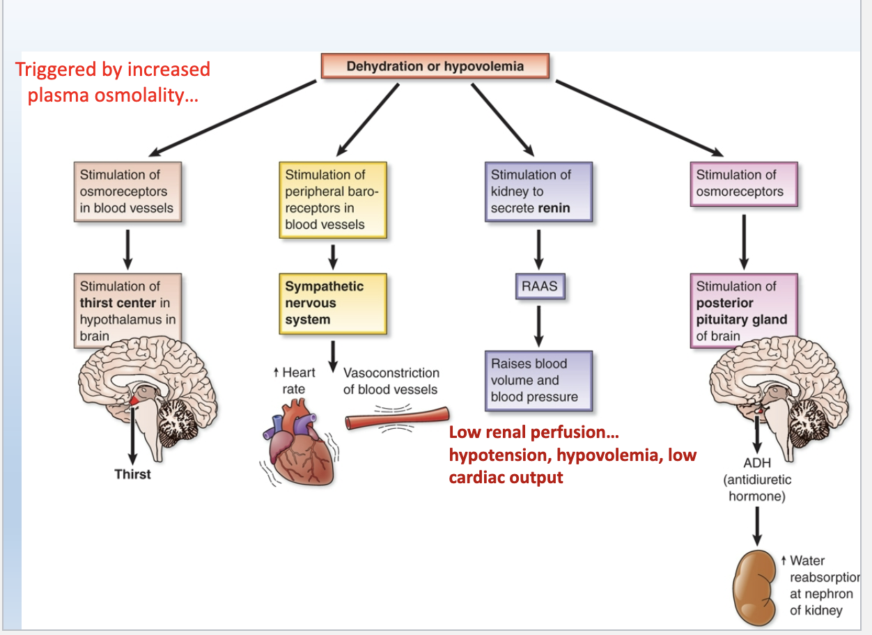
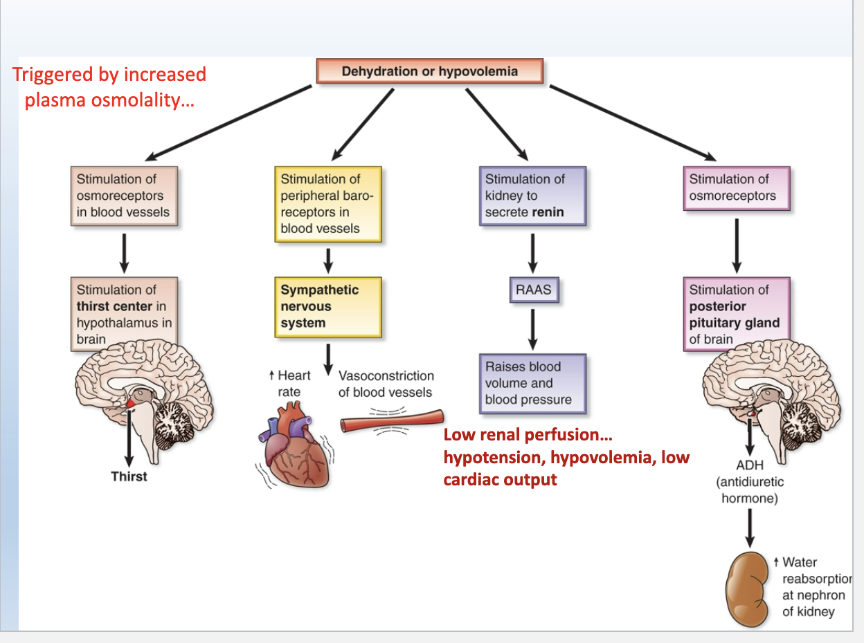
Dehydration / Hypovolemia chart
View img
How does Edema occur
excess of fluid in ICF + ISF
during increased HP or low OP
Things that can increase HP = may lead to edema
hypervolemia, inflammation, hypernatremia, weak venous valves
Things that can decrease OP = may lead to edema
Hypoalbuminea
Dehydration = aka. Hypovolemia
= diminished water vol. in body
Fluid moves from ICF → cells shrink
Decreased circulating blood vol. leads to tachycardia + hypotension
Response to dehydration
Osmoreceptors stimulate thirst, ADH release
Vasoconstriction + Increased HR
RAAS activated
Things that can cause dehydration
Reduced fluid intake
Reduced ADH / kidneys not responsive to ADH
Burns, Fever, Perspiration
Osmotic diuresis (occurs w/ elevated blood glucose)
Hypernatremia
Symptoms of Dehydration
Thirst
Dry, sticky mucous membranes
Lethargy/confusion
Sunken eyes
Lack of tears/sweat
Delayed cap refill, Poor turgor
Hypotension, Tachycardia
Dizziness/Falls
Low UOP
Dark, concentrated urine
Assessment of Fluid Vol. Status
Daily weights, I/O (intake, output), Vitals (HR/BP), Status of mucuous membranes, skin turgor, UOP
Normal Mg Value
1.5-2.5 mEq/L
Magnesium (Mg) is an intracellular cation (+)
main purpose of Mg: muscule + nerve RELAXATION
-high lvls of Mg = muscles relax
-low lvls of Mg = muscles are excited/irritated
Hypermagnesia
HIGH magnesium
> 2.5 mEq/L
Most common cause of Hypermagnesia
Kidney failure, bc kidneys won’t be able to remove Mg
Symptoms of Hypermagnesia
relaxed/weak muscles
decrease deep tendon reflexes (DTRs)
hypotension, arrthymias
cardiac arrest w/ very high lvls >10 mEq/L
Hypomagnesemia
LOW magnesium
< 1.5 mEq/L
Things that can cause Hypomagnesemia
-Prolonged diarrhea/laxative abuse 💩
-Increased renal excretion of Mg+ 🫘
-Sepsis
-Burns, serious wounds
Symptoms of Hypomagnesemia
“TOO EXCITED” = increased neuromuscular + cardiac excitability
-Muscular excitement
-Neuromuscular irritiliability: tetany, Chvostek’s sign, Trousseau’s sign
-increased deep tendon relfexes (DTRs)
-Tremors
-Tachycardia
-Confusion
-Irritability
-Seizures
Chvostek’s Sign
a sign of Hypomagnesemia OR Hypocalcemia
= facial spasm w/ tapping of facial nerve

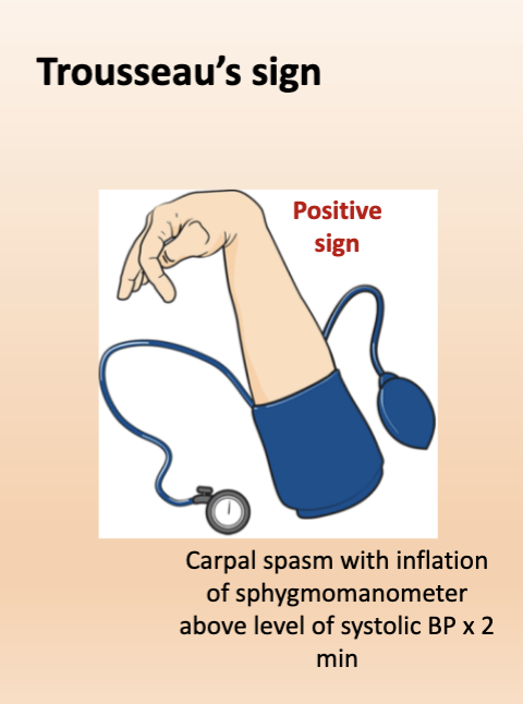
Trousseau’s Sign
a sign of Hypomagnesemia OR Hypocalcemia
= carpal spasm w/ inflation of BP cuff above lvl of systolic BP x2 min
Normal Potassium (K+) Level
3.5-5 mEq/L
Roles of Potassium (K+)
MOST IMPORTANT ELECTROLYTE w/ very sensitive lvls
-main electrolyte in ICF
-main role: muscle + nerve conduction (Cardiac & Skeletal Muscle)
Hyperkalemia
HIGH Potassium (K+)
Serum K+ >5.0 mEq/L
Things that can cause Hyperkalemia
-Burns
-Digoxin toxicity
-K+ sparing diuretics
-Metabolic acidosis
-Renal failure
Symptoms of Hyperkalemia
-Muscle cramping/twitching (early)
-Muscle weakness/paralysis (late)
-Cardiac dysrhythmias
-Tall, peaked T waves
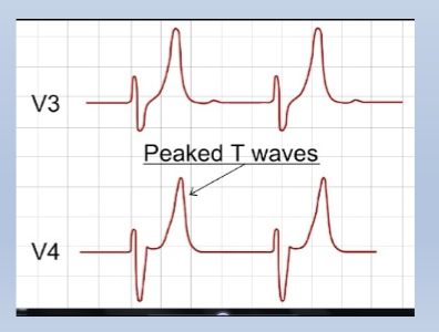
Hyperkalemia on an EKG/ECG
Peaked T waves

Hypokalemia
LOW Potassium (K+)
Serum K+ < 3.5 mEq/L
Things that can cause Hypokalemia
-Diuretic therapy *most common
-Anorexia
-N/V, diarrhea
Symptoms of Hypokalemia
-Weakness + Fatigue
-Muscle cramps
-Decreased DTRs
-Cardiac arrest, cardiac arrythmias, bradycardia
-Flattened T waves
Hypokalemia on an ECG/EKG
Flattened T waves

Acidosis leads to
Hyperkalemia
Alkalosis leads to
Hypokalemia
99% of calcium is in the _____ and 1% is in the ____.
bones, blood
Normal Calcium (Ca+) Level
8.6-10.2 mg/dl
Half of calcium is bound to _____ and the other half is ionized or “___”.
Albumin, Free
If ionized or “free” Ca+ levels become low, the pt. may become ______.
symptomatic
Main role of Calcium (Ca+)
Stabilizes the activity of motor neurons and controls how easily muscles contract
Hypercalcemia
HIGH Calcium (Ca+)
Serum Ca++ >10.2 mEq/L
=increases stability of motor neurons
Things that can cause Hypercalcemia
-Hyperparathyroidism
-Cancer