Nervous system
1/64
There's no tags or description
Looks like no tags are added yet.
Name | Mastery | Learn | Test | Matching | Spaced |
|---|
No study sessions yet.
65 Terms
Body’s communication systems
The nervous system and the endocrine system provide means by which organ systems communicate, maintaining homeostasis
The nervous system controls thoughts, movement, and emotion
The endocrine system controls growth, development, and digestion
The nervous system
Works quickly
Using chemical and electrical signals.
interconnected network of cells
signals move through cells
divide into:
Central nervous system (CNS)
Peripheral nervous system (PNS)
Divisions of the nervous system
CNS: interprets information (brain, spinal cord)
PNS: gathers and transmits information (Cranial nerves, spinal nerves)
The CNS and PNS pass signals between one another
Sensory receptor generates impulse
PNS passes impulse to CNS
CNS interprets impulse
CNS passes impulse to PNS
PNS stimulates a response
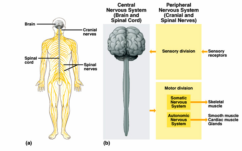
Divisions of peripheral nervous system
Sensory Division
• Picks up sensory information and delivers it to the CNS
Motor Division
• Carries information to muscles and glands
Divisions of the Motor Division
• Somatic – Carries information to skeletal muscle
• Autonomic – Carries information to smooth muscle, cardiac muscle, and glands
General functions of the nervous system
3 general functions:
Receiving stimuli = sensory function
Deciding about stimuli = integrative function
Reacting to stimuli = motor function
Sensory input, Integration and Motor output
Sensory input:
Sensory receptors gather information
Information is carried to the central nervous system
Integration:
Process and interpret sensory input and decide if action is needed
Sensory information used to create:
Sensations
Memory
Thoughts
Decisions
Motor output:
Decisions are acted upon
Impulses are carried to effectors
The response activates muscles or glands
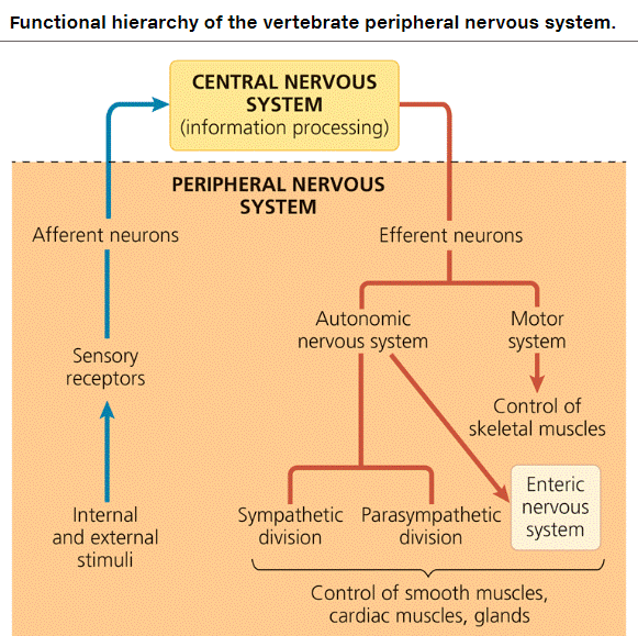
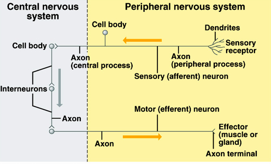
Functional hierarchy of the vertebrate PNS
Afferent Neurons (Sensory Neurons)
Carry sensory information from the body to the central nervous system (CNS: brain and spinal cord).
Detect stimuli such as touch, pain, temperature, sound, and light.
Efferent Neurons (Motor Neurons)
Carry motor commands from the central nervous system to muscles and glands.
Control muscle movements and glandular secretions.
Easy Way to Remember:
Afferent (A) → Arrives at the brain (Sensory input).
Efferent (E) → Exits the brain (Motor response).
Development aspects of the nervous system
The nervous system is formed during the first month of embryonic development
No more neurons are formed after birth, but growth and maturation continues for several years.
The brain reaches maximum weight as a young adult
However, we can always grow dendrites.
Cells of the nervous system
Cell types in neural tissue:
Neurons
Neuroglial cells
The signaling activity of the nervous system is made up of electrical activity within neurons and chemical flow between neurons
Neuron
A cell body: has nucleus and organelles
An axon: a long membrane-bound projection that transmits information away from the cell body in the form of electrical signals (impulses)
Dendrites: extend from the cell body and are covered by a membrane; receive impulses
Neurons have other structures to transmit signals
Terminal
Synapse
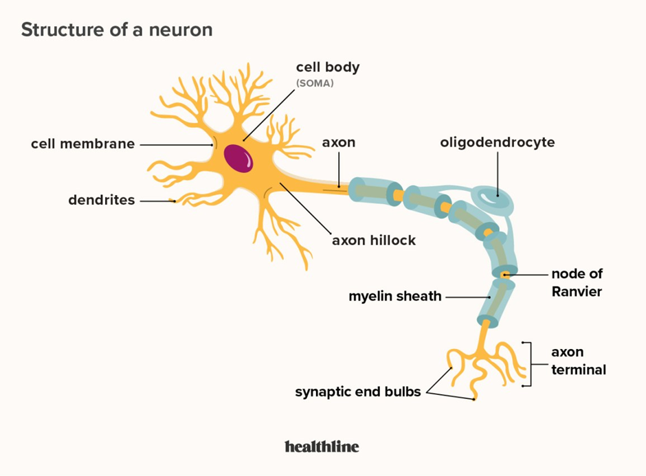
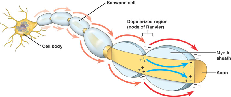
Axons and Synapses
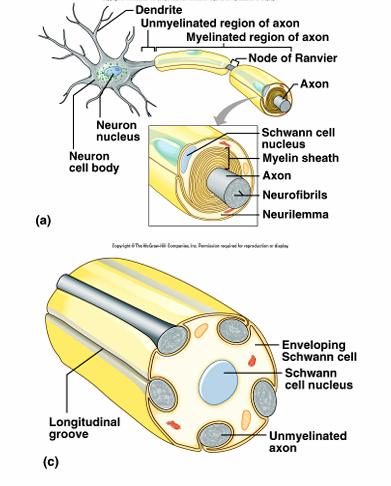
Axons are covered by a lipid layer called a myelin sheath
The myelin sheath insulates the neuron, which speeds up the transmission of action potentials along the axon.
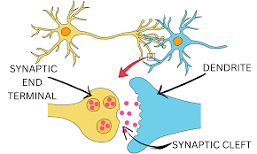
The end of an axon is called an axon terminal
Neurons communicate with each other at special junctions called synapses.
These synapses communicate by releasing neurotransmitters into synaptic cleft.
The synaptic cleft is a small gap between the axon terminal and the receiving cell.
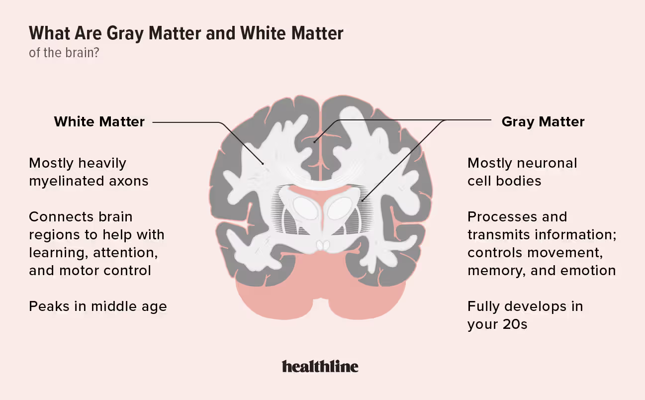
Myelination
White matter
Contains myelinated axons
Considered fiber tracts
Gray matter
Contains unmyelinated structures
Cell bodies, dendrites
Classification of Neurons - Functional Differences
Sensory neurons
Carry impulse to CNS
Most are unipolar
Some are bipolar
Integrative neurons (Interneurons)
Link neurons
Multipolar
In CNS
Motor neurons
Multipolar
Carry impulse away from CNS to effectors
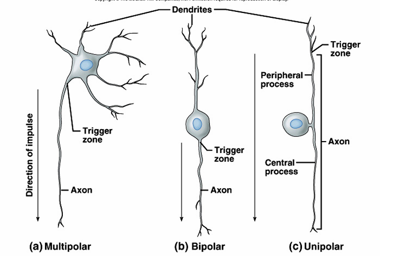
Classification of Neurons: Structural Differences
Bipolar neurons:
2 extensions (processes)
Eyes, ears, nose
Unipolar neurons
One extension
Ganglia of PNS
Sensory
Multipolar neurons:
99% of neurons
Many extensions
Most neurons of CNS
Neuroglial Cells in the PNS
1) Schwann Cells
Produce myelin found on peripheral myelinated neurons
Speed up neurotransmission
2) Satellite cells
Support clusters of neuron cell bodies (ganglia)
How Neurons Function - Nerve impulse
Neuron has a membrane potential
A membrane potential is a difference in the electrical charge across a cell membrane.
A membrane potential can change with an addition or removal of ions within the cell.
Ions move in and out of the cell by passing through protein act as ion channels.
Resting potential
A neuron is at rest when it is not sending or receiving a signal
Polarized membrane:
Inside is more negatively charged than the outside
More Na+ outside of cell
More K+ inside of cell
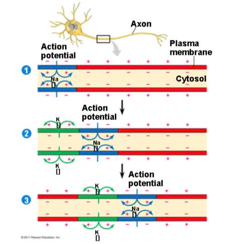
Action potential
When a dendrite or cell body is stimulated, the permeability of the neuron’s membrane changes suddenly
The stimulus depolarizes the neuron’s membrane, allows sodium to flow inside the membrane
The inside becomes more positively charged than the outside
The exchange of ions initiates an action potential in the neuron.
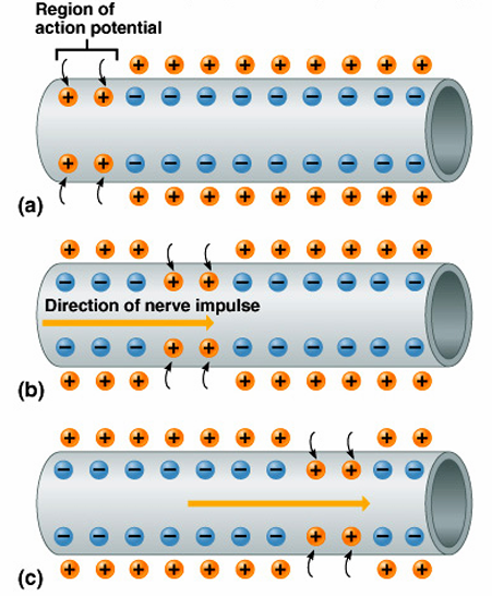
Nerve Impulse Propagation
An action potential is a moving electrical impulse
After the first segment of the neuron is stimulated, the next segment will become stimulated
=> If the action potential (nerve impulse) starts, it is propagated over the entire axon
Impulses travel faster when fibers have a myelin sheath
Refractory period
A neuron cannot generate another action potential until it has returned to its resting potential
The period in which a neuron cannot send a signal is called the refractory period
Returning the neuron to its resting potential requires energy
Potassium ions rush out of the neuron after sodium ions rush in => repolarizes the membrane
The sodium-potassium pump restores the original configuration => Requiring ATP
Conduction of Action Potentials
At the site where the action potential is generated, usually the axon hillock, an electrical current depolarizes the neighboring region of the axon membrane
Action potentials travel in only one direction: toward the synaptic terminals
Inactivated Na⁺ channels behind the zone of depolarization prevent the action potential from traveling backwards
Action potentials are formed only at nodes of Ranvier, gaps in the myelin sheath where voltage-gated Na⁺ channels are found
Saltatory conduction
Action potentials in myelinated axons jump between the nodes of Ranvier in a process called saltatory conduction
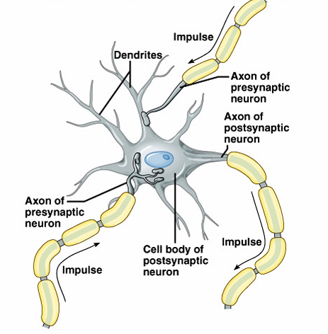
Continuation of the nerve impulse between neurons
When an action potential reaches the axon terminal, neurotransmitters are released into the synaptic cleft.
These neurotransmitters bind to receptors on the next neuron’s dendrites, opening ion channels.
If enough ion channels open, the action potential continues in the next neuron; otherwise, the signal stops.
Different neurotransmitters can either open or close ion channels, affecting signal transmission.
An action potential is started in the dendrite.
Video:
The synapse
Nerve impulses pass from neuron to neuron at synapses, moving from a pre-synaptic neuron to a post-synaptic neuron.
Central Nervous System
Develops from the embryonic neutral tube
The neutral tube becomes the brain and spinal cord
The brain is the control center of the nervous system
The spinal cord carries nerve signals between the body and the brain
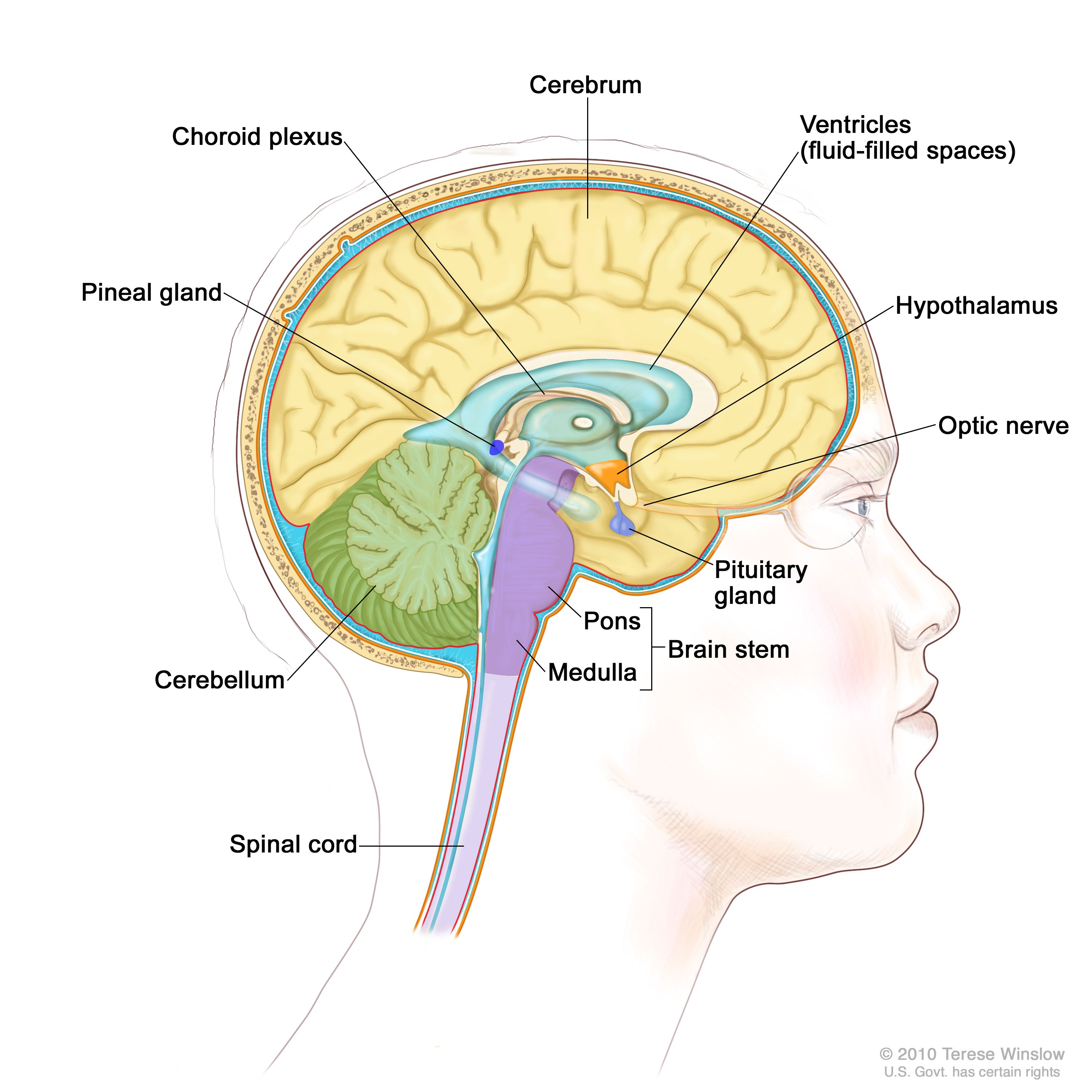
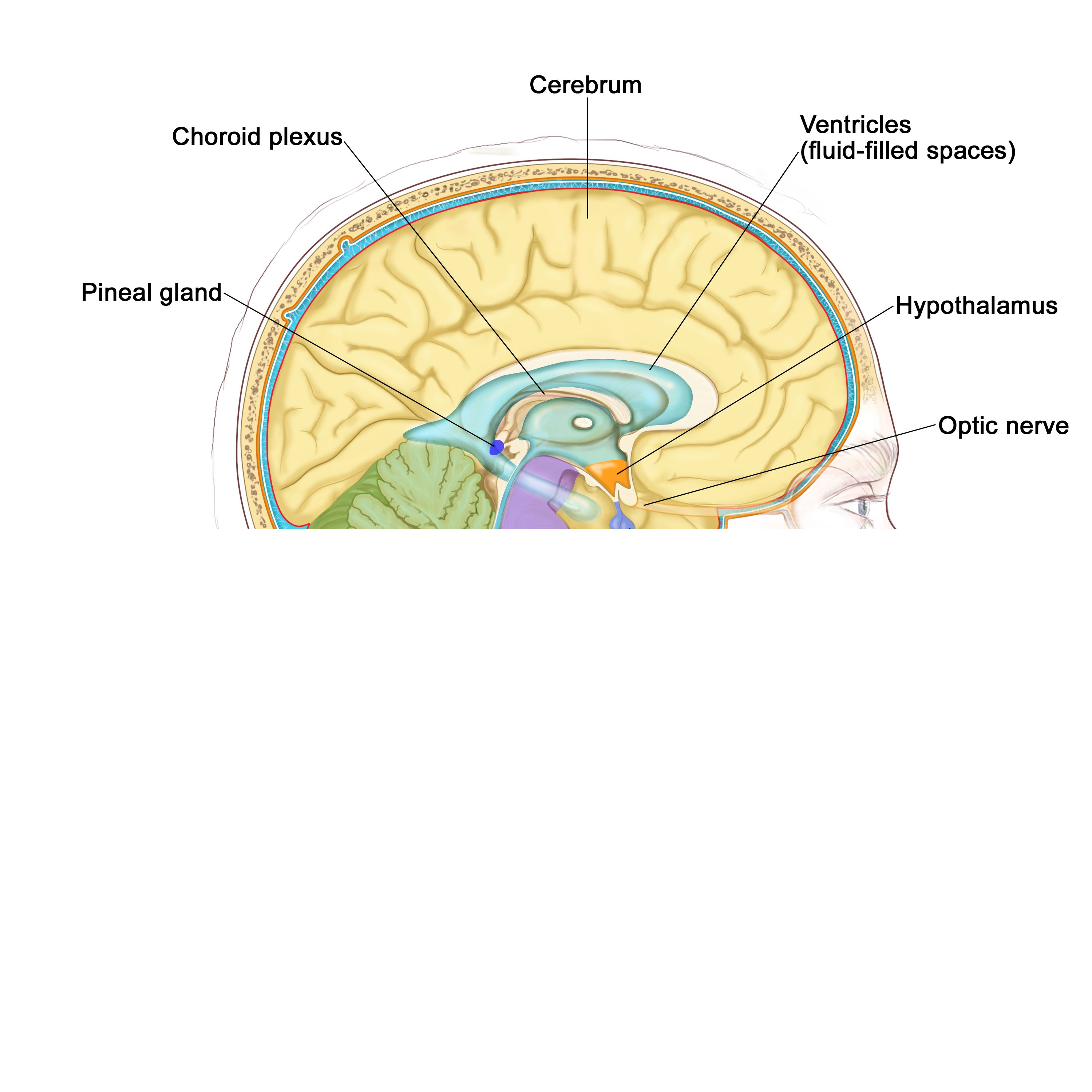
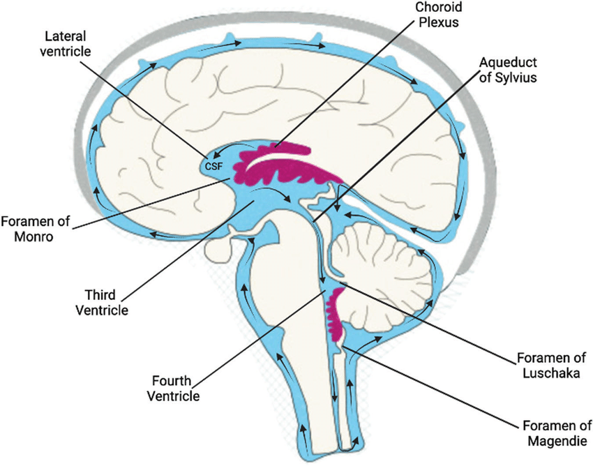
The opening of the neutral tube becomes the ventricles
Four chambers within the brain
Filled with cerebrospinal fluid
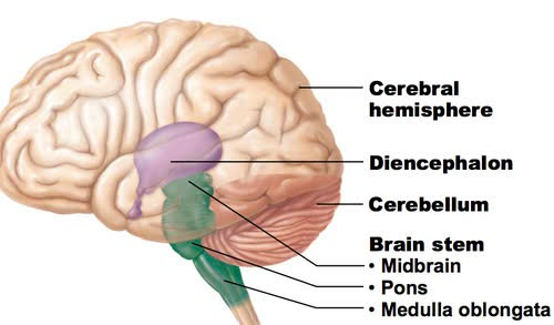
Regions of the brain
Cerebral hemispheres controls thought, movement, emotion
Diencephalon
Brain stem
Cerebellum
Cerebral Hemisphere (Cerebrum)
Paired (left and right) superior parts of the brain
Include more than half of the brain mass
The surface is made of ridges (gyri) and grooves (sulci)
Fissures (deep grooves) divide the cerebrum into lobes
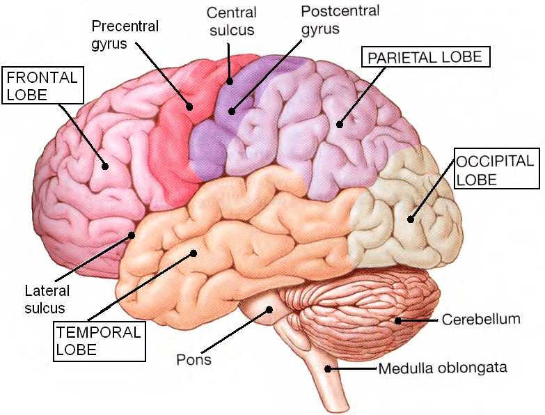
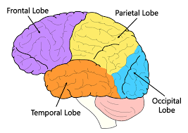
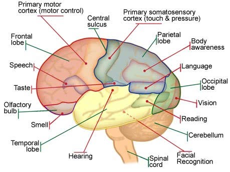
Lobes of the Cerebrum
Surface lobes of the cerebrum:
Frontal lobe
Parietal lobe
Occipital lobe
Temporal lobe
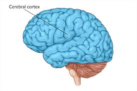
Cerebral cortex
The folded outer layer of the cerebrum
The cerebral cortex is the portion of the cerebrum that controls higher mental functions, general movement, organ function, perception, and behavioral reactions.
The many folds of the cerebral cortex allow the brain to have a large surface area and still fit into the skull.
Specialized areas of the cerebrum
Somatic sensory area – receives impulses from the body’s sensory receptors:
gustatory area (taste)
visual area
auditory area
olfactory area (smell)
Primary motor area – sends impulses to skeletal muscles
Broca’s area – involved in our ability to speak (speech/language region)
Language comprehension area
General interpretation area
Layers of the Cerebrum
The cerebral cortex is called gray matter. Beneath the gray matter is white matter.
Gray matter
Outer layer
Composed mostly of neuron cell bodies
Basal nuclei (basal ganglia) – internal islands of gray matter
White matter
Made up of myelinated axons, which link specific regions of the cortex with each other and with other neural centers
Example: corpus callosum connects hemispheres
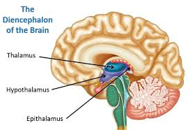
Diencephalon
Lies on top of the brain stem
Enclosed by the cerebral hemispheres
Made of three parts:
Thalamus
Hypothalamus
Epithalamus
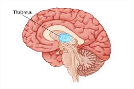
Thalamus
Surrounds the third ventricle
The relay station for sensory impulses
Transfers impulses to the correct part of the cortex for localization and interpretation
Hypothalamus
Under the thalamus
Important autonomic nervous system center
Maintains homeostasis and directly controls most of the body’s hormone production:
Helps regulate body temperature
Controls water balance
Regulates metabolism
An important part of the limbic system (emotions)
The pituitary gland is attached to the hypothalamus
Epithalamus
Forms the roof of the third ventricle
Houses the pineal body (an endocrine gland)
Includes the choroid plexus – forms cerebrospinal fluid
Brain Stem
Attaches to the spinal cord
Controls basic life functions
Parts of the brain stem:
Midbrain
Pons
Medulla oblongata
The brain stem also has a network of neurons called the reticular formation. This section helps control respiration and circulation and helps separate signals that are important from those that are not.
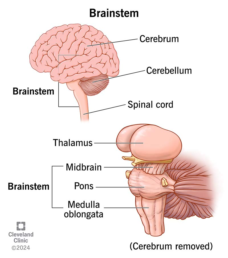
Midbrain
Mostly composed of tracts of nerve fibers
Reflex centers for vision and hearing
Cerebral aqueduct – 3rd to 4th ventricles
Pons
The bulging center part of the brain stem
Mostly composed of fiber tracts
Includes nuclei involved in the control of breathing
Relays (transmits) communications between the cerebral hemispheres and the cerebellum
Medulla Oblongata
The lowest part of the brain stem
Merges into the spinal cord
Includes important fiber tracts
Serves as both a relay center and a control center for:
Heart rate control
Blood pressure regulation
Breathing
Swallowing
Vomiting
Cerebellum
Lies below and behind the cerebral hemispheres
Two hemispheres with convoluted surfaces
Provides involuntary coordination of body movements, allowing for balance
Receives sensory impulses from muscles, tendons, joints, eyes, ears, and other brain centers
Protection of the Central Nervous System
Scalp and skin
Skull and vertebral column
Meninges
Cerebrospinal fluid
Blood-brain barrier
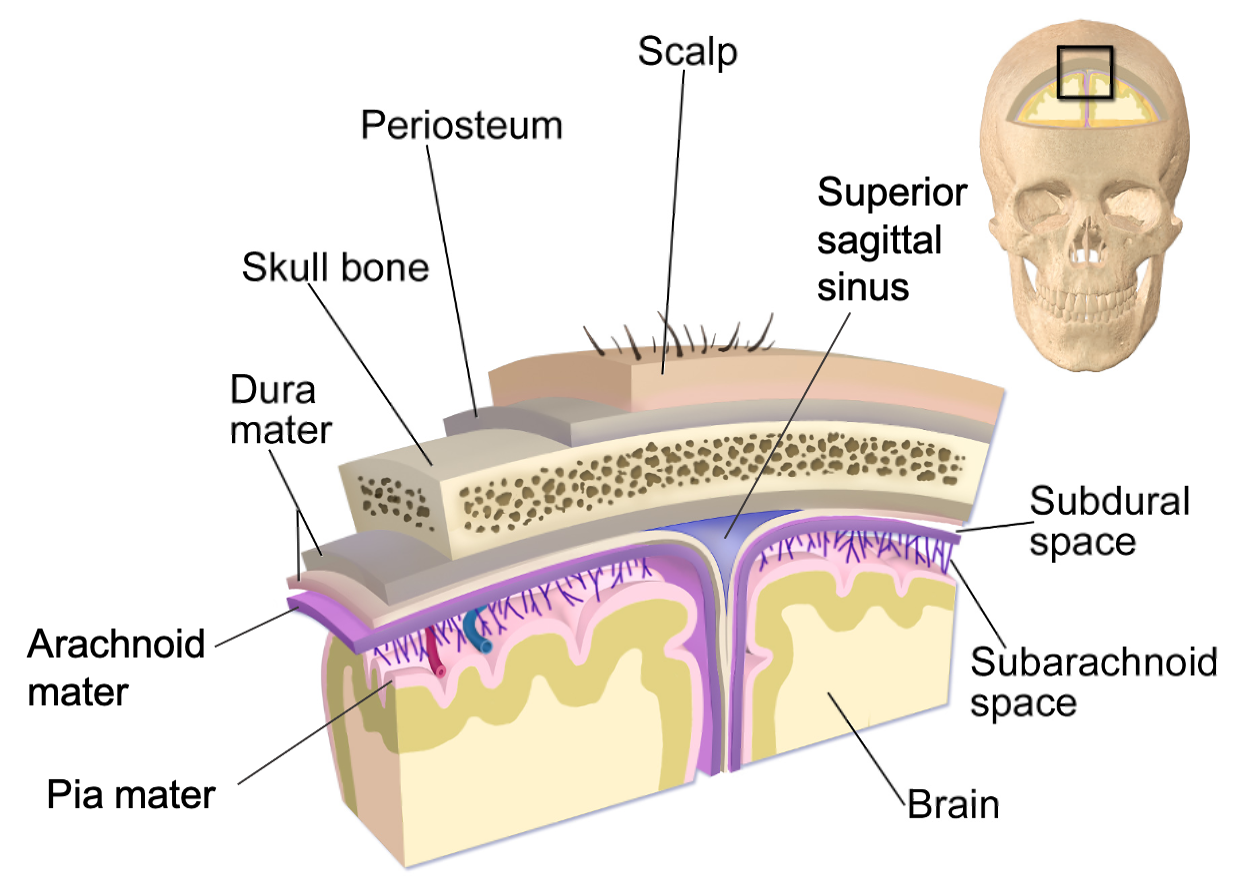
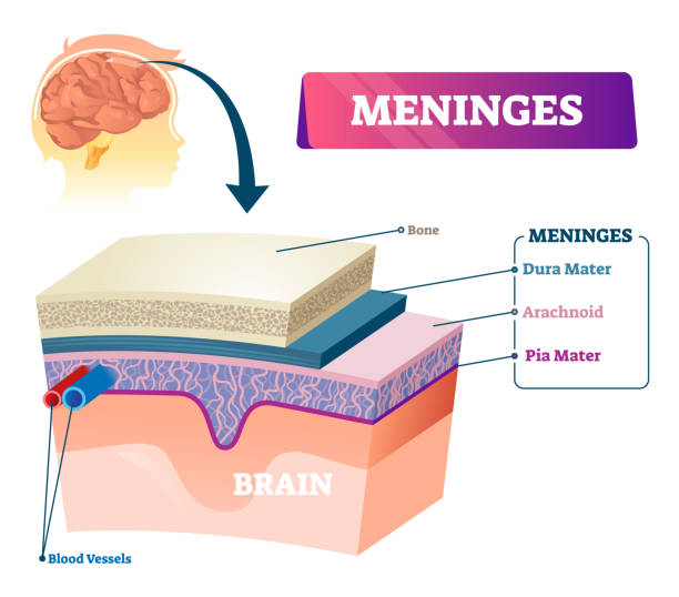
Meninges
Dura mater
Double-layered external covering
Periosteum – attached to the surface of the skull
Meningeal layer – outer covering of the brain
Folds inward in several areas
Arachnoid mater
Pia mater
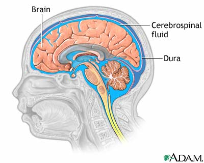
Cerebrospinal Fluid
Similar to blood plasma composition
Formed by the choroid plexus
Forms a watery cushion to protect the brain
Circulated in arachnoid space, ventricles, and central canal of the spinal cord
Ventricles and location of the cerebrospinal fluid
Cerebrovascular accident (CVA)
Commonly called a stroke
The result of a ruptured blood vessel supplying a region of the brain
Brain tissue supplied with oxygen from that blood source dies
Loss of some functions or death may result
Alzheimer’s Disease
Progressive degenerative brain disease
Mostly seen in the elderly, but may begin in middle age
Structural changes in the brain include abnormal protein deposits and twisted fibers within neurons
Victims experience memory loss, irritability, confusion, and ultimately, hallucinations and death
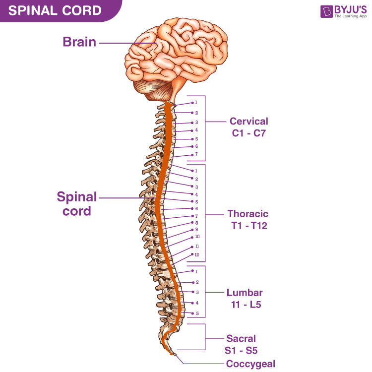
Spinal cord
The spinal cord is a column of nervous tissue that starts at the medulla oblongata and runs throughout the vertebral column to the region of T12 (12th thoracic vertebra)
Below T12 is the cauda equina (a collection of spinal nerves)
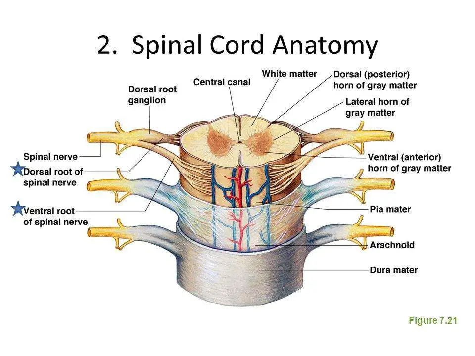
Spinal Cord Anatomy
Exterior white matter – conduction tracts
Internal gray matter – mostly cell bodies
Posterior (dorsal) horns
Anterior (ventral) horns
Central canal filled with cerebrospinal fluid
Meninges cover the spinal cord
Nerves leave at the level of each vertebra
Dorsal root: associated with the dorsal root ganglia – collections of cell bodies outside the central nervous system
Ventral root
Peripheral nervous system
Sensory Pathways
Picks up sensory information and delivers it to the CNS
Motor Pathways
Carries information to muscles and glands
Somatic nervous system – carries information to skeletal muscle
Autonomic nervous system – carries information to smooth muscles, cardiac muscles, and glands
Sympathetic divisions
Parasympathetic divisions
Nerves and ganglia outside the central nervous system
Nerve = bundled axon and dendrites of many neurons
Neuron fibers are bundled by connective tissue
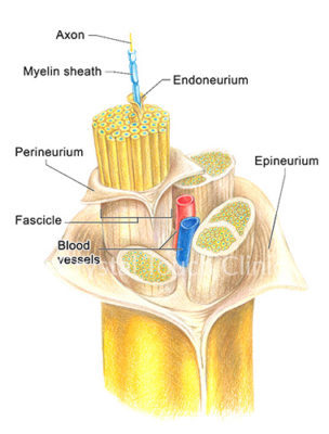
Structure of a Nerve
Endoneurium surrounds each fiber
Groups of fibers are bound into fascicles by perineurium
Fascicles are bound together by epineurium
Classification of Nerves
Mixed nerves – both sensory and motor fibers
Afferent (sensory) nerves – carry impulses toward the CNS
Efferent (motor) nerves – carry impulses away from the CNS
Spinal nerves
There is a pair of spinal nerves at the level of each vertebra
Spinal nerves are made up of both a dorsal and ventral root
The dorsal roots carry signals from sensory receptors
Ventral roots contain the axons of motor neurons
Interneurons can be found in both sections of the nervous system
Interneurons can relay information between other neurons
Sensory Division
Contains sensory receptors and the interneurons that connect them to the central nervous system
Sensory receptors receive information from the body’s external and internal environments
Motor Division
Allows the body to react to sensory information
The motor division is composed of the somatic nervous system and the autonomic nervous system
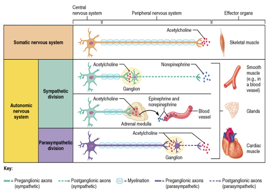
Comparison of Somatic and Autonomic Nervous Systems
Somatic Nervous System (SNS)
Generally voluntary
Controls skeletal muscles
Autonomic Nervous System (ANS)
Generally involuntary
Controls smooth muscle, cardiac muscle, and glands
Subdivided into sympathetic and parasympathetic divisions
Somatic Nervous System
Contains motor neurons that control the movement of skeletal muscles
Considered voluntary, but can operate without conscious control (e.g., reflexes)
Also relays signals in reflex pathways
The Reflex Arc
Reflex = rapid, predictable, and involuntary response to a stimulus
Reflexes are often self-protective
The patellar reflex is an example of a spinal reflex (the impulse bypasses the brain)
A reflex arc is the direct route from a sensory neuron to an interneuron to a motor neuron and then to the effector
Types of Reflexes and Regulation
Autonomic reflexes
Smooth muscle regulation
Heart and blood pressure regulation
Regulation of glands
Digestive system regulation
Somatic reflexes
Activation of skeletal muscles
Autonomic Nervous System
The involuntary branch of the nervous system
Controls internal body conditions by regulating smooth muscles in blood vessels and organs
Consists of only motor nerves
Divided into two divisions:
Sympathetic division
Parasympathetic division
Anatomy of the Autonomic Nervous system
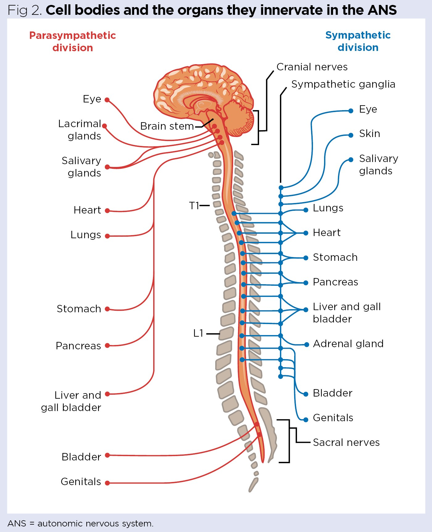
Autonomic Functioning
Sympathetic – "fight-or-flight," prepares the body when activated by physical or emotional stress (unusual stimulus). For example, pupils dilate and heart rate increases.
Takes over to increase activities.
Remember as the "E" division = exercise, excitement, emergency, and embarrassment.
Parasympathetic – housekeeping activities, controls the internal environment during routine conditions.
For example, pupils constrict and heart rate decreases.
Conserves energy.
Maintains daily necessary body functions.
Remember as the "D" division = digestion, defecation, and diuresis.
Physiological Effects of the Autonomic Nervous System
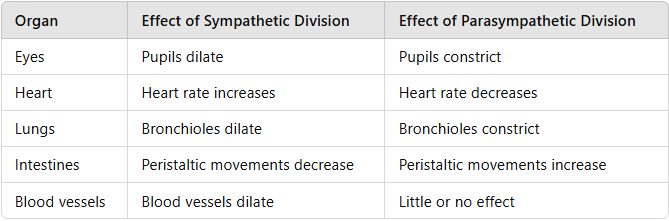
Sense Organs
In order to detect environmental changes
Sense organs receive stimuli and give rise to the senses such as sight, smell, taste, hearing, and pain.
Sense organs are part of the sensory division of the peripheral nervous system.
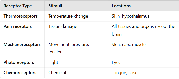
Receptors and Sense Organs
Sensory receptors are found in higher concentrations in the sense organs than in other parts of the body.
When stimulated, these sensory receptors convert the stimulus into electrical signals and send those signals to the brain.
Each signal that is sent to the brain is similar but may be sent to different parts of the brain to be interpreted.
Types of Sensory Receptors