MOLGEN 5607 - Cell Bio - Exam 1
 Call Kai
 Learn
Learn Practice Test
 Spaced Repetition
 Match
Match Flashcards
 Knowt Play
1/119

There's no tags or description
Looks like no tags are added yet.
Name | Mastery | Learn | Test | Matching | Spaced | Call with Kai | Chat |
|---|
No analytics yet
Send a link to your students to track their progress
120 Terms
Why does cellular organization matter?
The cytoplasm is a very crowded place; most proteins are made by cytoplasmic ribosomes.
What are the percentages of compartmentalization?
Internal compartments: 50% of cell volume
Plasma membrane: 5% of all cell membrane

What model best describes the cell membrane?
Fluid-mosaic model

Why is it not favorable for the planar bilayer to have uncovered ends?
Uncovered sections would uncover the non-polar, hydrophobic regions of the phospholipid

What determines the properties of the membrane?
The different types of phospholipids present
What do double bonds mean within the hydrocarbon tail of a phospholipid? What does this do?
The double bonds adds kinks to the hydrocarbon tail; Increasing permeability

Out of the four different types of phospholipids, which has a net-negative charge?
Phosphatidylserine

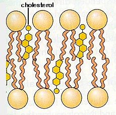
What is cholesterol?
A lipid and a steroid

What is cholesterol's purpose within the membrane?
It decreases permeability
What are liposomes?
Closed lipid bilayer spheres

What are microsomes?
vesicles formed from the endomembrane system (e.g the endoplasmic reticulum and Golgi complex)

What are the passive types of phospholipid mobility?
1. Lateral diffusion
2. Flexion of the hydrocarbon tails
3. Rotation
What is an active type of phospholipid mobility? Why is it not energetically favorable?
Flip-flopping: The polar heads would need to move through nonpolar regions.
T/F: The leaflets of cell membranes are typically always homogenous?
False: lipids can be asymmetrically distributed between the two leaflets of the bilayer. Depends on the membrane and its function
Most transmembrane proteins are ______.
alpha helical and non-polar

What are some of the different variations of transmembrane proteins:
1. Single alpha-helix
2. Multiple alpha-helices
3. Anchored by a lipid

What is an example of an anchored protein?
Myristoyl anchor

What are hydropathy plots?
It is a graph that can determine hydrophobic residues. Which in turn can define transmembrane regions.

Why are transmembrane proteins so important?
They convey information and signals from one side of the membrane to the other side.
What keeps a transmembrane protein folded together?
Hydrophilic amino acids
What is the role of detergent for cell biology?
It can help isolate and purify transmembrane proteins. Detergent monomers substitute with the lipid bilayer.

Why is protein purification important for cell biology?
It allows us to reconstitute functional membrane proteins in vitro.

What are biomolecular condensates?
Membraneless organelles formed from lipid droplets of multivalent proteins

What are variables that determine membrane transport?
1. Size
2. Polarity
3. Concentrations
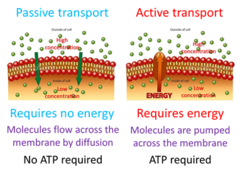
Examples of passive transport?
diffusion, osmosis, facilitated diffusion

Examples of active transport?
sodium potassium pump, endocytosis, exocytosis

What term describes a membrane protein's relationship with certain solute molecules?
Specificity
What are the different classes of membrane transport proteins?
1. Carrier/Transport Proteins
2. Channel Proteins
What are the mechanisms of carrier proteins? (i.e permeases)
- They undergo conformational changes by binding to the molecule they are transporting.
- SLOWER rate of transfer
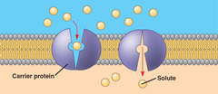
What are the mechanisms of channel proteins?
- The formation of hydrophilic pores
- They do not bind to the solutes
- FASTER rate of transfer

What is active transport?
the movement of ions or molecules across a cell membrane against their concentration gradient.

What protein is associated with active transport?
Carrier/Transporter Proteins (Or Permeases)

What are the different types of active transport?
1) ATP-driven
2) Coupled ion-driven

What are the different types of coupled transporter proteins?
1. Uniport (one solute one-way)
2. Symport (two solutes same-way)
3. Antiport (two solutes interchanged-way)

What is an electrochemical gradient?
The difference in charge between the membrane- brought by ion concentration
Explain the Na+/Glucose Transporter
-The binding of glucose and Na+ is co-operative
-Passive import of Na+ leads to the active transport of Glucose
-Symport Transporter

How does the Na+ concentration stay sufficient outside of the cell?
The Na+/K+ Pump

What are P-type pumps?
The phosphorylation of ATP results in a conformational change within the transmembrane protein.
Explain the Na+/K+ Pump
-It is an antiport transporter
-It is a P-type transporter
-Requires ATP Hydrolysis to function
-Pump is electrogenic (3Na+ out; 2K+ in)

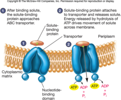
What are ABC Pumps?
-A Pump that acts an ATP-binding cassette
-Still uses ATP hydrolysis
-Prokaryotes (Brings things in); Eukaryotes (Brings things out)
Example: Overexpression of ABC can lead to an efflux of chemo-therapeutic drugs in cancer patients
Explain the importance of the Magainins experiment?
Showed that uncontrolled concentrations can lead to cell death (apoptosis).

T/F: Ion channels bind to the ionic solutes they bring in
False: They are hydrophilic pores that display ion-specificity
Passive transport that regulate via gates
What are the different types of ion channels?
1. Voltage-Gate
2. Ligand-Gate
3. Mechanical-Gate
What is the difference of a voltage gate and a ligand gate channel?
Voltage gate channels respond to electrochemical gradient, whereas ligand gate channels respond to extracellular ligands.
What is a selectivity filter within an ion channel? Why is it important?
A narrow region within the transmembrane ion channel to modulate which ions are recruited and which ones are not. If a channel gate is opened, that does not mean all ions should come through.
Explain how K+ channels work? Why can't Na+ get through? Why would that be detrimental if it did?
1. Within the cell, K+ ions are hydrated (surrounded by water molecules)
2. The charge within the channel pulls the hydrated-K+ ion towards it.
3. The charged helix stabilizes this hydrated-K+ and because the specificity pore is too small, it sheds the water off evenly.
4. After shed, it is able to leave the cell through the pore
- Na+ molecules are too small to do this. But if it was not, it would be detrimental to the Na+/Glucose symport transporter

What is the purpose of a specificity filter?
To regulate the influx of certain ions over other ones within a channel
What is the K+ leak channel? What is its purpose?
A K+ ion channel that is slightly opened state. Its purpose is to establish a membrane potential and thus create excitability from small ionic changes.
- Inside: 140mM
-Outside: 5mM
What are nerve signals?
changes within electrical potential across plasmic membranes
What is the resting potential inside a neuron? Why?
-Around -20mV to -200mV
-Na+/K+ gets Na+ out and K+-leak channels get K+ out. Leads to a net negative internal polarization

What is a voltage sodium gate in neurology?
-Voltage gate
-Gate closed: Polarization across membrane
-Gate opened: Depolarization occurs across the membrane (less negative inside)
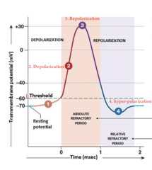
How does repolarization happen for sodium gates?
-The inactivation state
-The outside is still depolarized
-Different from the closed state- allows for repolarization to occur

What is action potential in a voltage sodium gate?
The triggering event from the rapid polarization to depolarization creates action potential.
What are chemical synapses?
Synapses specialized for the release and reception of neurotransmitters. These are through ligand-gated channels.

What triggers Action Potential to go down the axon?
Depolarization

If a sodium pump was mutated and couldn't go into inactive stage, what would occur?
The cell would not be able to repolarize itself. Thus, it would be sending a constant signal
If an axon could receive a signal in the middle of the axon?
The signal would not go from left to right, but bidirectionally (both ways).

What are delayed K+ channels?
The delayed voltage-gated K+ channels respond slower than that of Na+ channels. It opens the moment the Na+ pump closes. Helps to repolarize the cell after depolarization

T/F: The K+ leaking channel is stimulated to open.
False: It is a channel that is open even when unstimulated
How do proteins know where to go?
Signal Sequences

T/F: A protein can go through a membrane
False: Proteins need to be translocated single file through a translocation pore

What are the two methods that translocation of proteins happen?
1. Co-translational Import (ER)
2. Post-translational Import (Mitochondria)
If a sorting signal is not present on a protein, where does it go?
It will stay in the cytoplasm, as a majority of proteins are made by cytoplasmic ribosomes.
What is the function of the Endoplasmic Reticulum?
Protein Synthesis; Folding; Lipid/Hormone Synthesis

What is the Secretory pathway?
Rough ER -> Golgi -> secretory vesicles -> cell exterior

T/F: The ER takes up around 55% of the cell's total membrane
True
What is the ER's role in the secretory pathway?
It is the entry point- the glycosylation process
What was the experiment used to differentiate the smooth ER from the rough ER?
A tube of gradient glucose solution and centrifugation separated the two
How was the signal sequence hypothesis developed?
The addition of rough microsomes helped protect proteins from getting cleaved from protease

T/F: There are structural differences between membrane-bound ribosomes and cytoplasmic ribosomes
False: Both are structurally the same

What is the first step of ER protein transport?
A hydrophobic signal-sequence of the protein directs the ribosome towards the ER membrane.
What receives the signal sequence?
The Signal Recognition Particle (SRP)

What two things occur once the signal sequence portion fuses with the SRP?
1. Translation will come to a pause
2. The SRP will conformationally change to bind to the ER membrane
After the SRP changes its domain, what happens next?
It will bind to the SRP-receptor and transfer the ribosome to the translocon
What is the ribosomal translocon on the ER called?
Sec61
What does Sec61 identify on the ribosome?
The Hydrophobic Signal Sequence
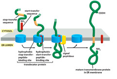
T/F: Once the Ribsomal-Sec61 complex forms, translation resumes.
True, this process is co-translational
Once translation ends, what happens to the signal sequence?
It is cleaved by signal peptidase
What are the structural components of Sec61?
A plugged pore within the ER membrane

What is an example of a post-translational protein insertion?
Transmembrane proteins
For transmembrane proteins, what does the signal peptide act as?
It is the first hydrophobic start, transfer sequence. This means the protein will begin to be translated through the translocon Sec61 complex- with the signal sequence attached to the lateral gate.
What indicates a stop-transfer sequence? What occurs when the Sec61 complex reaches it?
1.) A second hydrophobic peptide chain
2.) Upon reaching it, signal peptidase will cleave the first transfer sequence. Secondly, the second transfer sequence will move through the lateral gate into the membrane. Translation will continue outside of the membrane (cytosol)

For typical signal pass transfer sequences, where is the transfer sequence usually at?
The N-terminus
Is it possible to have this orientation flipped where the carboxyl-end is inside the lumen?
Yes, but the start signal patch would have to be in the middle of the peptide sequence
Explain multi-pass transmembrane proteins?
1.) Multiple hydrophobic regions within the center areas of the protein
2.) The first hydrophobic region reaches the Sec61 complex, opening it and translation continues through the pore. (START)
3.) The second hydrophobic region reaches the Sec61 complex and the pore closes and translation continues outside of the membrane. (STOP)
4.) The hydrophobic peptides are moved laterally into the membrane.

Explain the Two-Start Conumdrum
The first start signal acts as terminal signal sequence and thus is recognized by the SRP. It is immediately brought to the Sec61 complex and translocated within the membrane. Since the nascent chain is translated within the cytosol, that means the second hydrophobic stretch is a START-transfer sequence (as nothing was translocated into the lumen so it could not be a STOP).

What were the genetic approaches to studying proteins translocation?
Histidine enzyme works correctly in the cytoplasm- the cell lives. When the histidine enzyme was targeted to the ER- the cell dies.
What type of lipid is made in the Rough ER membrane?
Lipid-liked oligosaccharide
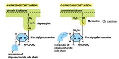
What is the function of N-linked glycosylation?
It helps to fold proteins correctly
What are the steps of glycosylation- if folded correctly?
1.) Protein enters the Sec61 complex and needs folded.
2.) Precursor oligosaccharide attached to the protein.
3.) Immediately two glucose molecules are trimmed- leaving one molecule
4.) The last glucose in the chain attaches and powers the calnexin protein
5.) The protein drops the N-linked oligosaccharide if folded properly.
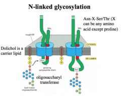
What is calnexin?
Calnexin is a chaperone protein that binds to glycoproteins.
What happens if the protein is misfolded after leaving the calnexin? Give two options
Option A.)
1.) If the protein is misfolded after calnexin, glucosyl transferase will reattach a glucose molecule.
2.) This reattachment of glucose will reinitiate calnexin recruitment and thus refolding.
Option B.)
1.) A chaperone will recognize the surface-exposed hydrophobic patch and thus attach to it.
2.) The misfolded protein will be carried outside of the ER and targeted for degradation via a proteosome.

What part of the ER is responsible for lipid synthesis?
The cytoplasmic half of the ER membrane is responsible for phospholipid synthesis.
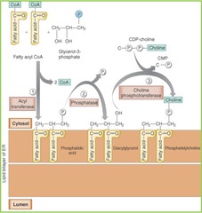
What are the two different types of
phospholipid translocating enzymes? What differentiates them?
Flippase - Enzyme specifically catalyzes the transfer of phospholipids. It creates an asymmetric bilayer leaflet. (Golgi and CM)
Scrablase - Enzyme that is not specific when catalyzing the transfer of phospholipids. It homogenizes the bilayer leaflets. (ER)
T/F: The mitochondria participates in the secretory pathway
False- lipids go through tight junction sites between the ER and the Mitochondria
T/F: The mitochondrial ribosomes make the majority of the proteins.
False- cytoplasmic ribosomes make the majority of proteins.
What is the mitochondrial signal sequence?
An amphipathic alpha-helix (+/- charge)
What are the outer and inner membrane translocation complexes?
Outer: TOM Complex
Inner: TIM23 Complex
What are the functions of hsp70 chaperone proteins? What is their function and why are they needed for mitochondrial translocation?
a.) hsp70 proteins are chaperones that bind to surface, uncovered hydrophobic residues.
b.) Their function is to keep the protein from folding so they can be translocated within the two membranes of the mitochondria.
c.) Mitochondrial translocation is post-translational, so thus the proteins need to keep from folding.
As soon as the protein enters the mitochondria, what happens next?
The amphipathic signal sequence is cut by mitochondrial signal peptidase.