Biopsych EXAM 1
 Call Kai
 Learn
Learn Practice Test
 Spaced Repetition
 Match
Match Flashcards
 Knowt Play
1/115

There's no tags or description
Looks like no tags are added yet.
Name | Mastery | Learn | Test | Matching | Spaced | Call with Kai | Chat |
|---|
No analytics yet
Send a link to your students to track their progress
116 Terms
Multipolar neuron
Many dendrites one axon, function as motor neurons, in the autonomic nervous system, and as interneurons
Bipolar neuron
2 processes come out of the soma, function as sensory neurons in the retina and ear, olfactory sense, cochlea, vestibular sense
Unipolar neuron
Single process, found in the CNS of invertebrates, may be found in human brains in cerebellum
Pseudounipolar neuron
One process that immediately branches into 2, function in the spinal cord and as sensory neurons
Anaxonic neuron
Multipolar without an axon, cannot create an action potential, function as moderators in retina and olfactory bulb
Afferent
Arrives to CNS
Efferent
Exits CNS
Somatic nervous system
Motor (skeletal muscles) and sensory
Autonomic nervous system
Sympathetic and parasympathetic nervous systems
Reflex arc
Stimuli → sensory neuron → interneuron in the spinal cord → motor neuron → move
Interneuron
A neuron in the spinal cord that connects sensory neurons to motor neurons to form the reflex arc
Schwann cell
Glia found in the PNS, wrap around axons to form myelin sheaths, many are needed for one axon, can guide their axons to reconnect after damage
Oligodendrocyte
Glia found in the CNS, wrap around axons to form myelin sheaths, one of these covers many axons. Linked to learning, memory, and motor skills
Microglia
Glia found in the CNS, function as the immune system of the brain by destroying any pathogens, phagocytosis of damaged/dying neurons, remodel or prune unused synapses. Survey environment, will regenerate if depleted, release chemicals to communicate with neurons, promote repair. linked to schizophrenia, autism, depression, and anxiety
Astrocyte
Glia found in CNS, provides nutrients to neurons from blood, repairs damage to nervous tissue, protects blood brain barrier. connected to neuron communication and memory, replicate often so are more likely to cause tumors than other cells
Ependymal cell
Glia that makes up the choroid plexus and cerebrospinal fluid, and lines the ventricles
Satellite cell
Glia found in PNS, absorb harmful toxins to protect neurons, regulate environment of neurons, provide nutrient support, cling to somas
Dorsal
Top
Ventral
Bottom
Anterior
Front
Posterior
Back
Ipsilateral
Same side
Contralateral
Opposite side

What plane is this?
Coronal plane

What plane is this?
Horizontal plane

What plane is this?
Saggital plane
Midsaggital
View when the brain is sliced directly down the middle into the 2 hemispheres
What are the three meninges?
Pia mater, Arachnoid mater, Dura mater

What is #15?
Pia mater
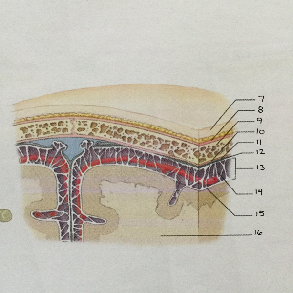
What is #13?
Arachnoid mater

What is #14?
Subarachnoid space
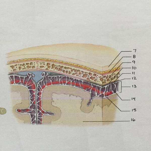
What is #11?
Dura mater
What does the dura mater do?
What does the pia mater do?
What does the arachnoid mater do?
What do the meninges do?
Telencephalon
Diencephalon
Mesencephalon
Metencephalon
Myelencephalon
What are the embryological divisions?
Telencephalon, Diencephalon, Mesencephalon, Metencephalon, Myelencephalon
White matter
Myelinated axons, makes up the inner cortex
Grey matter
Somas and glia, make up the external cortex
Gyri
Hills
Sulci
Valleys
What are the lobes of the brain?
Frontal, temporal, parietal, occipital
Frontal lobe functions
Temporal lobe functions
Parietal lobe functions
Occipital lobe functions
What structures make up the limbic system?
Hippocampus, amygdala, cingulate cortex, fornix
Hippocampus
Amygdala
Cingulate cortex
Fornix
Limbic system functions
Basal ganglia function
Coordinating smooth movement, preventing unwanted movement, reward, motivated behaviors, addiction
Thalamus function
Sensory relay
Hypothalamus function
Regulating homeostasis, hunger, sleep,
Midbrain function
Pons function
Medulla function
Cerebellum function
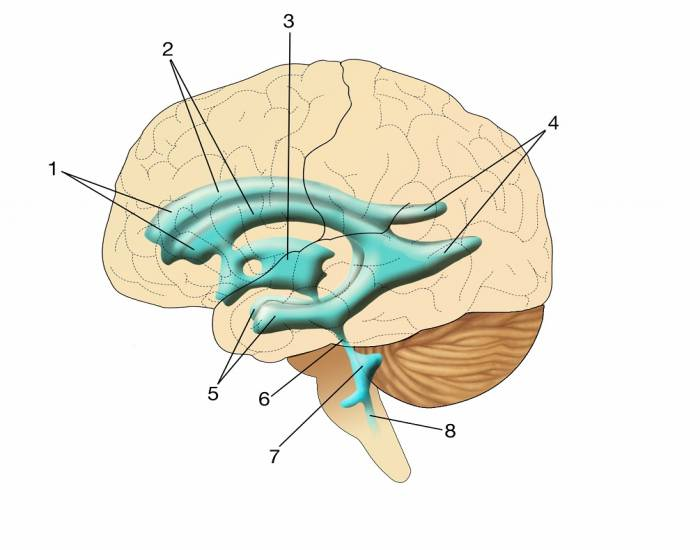
What is #1?
Lateral ventricles

What is #3?
Third ventricle
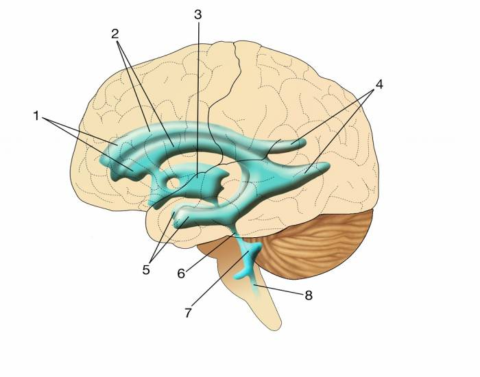
What is #6?
Cerebral aquaduct
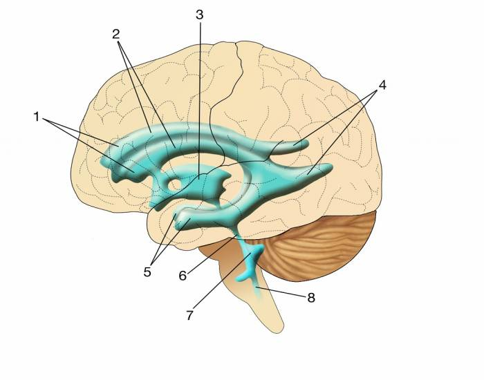
What is #7?
Fourth ventrical
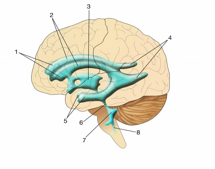
What is #8?
Central canal
What is inside the ventricals?
Cerebrospinal fluid
Choroid plexus
Part of the ventricular system, this structure is made up of ependymal glia cells and filters blood to cerebrospinal fluid and cerebrospinal fluid back to blood
What is a postsynaptic potential?
The membrane potential of the postsynaptic neuron in the specific location that was part of the synapse. It’s caused by neurotransmitters binding to chemically gated ion channels, allowing ions into the neuron, which either raises or lowers the membrane potential slightly and locally (not whole neuron)
Excitatory postsynaptic potential (EPSP)
The local membrane potential of the postsynaptic neuron (area at the synapse) becomes depolarized (intracellular space is less negative)
Inhibitory postsynaptic potential (IPSP)
The local membrane potential of the postsynaptic neuron (area at the synapse) becomes hyperpolarized (intracellular space is more negative)
Summation
The culmination of all EPSPs and IPSPs into one overall signal
In a neuron at rest, where are K+ ions most concentrated?
Inside the cell
In a neuron at rest, where are Na+ ions most concentrated?
Outside the cell
In a neuron experiencing an action potential, what are Na+ ions doing?
Moving into the cell
In a neuron experiencing an action potential, what are K+ ions doing?
Exiting the cell
In a neuron experiencing depolarization, what is happening?
Na+ ions are moving into the cell, K+ ions are beginning to move out of the cell, the intracellular space is rapidly becoming less negative
In a neuron experiencing repolarization, what is happening?
Na+ channels have closed, K+ is exiting the cell, the intracellular space is becoming more negative
In a neuron experiencing hyperpolarization, what is happening?
K+ channels close slowly as the membrane potential returns to resting, causing the voltage to overshoot and become more negative than resting
What is the absolute refractory period?
This period is when it is physically impossible for the neuron to have another action potential, and refers to the time when the neuron is in depolarization and repolarization
What is the relative refractory period?
This period is when it is difficult but not impossible for the neuron to have another action potential, and refers to the time when the neuron is in hyperpolarization
When do K+ channels close?
When the membrane potential returns to resting
What is the threshold?
The voltage needed to open voltage gated Na+ channels
Sodium
Na+
Potassium
K+
Chloride
Cl-
Calcium
Ca++
Average resting potential of a neuron
-70mV, range from -40mV to -90mV
When are leak channels open?
All the time
When are voltage gated channels open?
When the electrical threshold is reached
When are chemically gated channels open?
When neurotransmitters bind the the receptors
What does the sodium potassium pump do?
This protein uses active transport to put 3 Na+ outside of the cell and 2 K+ inside of the cell. This returns the ions to their resting places so the neuron is ready for another action potential
Conduction of an action potential down a myelinated axon is called:
Saltatory conduction
The intracellular space of a neuron at rest is
MORE negative than the extracellular space
Why does increased myelin make an action potential travel faster?
Because the segments covered in myelin don’t have to open ion channels
The words inhibition/inhibitory and excitation/excitatory refer to what?
Neurotransmitters
Which ion is involved in the release of neurotransmitters into the synapse?
Calcium (Ca++)