Module 3
 Call Kai
 Learn
Learn Practice Test
 Spaced Repetition
 Match
Match Flashcards
 Knowt Play
1/115

There's no tags or description
Looks like no tags are added yet.
Name | Mastery | Learn | Test | Matching | Spaced | Call with Kai | Chat |
|---|
No analytics yet
Send a link to your students to track their progress
116 Terms
Skeletal System
Includes the bones of the skeleton in addition to Cartilages, Ligaments, and other Connective tissues
They help to hold bones together and allow for movement of the bones at joints
Cartilage
A type of connective tissue, but the matrix is flexible
Have a unique appearance due to Chondroitin Sulfates.
Embedded within the cartilage matrix are Chondrocytes.
Surround by the Perichondrium.
Is Avascular.
All of these functions are carried out by diffusion through the matrix. This is why damaged cartilage does not repair itself as most tissue do.
Modes of Growth: Interstitial and Appositional
Ex. Hyaline, Fibrocartilage, and Elastic
Chondroitin Sulfates
A polysaccharide that binds with a ground substance protein to form Proteoglycans.
Chondrocytes
Cartilage cells
Found inside Lacunae which is embedded inside the Cartilage Matrix.
Perichondrium
A layer of Dense Irregular Connective Tissue that surrounds cartilage.
Avascular
It has no blood vessels supplying nutrients and removing metabolic wastes.
Hyaline Cartilage
The most common type of cartilage found in the body
Is located in the Rib cage and Nose.
Ex. Articular Cartilage
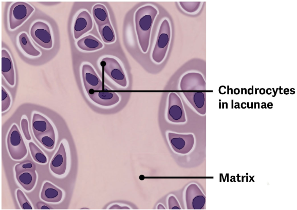
Articular Cartilage
Has the structure of Hyaline Cartilage but no Perichondrium
Is located especially at joint articulations to help cushion joints and enable then to move freely.
Serves to prevent bone to bone contact.
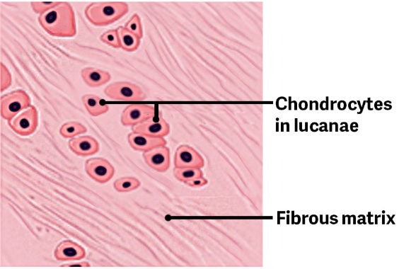
Fibrocartilage
Is tough due to it’s thick bundles of collagen fibers dispersed through it’s matrix.
Ex. Menisci in the knee joint the Intervertebral Discs.
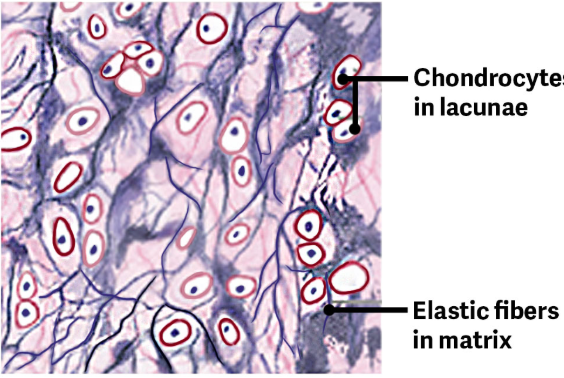
Elastic Cartilage
Contains elastic fibers, as well as collagen and Proteoglycans.
This tissue gives rigid support, as well as elasticity.
Ex. Ear - When tugging at the ear the lobe returns to it’s initial shape because the external ear contains elastic cartilage.
Interstitial Growth
Occurs as chondrocytes in the matrix grow and divide.
The daughter cells then produce additional matrix, so this enlarges the cartilage from the inside.
This occurs mostly during childhood and adolescent development and begins early in embryonic tissue.
Does not occur in adults

Appositional Growth
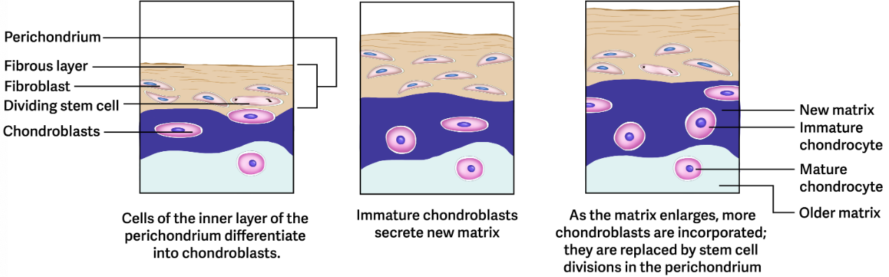
Occurs as new layers of cartilage are added on the cartilage surface as the inner layer of the perichondrium divides and grows.
The cells that are closest to the inner layer then develop into immature chondrocytes, which care found near the perichondrium.
Chondroblasts then start to secrete cartilage matrix around themselves, eventually becoming chondrocytes.
Eventually they secrete enough matrix that chondrocytes become fixed within their own matrix and begin to mature.
Appositional growth slowly adds the outer surface of the cartilage.
Does not occur in adults.
Can occur in very unusual circumstances to repair a minor surface Cartilage injury.
What happens to severe injury in Cartilage
It is replaced with fibrous tissue and the cartilage will not retain original function.
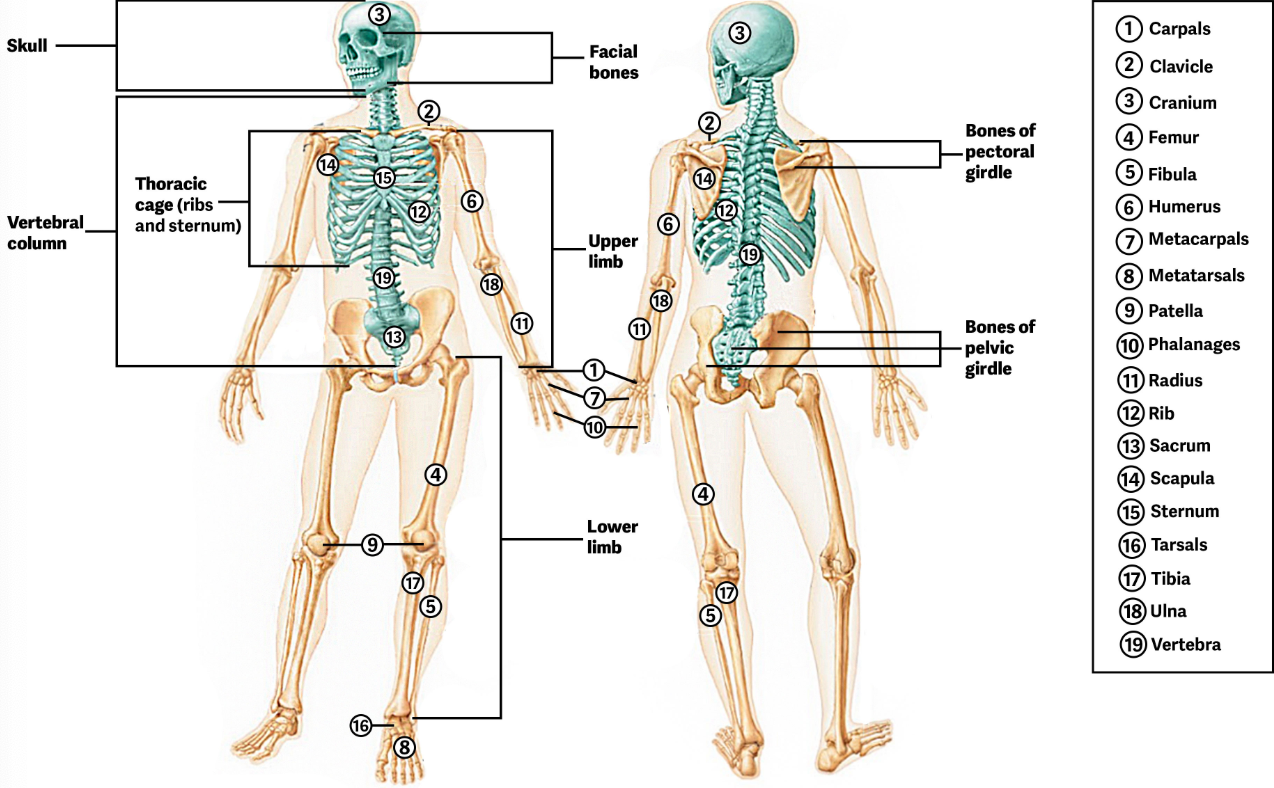
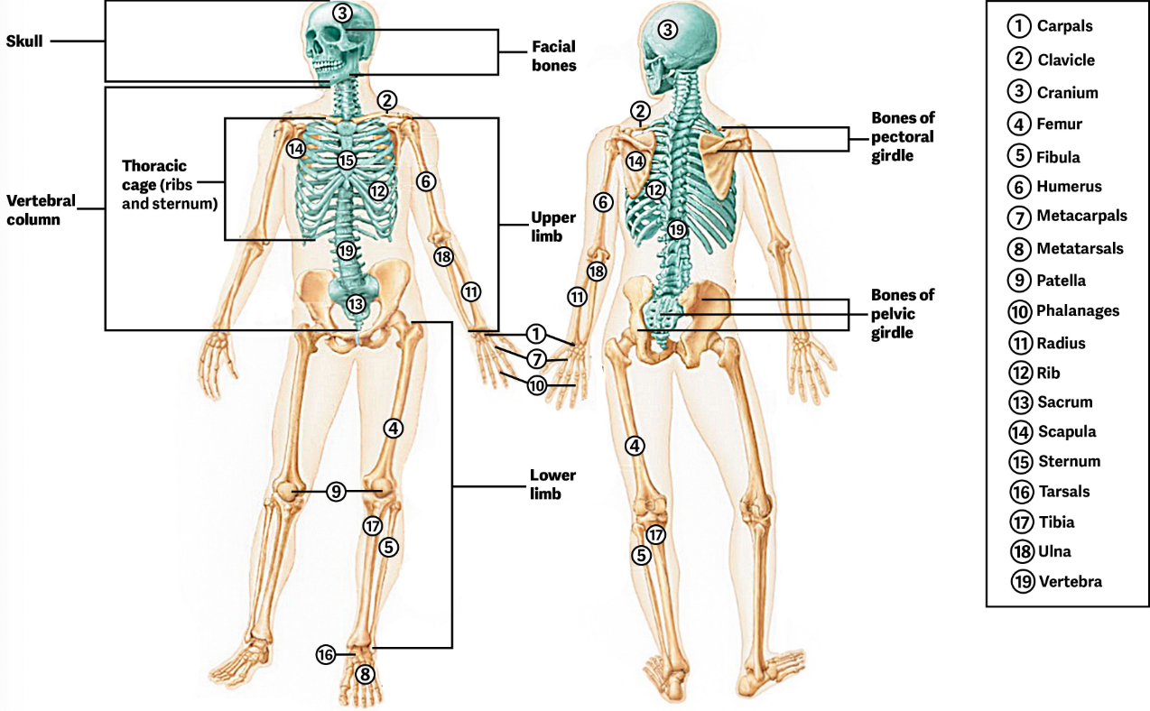
Axial Skeleton
Lies on the midline of body and consists of the skull, Vertebral Column, Sternum, Laryngeal skeleton, and Thoracic.
Appendicular Skeleton
Consists of the bones within the pectoral and pelvic girdles and attached limbs.
The pectoral girdle (shoulder) and upper limbs (arms) are specialized for flexibility and increase range of motion, while the pelvic girdle and lower limbs are speciaLized for strength.
Types of Bones
Bones are classified by their structure and shapes
The shape determines it’s function
Ex. Long Bones, Flat Bones, Sutural Bones, Irregular Bones, Sesamoid Bones.
Long Bones
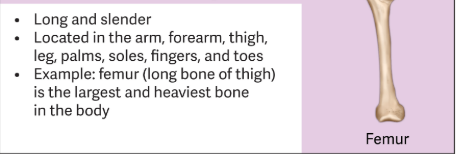
These bones have a structure that supports body weight and enables movement.
Are long and thin
Ex. Humerus, Ulna, Radius, Tibia, Fibula, Metacarpals, and Metatarsals
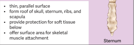
Flat Bones
Are very thin and have surfaces that are parallel with each other.
Within the forms the roof of the skull to protect the brain.
Ex. Sternum, Ribs, adn Scapula
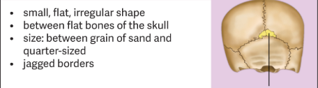
Sutural Bones
They are found in between the flat bones of the skull.
The number of this type of bone varies from person to person.
Are small and flat
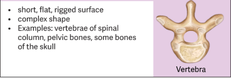
Irregular Bones
Are varied in structure with ridges or complex shapes.
The vertebrae are irregular bones designed to protect the spinal cord as well as enable spinal movements.
Ex. Pelvic bones (Ilium, Ischium, and Pubis)
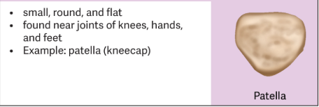
Sesamoid Bones
They develop inside tendons to help reinforce the tendon and give it strength.
They can also be found near the joints of the hands and feet.
But not all individuals have sesamoid bones in other joint locations.
Are small and round.
Bone (Osseous Tissue)
A hard, dense connective tissue that forms most of the adult skeleton, the support structure of the body.
Contains relatively small amount of cells entrenched in a matrix of collagen fibers that provide a surface for inorganic salts crystals to adhere.
These crystals form when calcium phosphate and calcium carbonate combine to create hydroxyapatite.
The hydroxyapatite crystals gives bones their hardness and strength, while collagen fibers give them flexibility so that they are not brittle.
Without a calcified matrix bone will have a normal appearance but will be too flexible and unable to support the weight of the body.
Hydroxyapatite also incorporates other inorganic salts on the collagen fibers.
Although bone cells compose a small amount of the bone volume, they are crucial to the function of bones.
Is continually being broken down and built up again by the work of specialized cells in adults.
Types: Osteoblasts, Osteocytes, Osteogenic Cells, and Osteoblasts
Skeleton System
Is the body system composed of bones and cartilage that performs critical functions of the human body.
Supports the body
Facilitates movement
Protects internal organs
Stores and releases minerals and fat
How do bones support, Leverage and Protect the body
The bones and cartilage of the skeletal compose the scaffold that supports the rest of your body.
Bones also facilitate movement by serving as points of attachment for your muscles
While some bones only serve as a support for the muscles, others also transmit the forces produced when your muscles contract.
Form a mechanical point of view, bones act as levers and joints serve as fulcrums. Unless a muscle spans a joint and contracts, a bone is not going to move.
Bones also protect internal vital organs from injury by covering or surrounding them.
Ex. Ribs to the lungs and heart, bones of the vertebral column to spinal cord
How do bones use Mineral Storage and Energy Storage
One a metabolic level bone tissue performs several functions.
Bone matrix stores many critical minerals important to the functioning of the body. Ex. Calcium and Phosphorus
These minerals are incorporated into bone tissue and can be released back into the bloodstream to maintain homeostatic levels need to support physiological processes
Calcium Ions are essential fro muscle contraction and controlling the flow of other ion involved in the transmission of nerve impulses.
Bone also serves as a site for storage and blood cell production
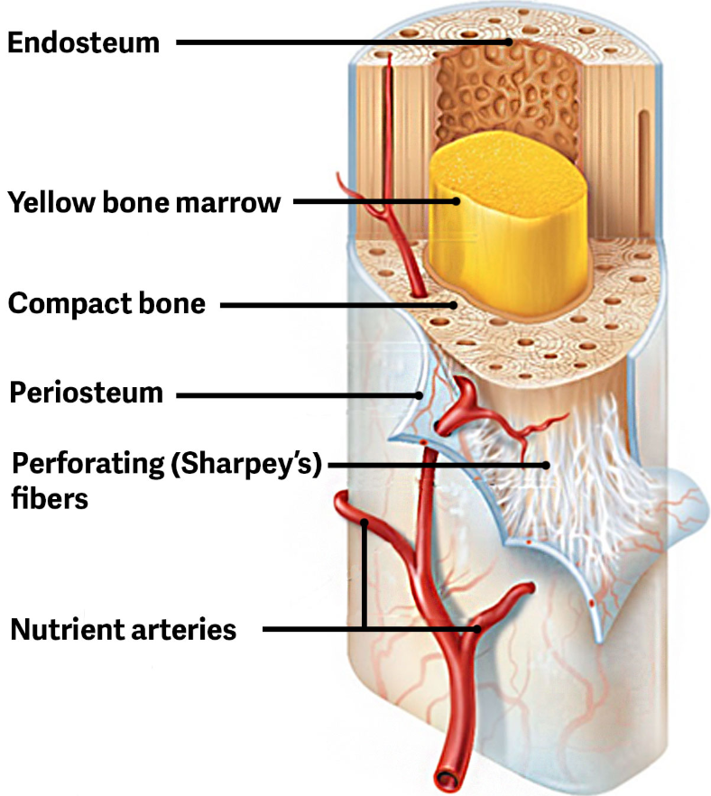
Bone Marrow
The softer connective tissue that fills the interior of most bone
Newborns have all red bone marrow and overtime it is converted to yellow bone marrow in long bones.
Contains blood vessels that supply nutrients and remove waste to and from astrocytes in the trabeculae
Ex. Yellow and Red Marrow
Yellow Marrow
A fat storage tissue found mainly in lung bones
Contains Adipose Tissue
The triglycerides stored in the Adipocytes of the tissue can serve as a source of energy.
Red Marrow
Is where the Hematopoiesis takes place.
Red blood cells, white blood cells, and platelets are all produced here.
Is primarily found in short and flat bones
Structure of a Long Bone
A longitudinal section of a typical bone is not solid but has medullary cavity filled with bone marrow.
Includes: Marrow Cavity, Diaphysis, Compact bone, Epiphysis, Spongy bone, Metaphysis, and Articular Cartilage.
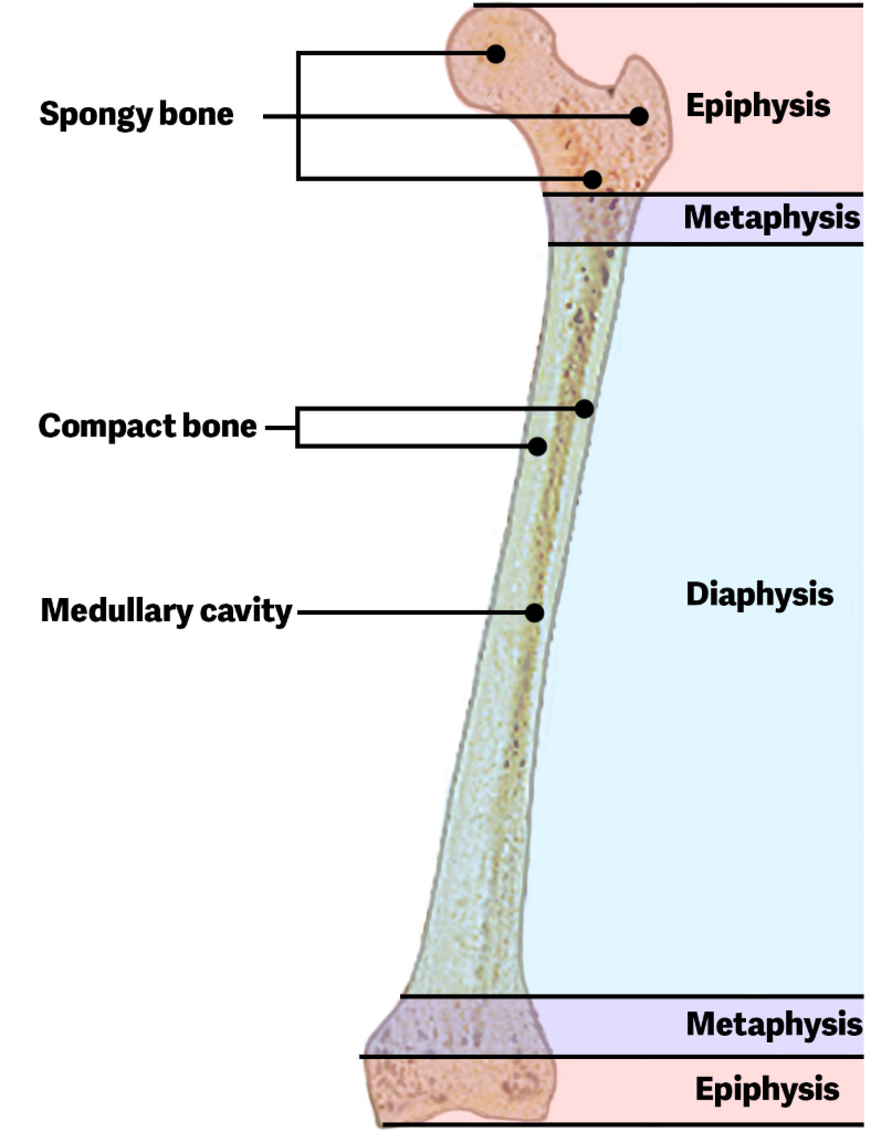
Marrow Cavity ( Medullary )
Extends throughout the Diaphysis
Surrounded by Compact Bone
Diaphysis
Shaft or center length of the bone
Compact bone
The solid outer layer of the bone
Is a layer of spongy bone that is thickest at the ends of the bone.
Ends of each bone is called Proximal and Distal Epiphysis.
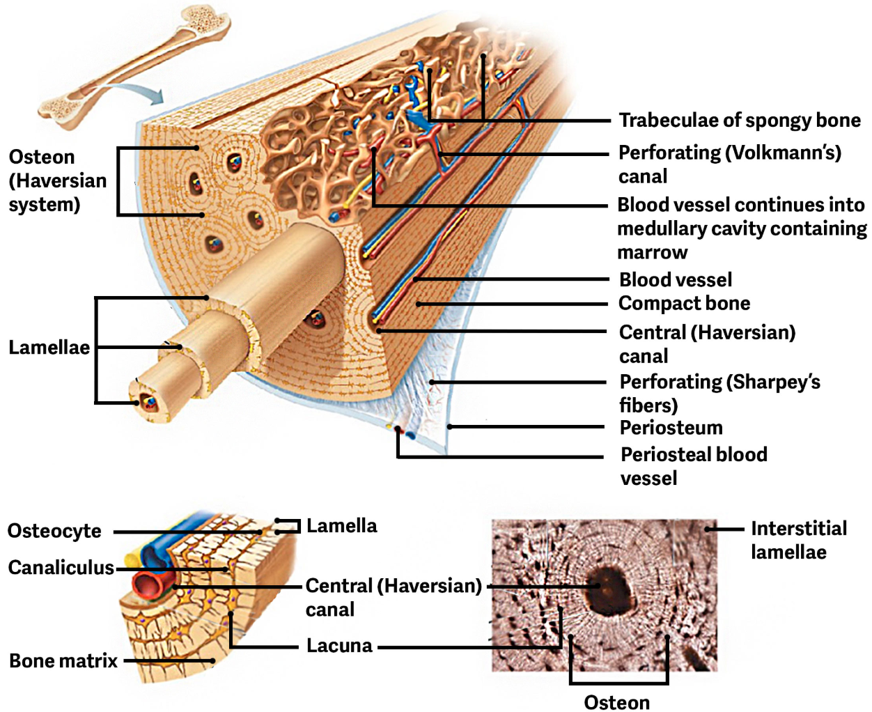
Contains many Osteons ( Haversian Systems )
Epiphysis
Widens at each end and is made mostly of Spongy Bone.
Spongy bone
Trabecular Bone
Metaphysis
Where the Epiphysis and Diaphysis meet
Compact bone is usually found on the surface of the bone and surrounds an interior of spongy bone.
The end of each long bone is covered with Articular Cartilage.
Articular Cartilage
Which helps to reduce friction when a bone articulates ( connect in a joint ) with another bone.
Covers the end of each long bone
Flat Bones
Have a parallel structure
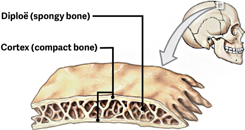
Cortex
Where spongy bone is found in between two layers of compact bone.
Diploe
The layer of spongy bone in the cranium
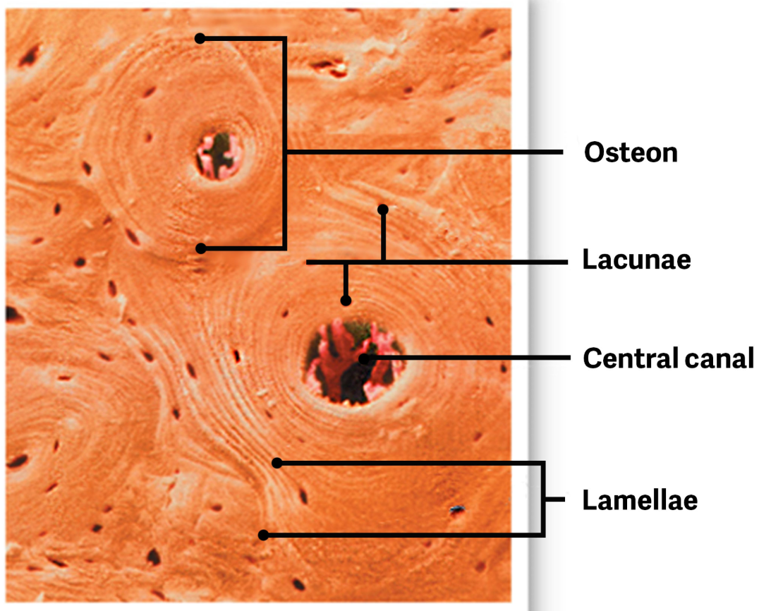
Osteons
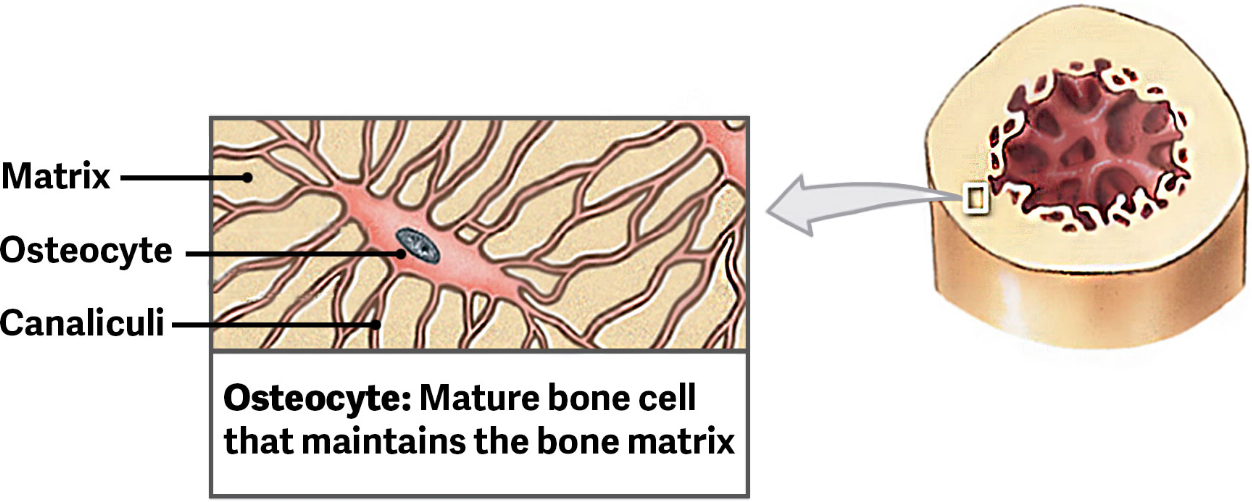
Where osteocytes can be found in tiny open chambers called lacunae.
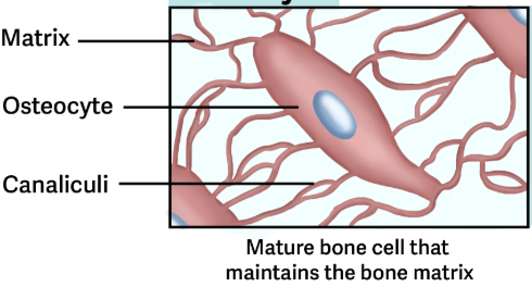
Canaliculi
Are small, narrow channels found between the lacunae.
They allow for osteocytes to receive nutrients, remove waste, and allow for communication between cells.
Lacuna ( Singular Lacunae )
A small open pocket surrounded by hard, dense matrix.
Where a single osteocyte is found
Since osteocytes can never divide a lacuna can only ever contain one osteocyte.
They are separated by matrix that contains the protein fibers of collagen and mineral deposits, primarily of calcium and phosphorus salts.
Include: Lamellae
Lamellae
The layers of matrix that separate the lacunae
Central canals
Contain blood vessels and nerves
The blood vessels bring the nutrients that allow the bone to renew itself.
Perforating Canals
Run perpendicular to the bone surface and carry blood vessels deep into the bone and bone marrow.
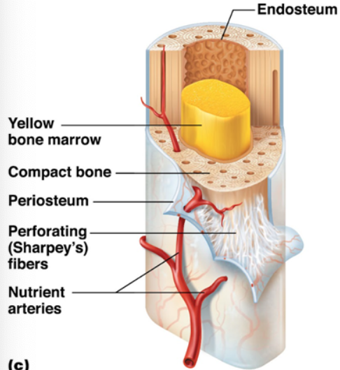
Periosteum
Covers the superficial layer of compact bone, except within joint cavities
A protective membrane that has tough fibrous outer and an inner cellular layer.
Functions to separate the bone from the surrounding structures and provides a structure for the vast amount of blood vessels that go into tissue.
Uses Perforating fibers
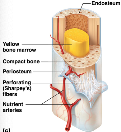
Perforating fibers ( Sharpey’s fibers )
Are made of strong collagen fibers tha thelp connect periosteum to the bone
Endosteum
Lines the marrow cavity and is a membrane lining that lines all the internal cavities inside the bone.
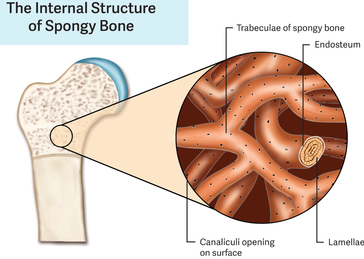
Spongy Bone
Contains numerous bony bars and plates separated by irregular spaces
Does not contain osteons
This matrix is an open network called trabeculae
Although lighter than compact bone, it is still designed for strength.
Just like braces, the solid portions follow lines of stress
Can be found in locations where stress comes from many different directions or where the bone do not have much stress applied.
Trabeculae
Lacks capillaries and venules
In the spaces between trabeculae is where red bone marrow and yellow bone marrow are stored.
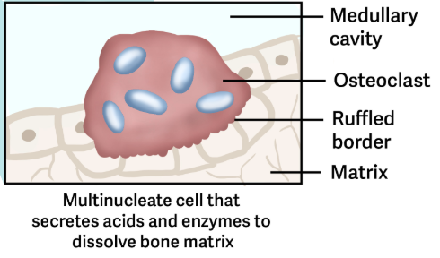
Osteoclasts
Are bone absorbing cells
Secrete acids that break down bone, remove worn cells, and therefore deposits calcium in blood, which has been removed from the bone matrix
They are found on bone surfaces are multinucleated, and originate from monocytes and macrophages.
Osteoblasts
immature bone cells that work to form new bone matrix, repairing the destruction caused by the work of osteoclasts.
As they form new bone, osteoblast take calcium from blood and deposit it into bone. → Once the calcium binds to the Osteoid, it hardens the matrix that eventually entraps osteoblasts to form osteocytes
Can not divide to form new cells.
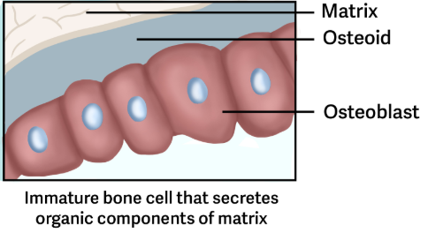
Osteoid
An unmineralized collagen matrix that bind calcium
Secreted by Osteoblasts
Osteocytes
Are mature cells that eventually form as some of the osteoblast cells get caught in the matrix they secrete and are converted to osteocytes.
Maintain the mineral concentration of the matrix via by the cytoplasmic process that extends through Canaliculi channels within bone matrix.
The cells found within the lacunae of osteons.
Through the process of remodeling, old bone tissue is eplaced by new bone tissue
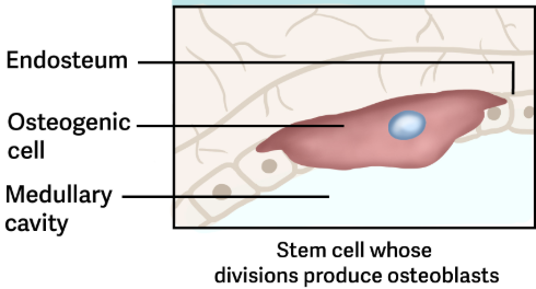
Osteogenic Cells
Are undifferentiated cells with high mitotic activity, and they are the only bone cells that divide.
Immature osteogenic cells are found in the deep layers of the periosteum and the marrow.
They differentiate and develop into osteoblasts.
Bone Formation
Most of the bone of the human skeleton are cartilaginous during prenatal development.
Since the cartilaginous structure are shaped like the future bones, they provide models of these bones.
Throughout fetal development and into childhood growth and development, bone forms on the cartilaginous matrix.
By the time a fetus is born, most of the cartilage has been replaced with bone.
Some additional cartilage will be replaced throughout childhood, and some cartilage remains in the adult skeleton.
Ex. Ossification
Ossification
The process of converting tissue to bone
Calcification occurs during ossification.
Ex. Endochondral and Intramembranous
Calcification
The process of covering tissue to bone
Occurs during Ossification, but it can also occur in other tissue that are not bone.
Endochondral Ossification
Is the ossification of bones via replacement of the mini hyaline cartilage models in bones.
Long bones develop by replacing hyaline cartilage
Cartilage does not become bone. Instead, cartilage serves as a template to be completely replaced by new bone.
About 6-8 weeks after conception, some mesenchymal cells differentiate into chondrocytes that form the cartilaginous skeletal precursor of the bones.
Takes much longer than Intramembranous Ossification
Soon after, the perichondrium, a membrane that covers the cartilage, appears. Bone then develops in several steps
Ex. Bones at the base of the Skull and Long bones
Step 1 of Endochondral Ossification
Chondrocytes Enlarge and then die as the matrix calcifies
As more matrix is produced, the chondrocytes in the center of the cartilaginous model grow. As the matrix calcifies, nutrients can no longer reach the chondrocytes.
This results in their death and disintegration of the surrounding cartilage.
Step 2 of Endochondral Ossification
Blood vessels surround the outside of the cartilage
Blood vessels grow around the outside of the cartilage, which converts the perichondrium into osteoblasts.
This develops the periosteum, which gives the shaft a superficial layer of bone
Step 3 of Endochondral Ossification
Primary Ossification Center
Blood vessels invade the spaces inside the shaft. This enlarges the cavities and brings osteogenic cells, many of which will become osteoblasts.
The blood supply to the shaft becomes the primary ossification center and the spaces combine to become medullary cavity.
Step 4 of Endochondral Ossification
The medullary cavity expands, bone increases in length
The medullary cavity is formed as growth and remodeling continues.
Chondrocytes and cartilage continue to grow at the ends of the bone (the future epiphyses), which increases the bone’s length, at the same time bone is replacing cartilage in the diaphysis.
Step 5 of Endochondral Ossification
Secondary Ossification Centers
Are formed on each end of the bone as vessels penetrate the epiphyses regions.
These vessels deliver osteoblasts and develop bone in the end of each bone.
Step 6 of Endochondral Ossification
Chondrocytes and Osteoblasts work at the same rate grow the bone by epiphyseal cartilage.
Spongy bone is created in the epiphyses on each end of the long bone
In the metaphyseal region, the Epiphyseal Cartilage is then formed between the Diaphysis and the Epiphysis.
On the epiphyseal side, the cartilage continues to grow and divide, while on the diaphyseal side, the chondrocytes degenerate.
During childhood, osteoblasts work at the same rate as the chondrocytes.
Osteoblast move upwards towards the epiphysis, and cartilage is gradually replaced by bone.
Step 7 of Endochondral Ossification
Cartilage activity slows, Osteoblast activity increases during puberty, replace cartilage with bone.
At puberty, the rate of epiphyseal cartilage production slows down, but osteoblast activity increases.
This makes the epiphyseal cartilage get smaller and smaller until eventually there is no cartilage left.
After growth is complete, there is a remnant of the epiphyseal cartilage in mature adult bones called the Epiphyseal line.
Epiphyseal Line
A thin covering of cartilage remains on the end of the bones
Intramembranous Ossification
Is the formation of flat bones from connective tissue that begins around week 8 of fetal development.
Compact and spongy bone develops directly from sheets of mesenchymal (undifferentiated) connective tissue.
Is also called dermal ossification because this occurs in the deepest layers of the dermis forming Dermal Bones.
Ex. The flat bones of the face, most of the cranial bones, and the clavicles.
Step 1 of Intramembranous Ossification
The process begins when mesenchymal cells in the embryonic skeleton gather and begin to differentiate into various specialized cells.
Some of these cells will differentiate into capillaries, while others will become osteogenic cells and then osteoblasts. Although they will ultimately spread out by the formation of bone tissue, early osteoblasts appear in a cluster ( Ossification center ).
The osteoblasts secrete osteoid, uncalcified matrix, which calcifies within a few days mineral salts are deposited on it, theory, entrapping the osteoblasts within.
Step 2 of Intramembranous Ossification
Osteoblasts become entrapped and develop into osteocytes.
One entrapped, the osteoblasts become osteocytes as osteoblasts transform into osteocytes, osteogenic cells in the surrounding connective tissue differentiate into new osteoblasts.
Spicules grow out of the ossification center.
Spicules
Are the developing bone growing outwards from the ossification center.
Step 3 of Intramembranous Ossification
Blood vessels become entrapped and spicules connect
Blood vessels begin to become trapped in the bone as it grows, and spicules connect.
Step 4 of intramembranous Ossification
Inner capillaries become the spongy bone trabecular matrix
Osteoid ( unmineralized bone matrix ) secreted around the inner capillaries results in a trabecular matrix.
Step 5 of Intramembranous Ossification
Remodeling of the spongy bone around outer capillaries becomes the osteon formation of compact bone osteoblasts on the surface become the periosteum
Remodeling of spongy bone around blood vessels on the surfaces produce the typical osteon formation of compact bone on both sides of the inner spongy bone.
Osteoblasts on the surface of the spongy bone become the periosteum.
Blood and Nerve Supply
The spongy bone and medullary cavity receive nourishment and remove waste from arteries and veins that carry blood and pass through the compact bone.
The arteries and vessels travel through the Nutrient Foramen
The osteocytes in spongy bone are nourished by blood vessels of the periosteum that penetrate spongy and blood that circulates in marrow cavities.
As the blood passes through the marrow cavities, it is collected by veins, which then pass out of the bone through the framing.
Nerves follow the same paths into bone where they tend to concentrate in the mre metabolic active regions of the bone. The nerves sense pain and play roles in helping to regulate blood supply and bone growth.
Bone remodeling
When resorption of old or damaged bone takes place on the same surface where osteoblasts lay new bone to replace that which is reabsorbed by Osteoclasts.
Bone is a living tissue and able to repair itself when damaged or worn out
Occurs un adults and children.
Osteoblast and Osteoclast balance.
Remains in homeostatic balance.
When happens on different surface we get different disease states.
Minerals of the bone matrix are continually being resorbed, recycled and placed back into the bone matrix
Due to continual remodeling the thickness of bones can change
Results from forces or lack of forces placed on bone.
Physical use, Hormones, Injury or Exercise
Even without injury or exercise the bones can change.
10-20% of an adult skeleton is replaced annually
This is done by the work of destroying old bone renewing it with fresh bone.
Osteolysis
Osteoclasts removes old osteons
One osteoblast forms another osteon to replace the lost one.
Appositional Bone Growth
Is when a bone grows in diameter and can continue even after longitudinal growth cases.
What does the lack of mechanical stress do to Bones
Bones lose mineral salts, collagen fibers, and decrease in bone strength.
Ex. Broken bone in a cast or a sedentary lifestyle → Bone Atrophy.
What does Mechanical stress do to bones
Makes bones thicker and stronger due to the stimulation of osteoblasts depositing mineral salts and collagen fibers in places of applied forces.
Ex. Exercise or resistance training ( muscle tendon pulling on bone ).
Dietary Needs for Bones
Vitamins are also necessary in the diet for proper bone growth and development.
Needed for adults and children.
Calcium and Phosphate promote work of osteoblasts.
Ex. Vegetables and Diary.
Ex. Vitamin D, Vitamin A, and Vitamin K / B12.
Vitamin D
Synthesis of hormone calcitriol and essential for normal calcium and phosphate absorption.
Vitamin A
Stimulates osteoblast activity.
Vitamin K, B12
Synthesis of proteins in bone.
Hormone Regulation of Bone TIssue
The endocrine system produces and secretes hormones, which interact with the skeletal system
These hormones interact with the skeletal system → Control bone growth, maintain bone are formed, and remodel it.
Hormones can influence osteoblast activity and/ or maintain bone matrix.
Ex. Growth Hormone (GH), Thyroxine, Sex Hormones.
Growth Hormone (GH)
Controls bone growth in several ways.
Secreted by the Pituitary Gland.
Triggers chondrocyte proliferation in epiphyseal plates → Increase length of bones.
Increases calcium retention → Enhances mineralization.
Stimulates osteoblastic activity → Which improves bone density.
Thyroxine
Stimulates bone growth and promotes synthesis of bone matrix.
Secreted by the Thyroid gland.
Promotes osteoblastic activity.
Sex Hormones
Promote osteoblastic activity and production of the bone matrix.
Responsible for adolescent growth spurt.
Promote conversion of epiphyseal plate to epiphyseal line.
Ex. Estrogen → Ovaries and Testosterone → Testes.
Calcitrol
Stimulates absorption of calcium and phosphate from digestive tract.
Produced by the kidney.
Calcium Balance and Hormonal Control
Bone remodeling and modeling requires balance.
Osteoclasts to resorb unneeded, damaged, or old bone.
Osteoblasts to lay new bone.
Hormones: PTH and Calcitonin
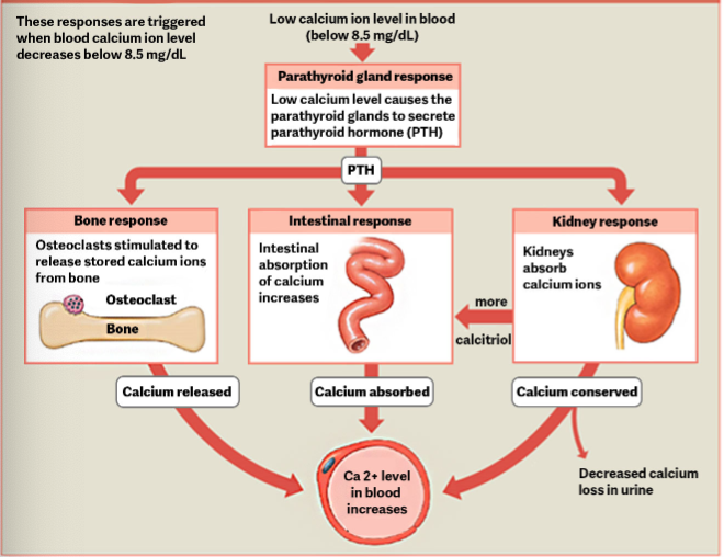
Parathyroid Hormone (PTH)
Simulates osteoclast proliferation and activity.
Secreted by parathyroid gland.
Located on the posterior side of the thyroid gland in the neck.
Are released when calcium levels are low in the blood. (under 8.5mg/dL)
Not enough calcium binding to cell receptors.
It’s goal is to increase calcium in the blood.
Stimulate osteoblast proliferation.
Promote reabsorption of calcium from the urine.
At the kidneys.
Stimulate synthesis of Vitamin D
Stimulates calcium absorption from digested food in the small intestine.
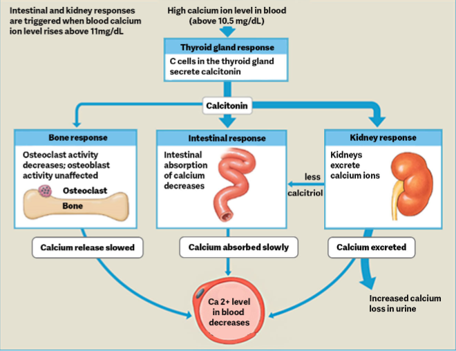
Calcitonin
Inhibits osteoblast activity and stimulates calcium uptake by bones.
Goal is to decrease calcium in the blood.
Secreted by Thyroid gland.
Inhibits osteoclast activity.
Stimulates calcium uptake.
Kidneys secrete more calcium in the urine.
Decreased absorption of calcium in the intestines.
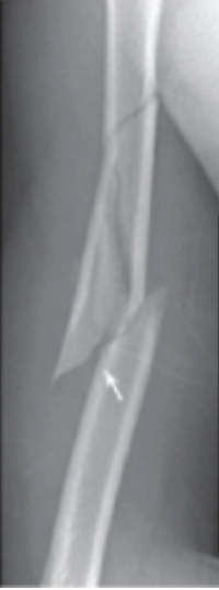
Bone Fracture
Ranged of Injury minor to severe.
Some fractures have life-threatening complications.
Ex. Fractured diaphysis of the femur.
Release fat globules into the bloodstream.
May become lodged in a blood vessel, blocking blood flow (fat emboli, FE).
May occur in capillaries beds of the lungs.
Respiratory distress or death if not treated.
Fracture
Broken bone
Will heal even with serve damage if blood supply and cells of endosteum and periosteum survive.
Ex. Open (Compound) and Closed (Simple).
Closed Reduction
Broken bone is set into it’s natural position without surgery
Open Reduction
Requires surgery to expose the facture and reset the bone.
Types of Bone Fracture
Classified by complexity, location and other features present.
May be described using more than one term.
If fracture has features of more than one type.
Ex. Compound and Spiral Fracture.
Ex. Compound, Simple, Displaced, Nondisplaced, Transverse, Spiral, Comminuted, Impacted, Compression, Greenstick, Depressed.
Open (Compound)
One end of the broken bone tears through the skin.
Carries high risk of infection
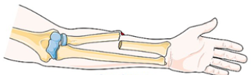
Closed (Simple)
Skin remains intact
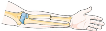
Displaced
Produces a new or abnormal bone alignment.