Lab 1: Microscopy, Histology and Integumentary System
 Call Kai
 Learn
Learn Practice Test
 Spaced Repetition
 Match
Match Flashcards
 Knowt Play
1/303

There's no tags or description
Looks like no tags are added yet.
Name | Mastery | Learn | Test | Matching | Spaced | Call with Kai | Chat |
|---|
No analytics yet
Send a link to your students to track their progress
304 Terms

1 (compound light microscope)
ocular lens
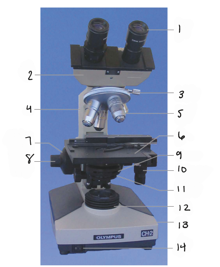
2
body tube

3
nosepiece

4
arm
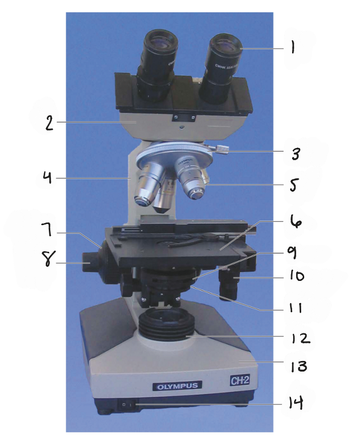
5
objective lenses

6
stage
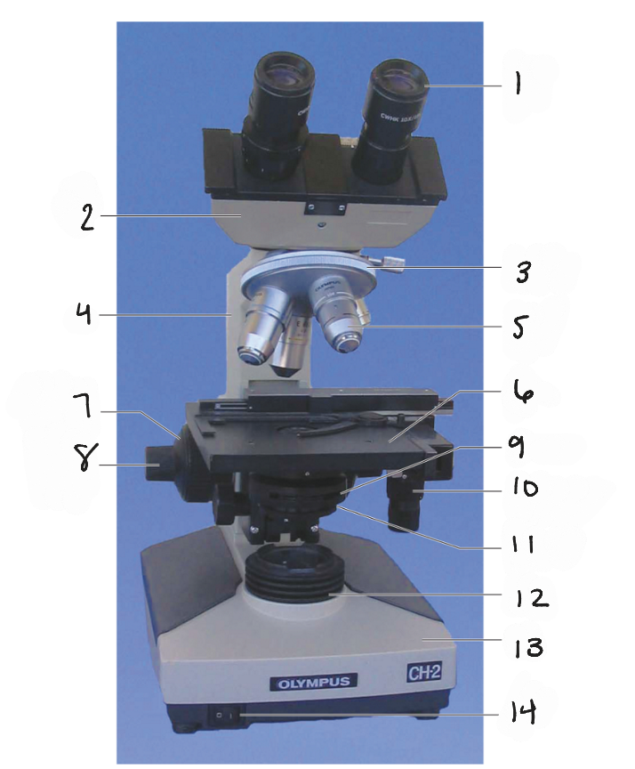
7
coarse-focus knob

8
fine focus knob
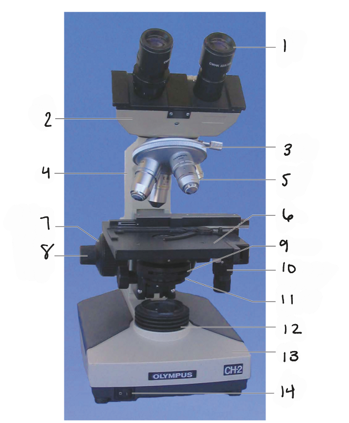
9
condenser

10
mechanical stage control knob
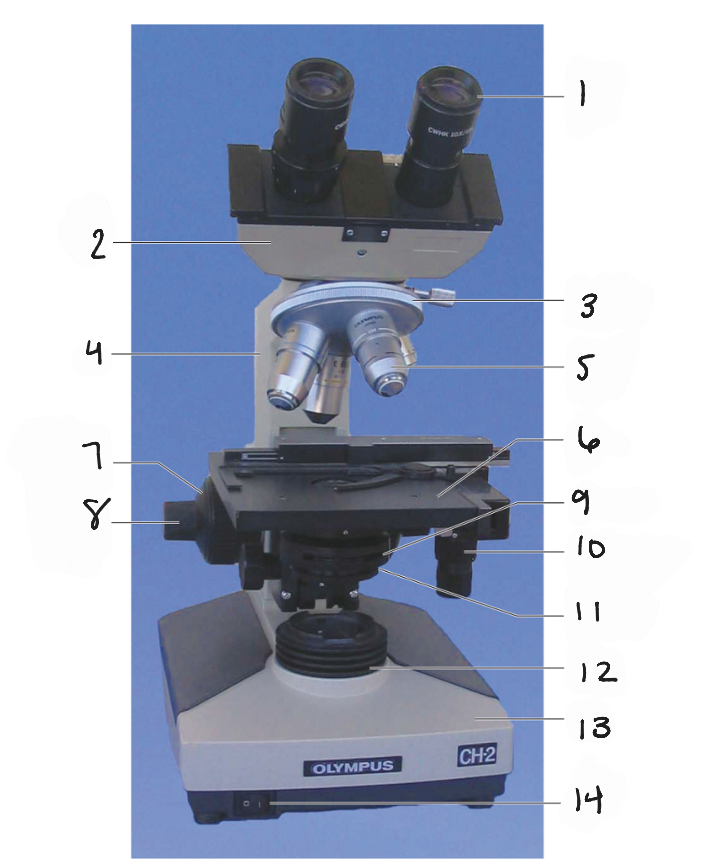
11
iris diaphragm lever
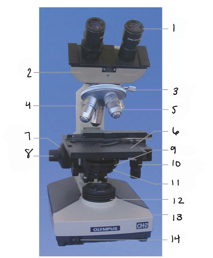
12
light source
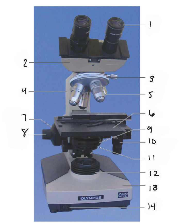
13
base
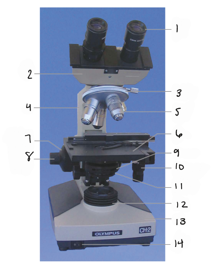
14
on/off switch
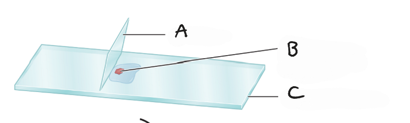
A
coverslip

B
specimen in drop of water
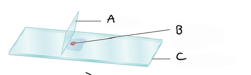
C
microscope slide
Problem with microscope: nothing is visible in the lense
Solutions:
plug in microscope
turn the power supply on
rotate the objective lens until you hear it click into place
the bulb is burned; replace the bulb
Problem with microscope: you see a dark crescent
solution: The objective lens is not in the proper position; click the lens into place
Problem with microscope: All you see is a light circle
solution:
The microscope is out of focus; adjust the coarse-focus knob
The light is up too high; turn down the light
the iris diaphragm is open too much; close it down

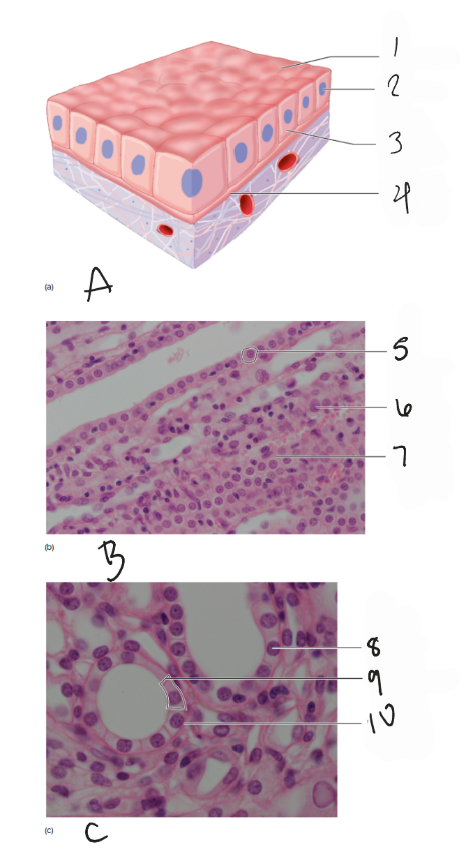
A
diagram of simple squamous epithelium
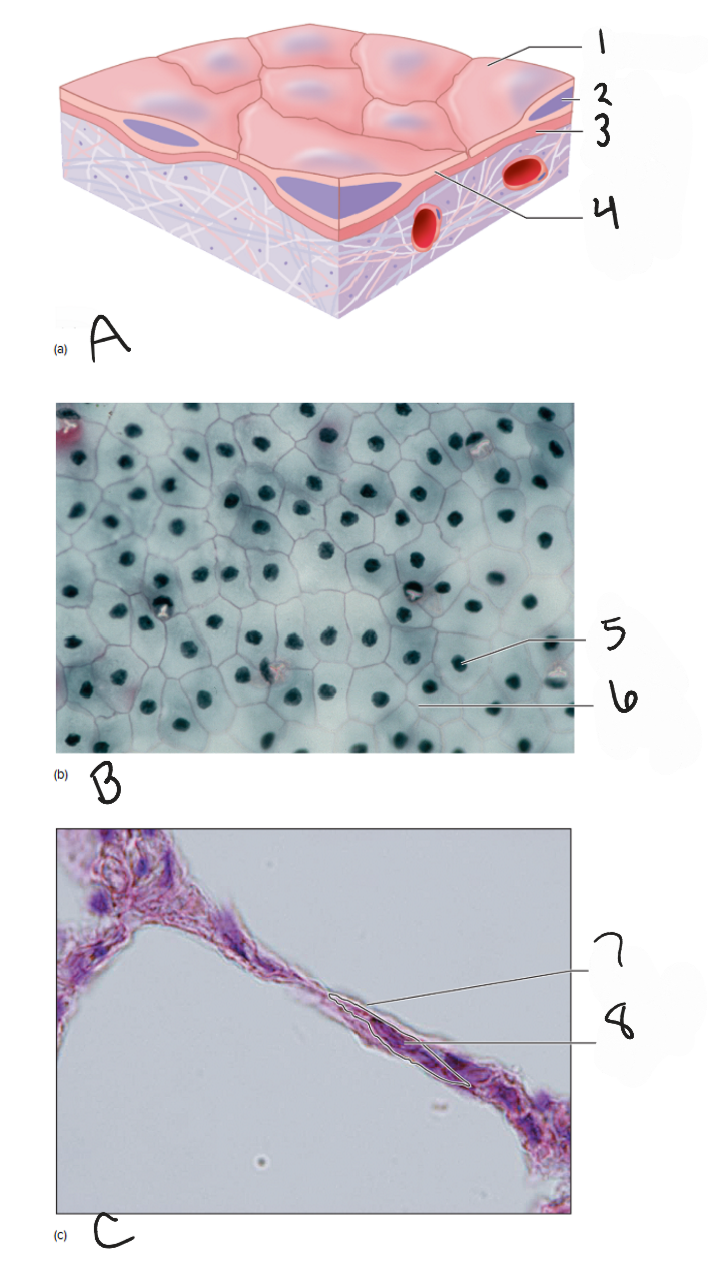
1
free surface
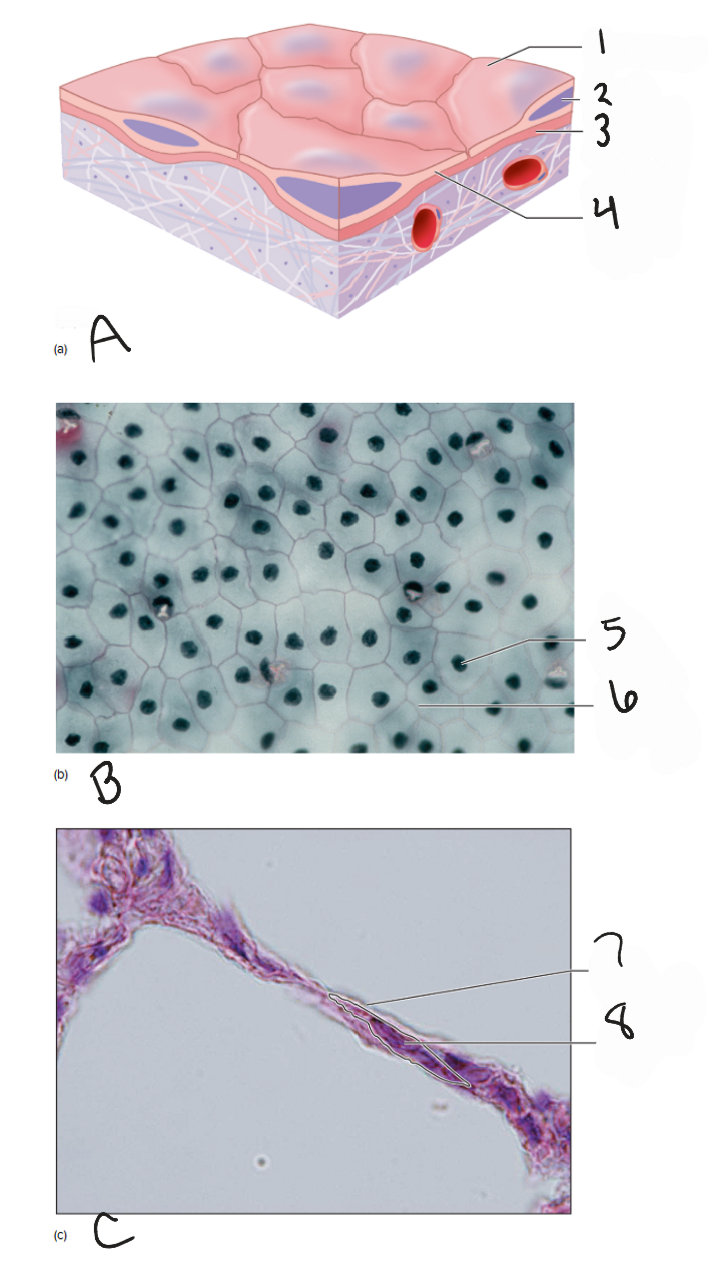
2
nucleus
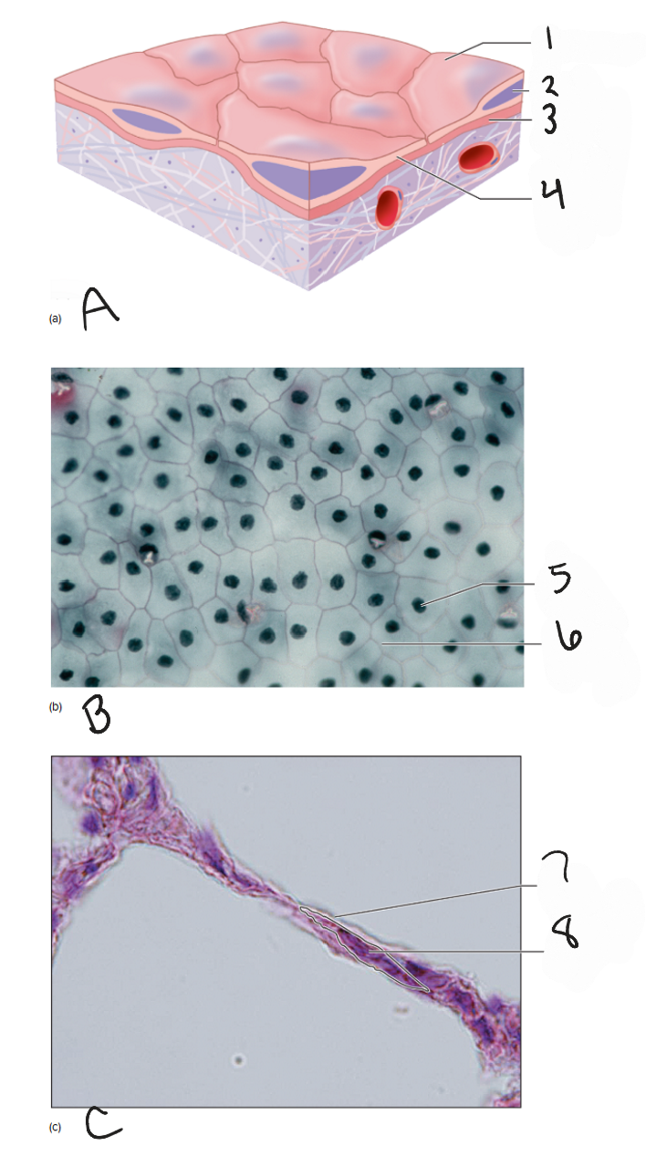
3
basement membrane
4
simple squamous epithelial cell

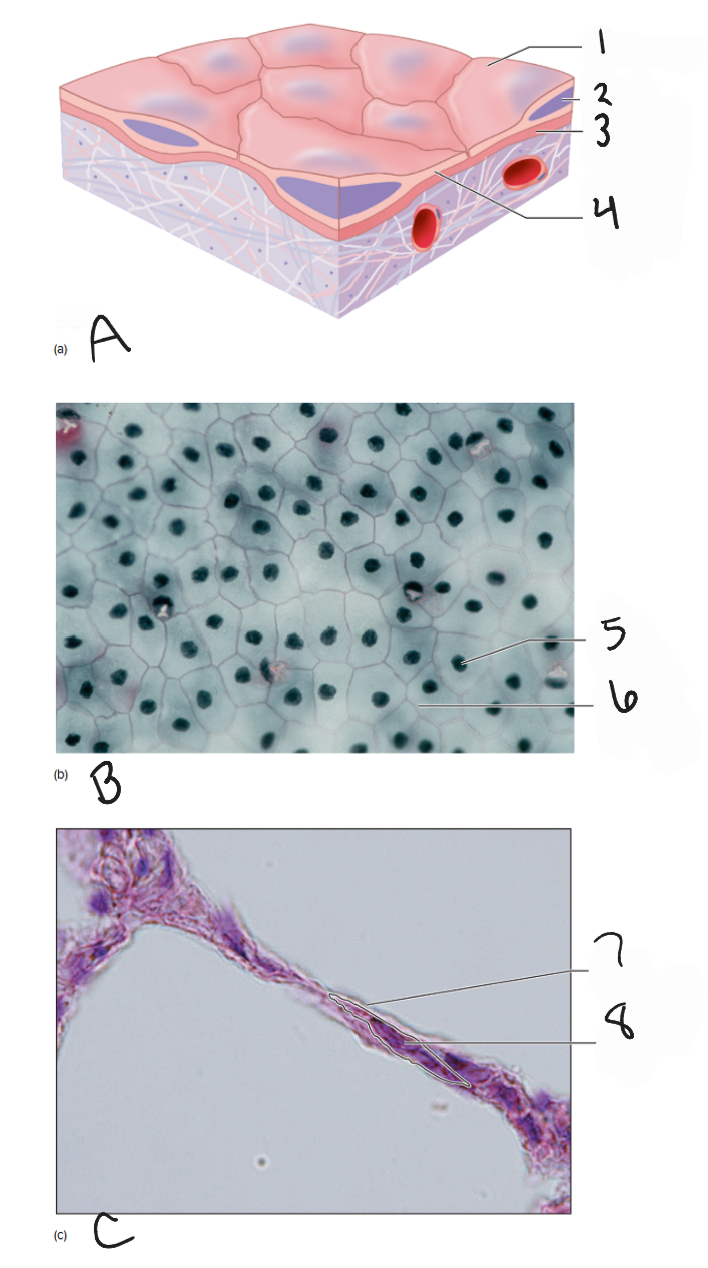
B
photomicrograph of mesothelium, surface view 400x

5
cell nucleus
6
cell membrane
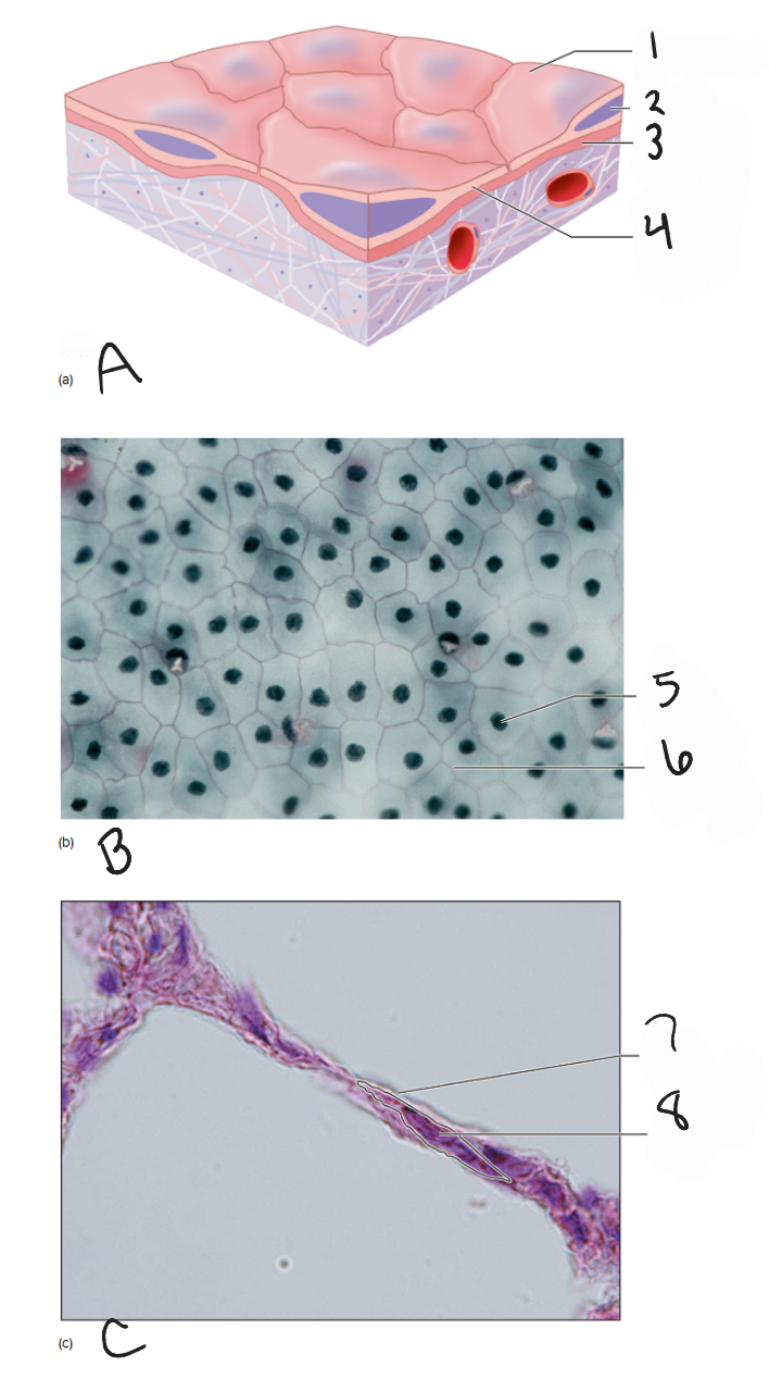
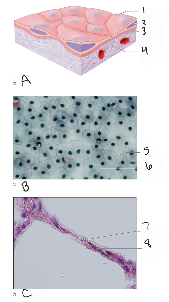
C
photomicrograph of lung tissue, side view 1000x

7
cell membrane

8
nucleus
A
diagram of simple cuboidal epithelium
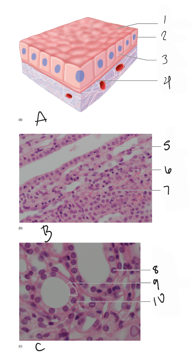
1
Free surface
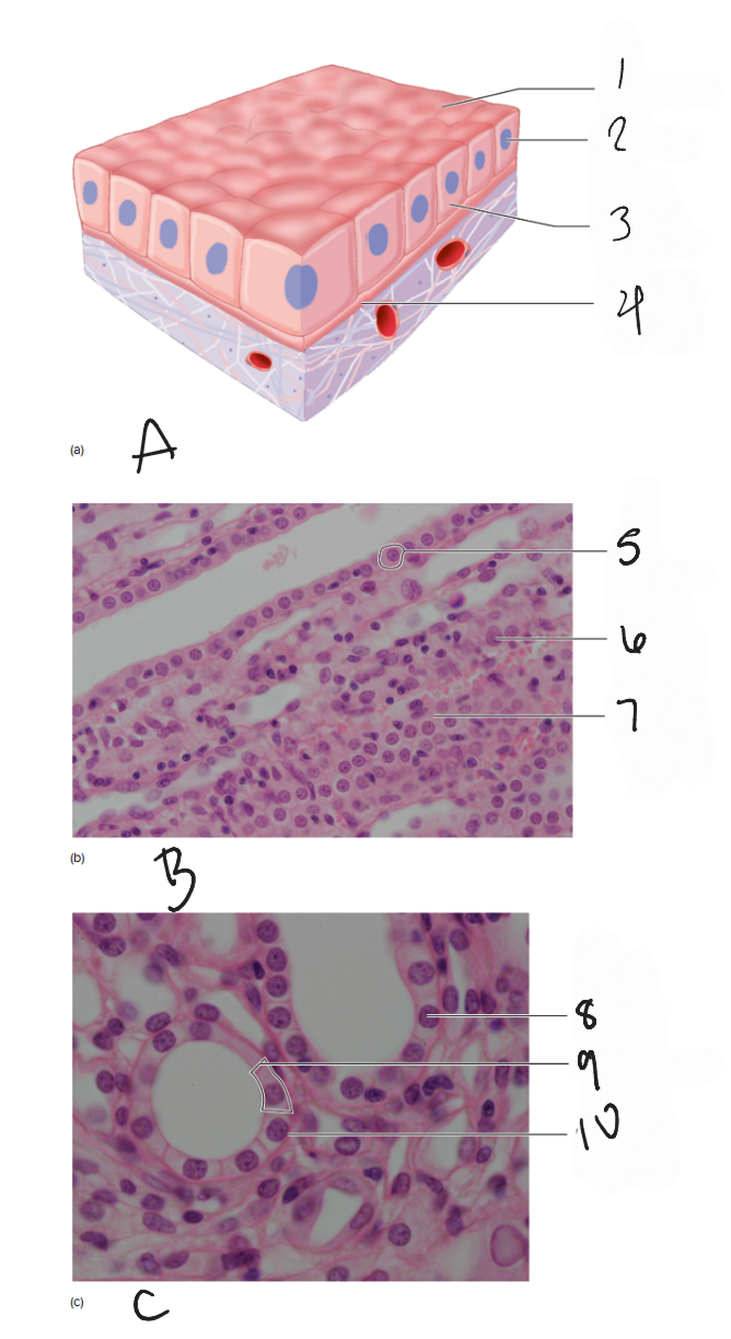
2
nucleus
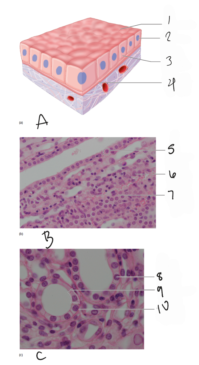
3
simple cuboidial epithelial cell
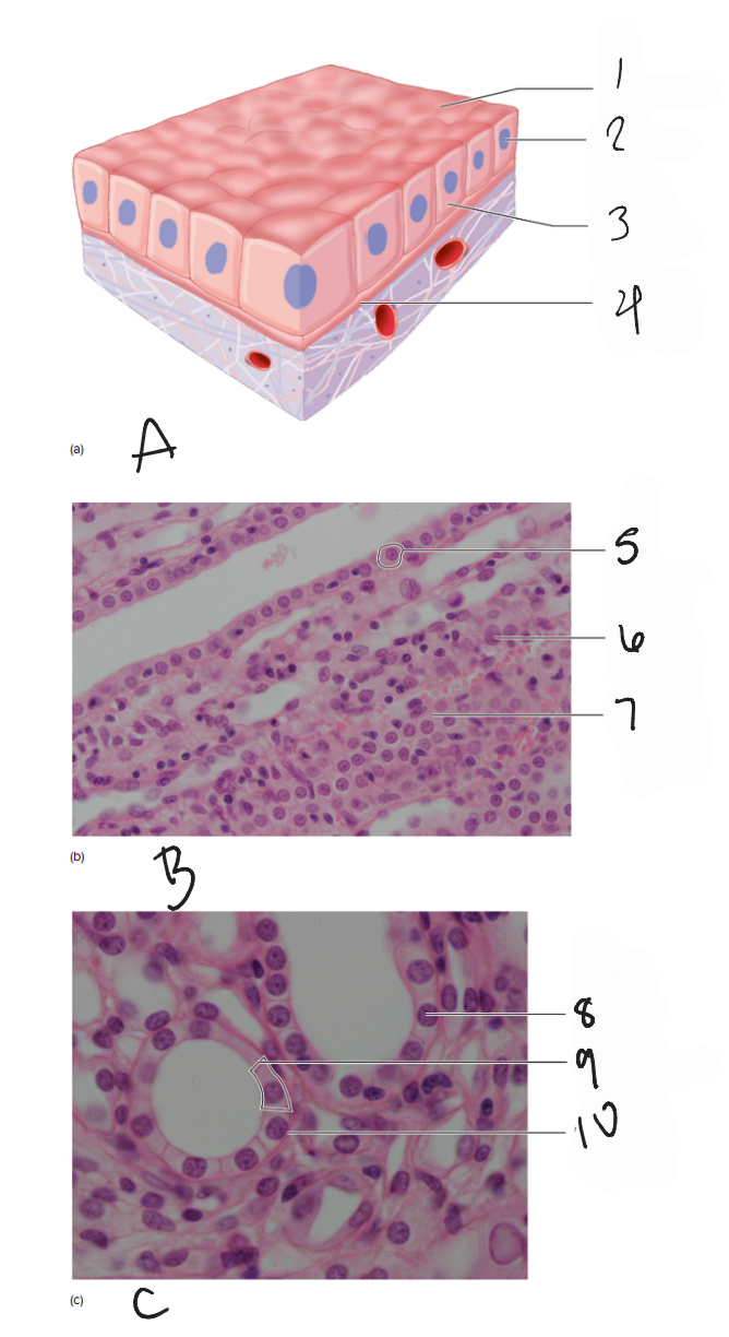
4
basement membrane
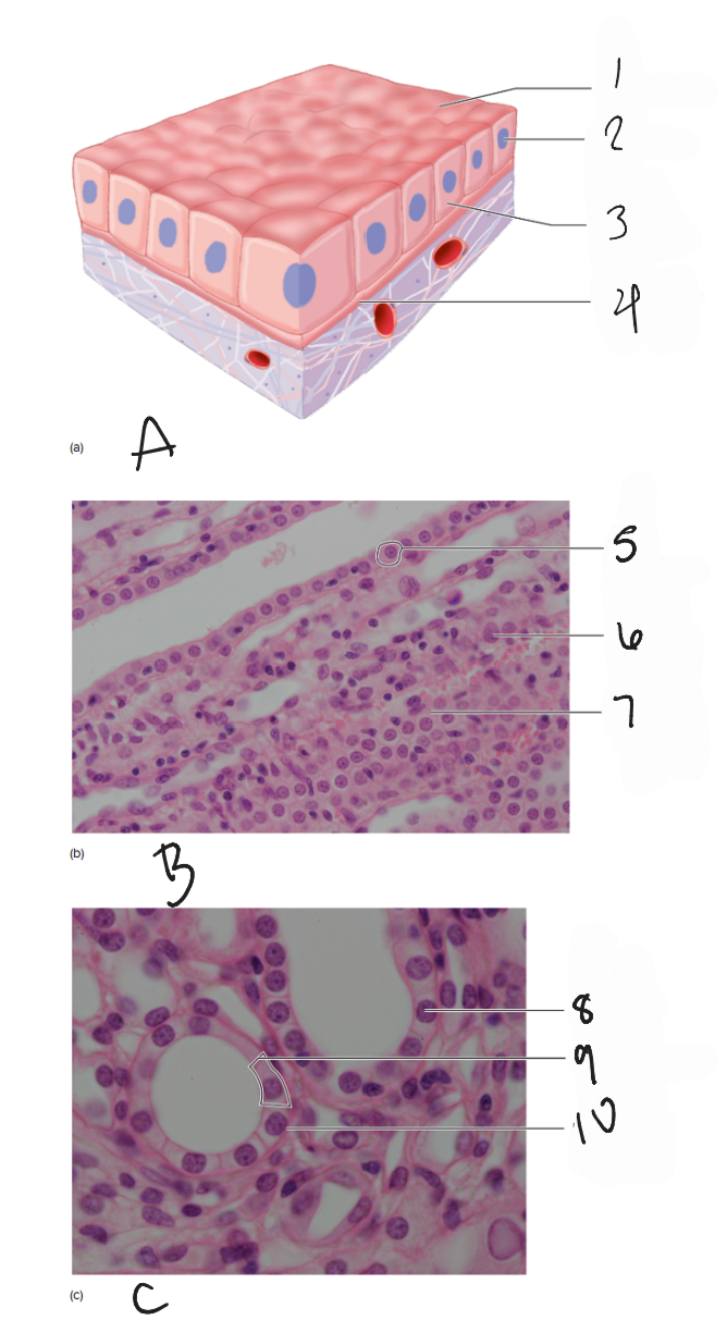
B
photomicrograph of kidney, long section (400x)
5
cell membrane
6
nucleus
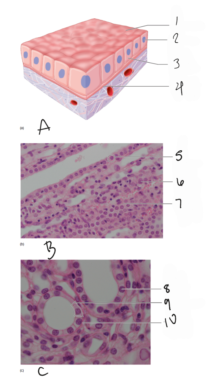
7
basement membrane

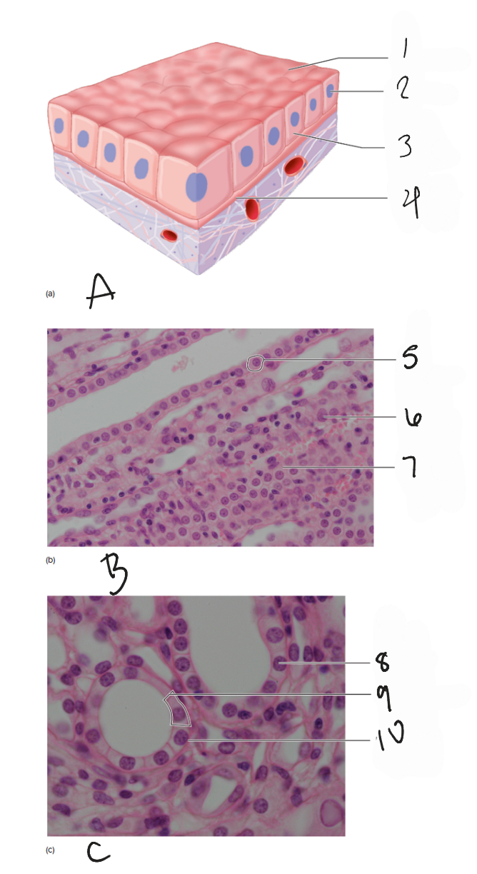
C
photomicrograph of kidney, cross section (400x)

8
nucleus

9
cell membrane
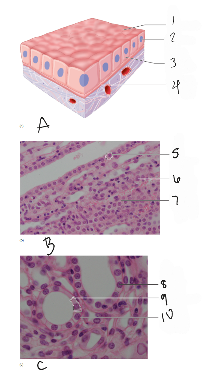
10
basement membrane
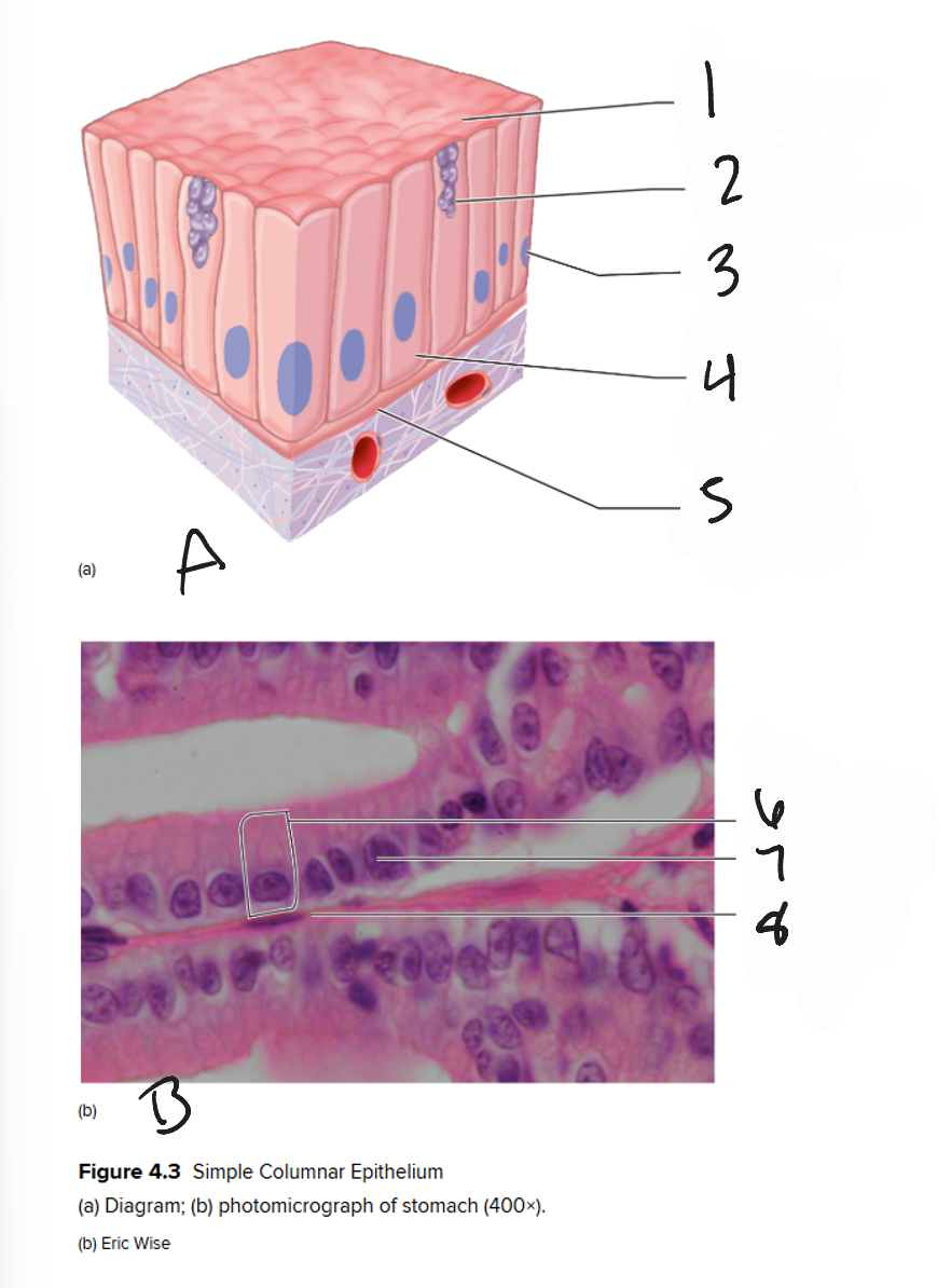
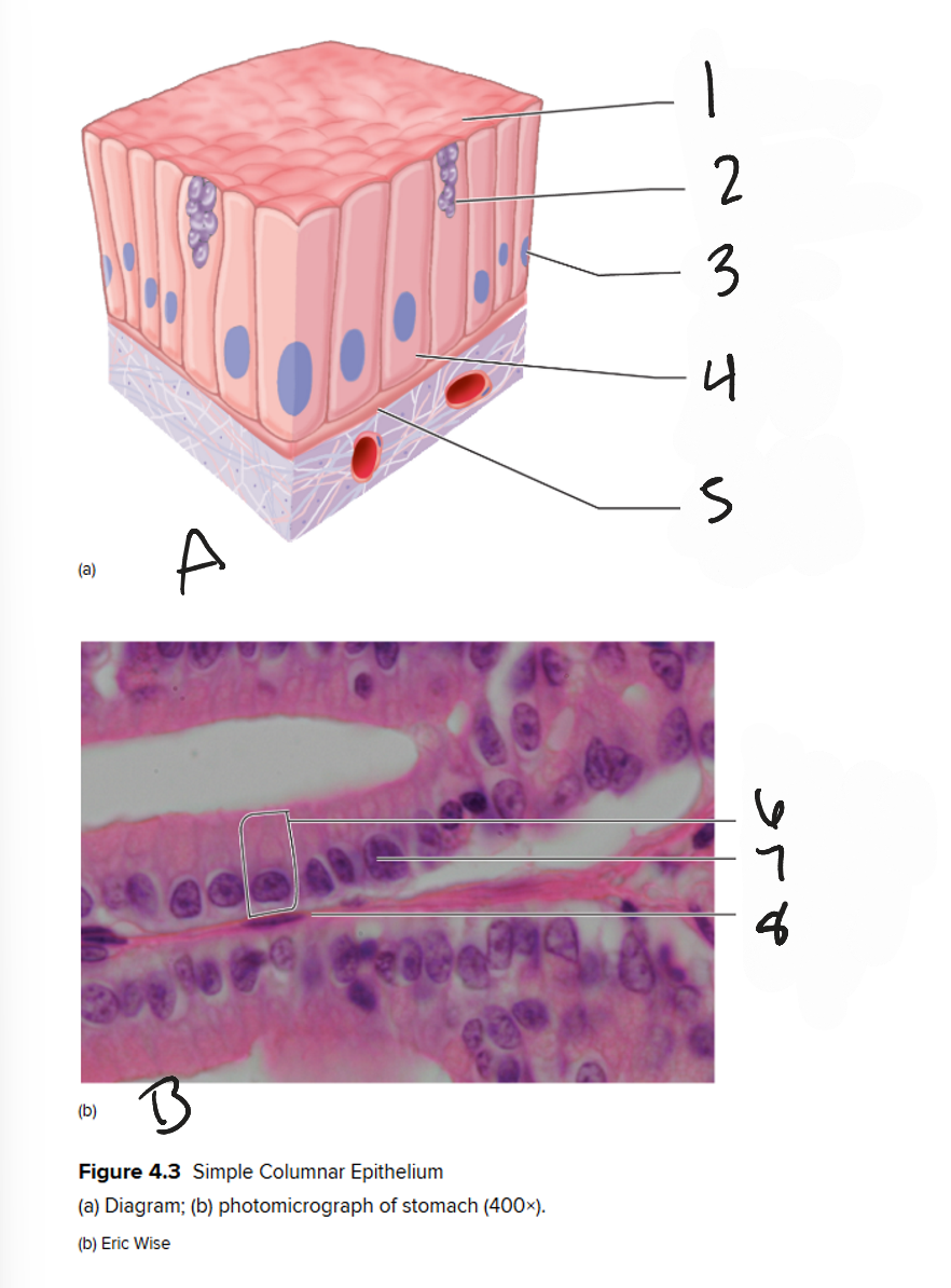
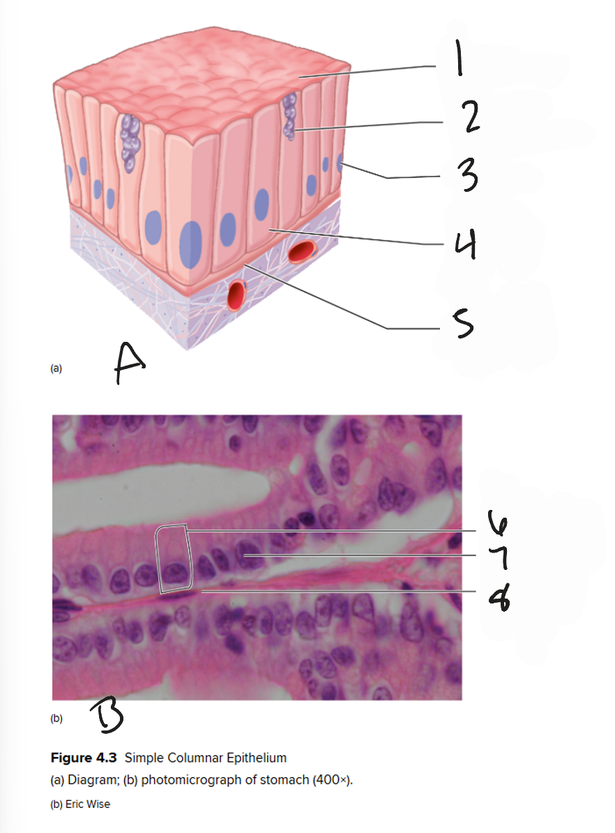
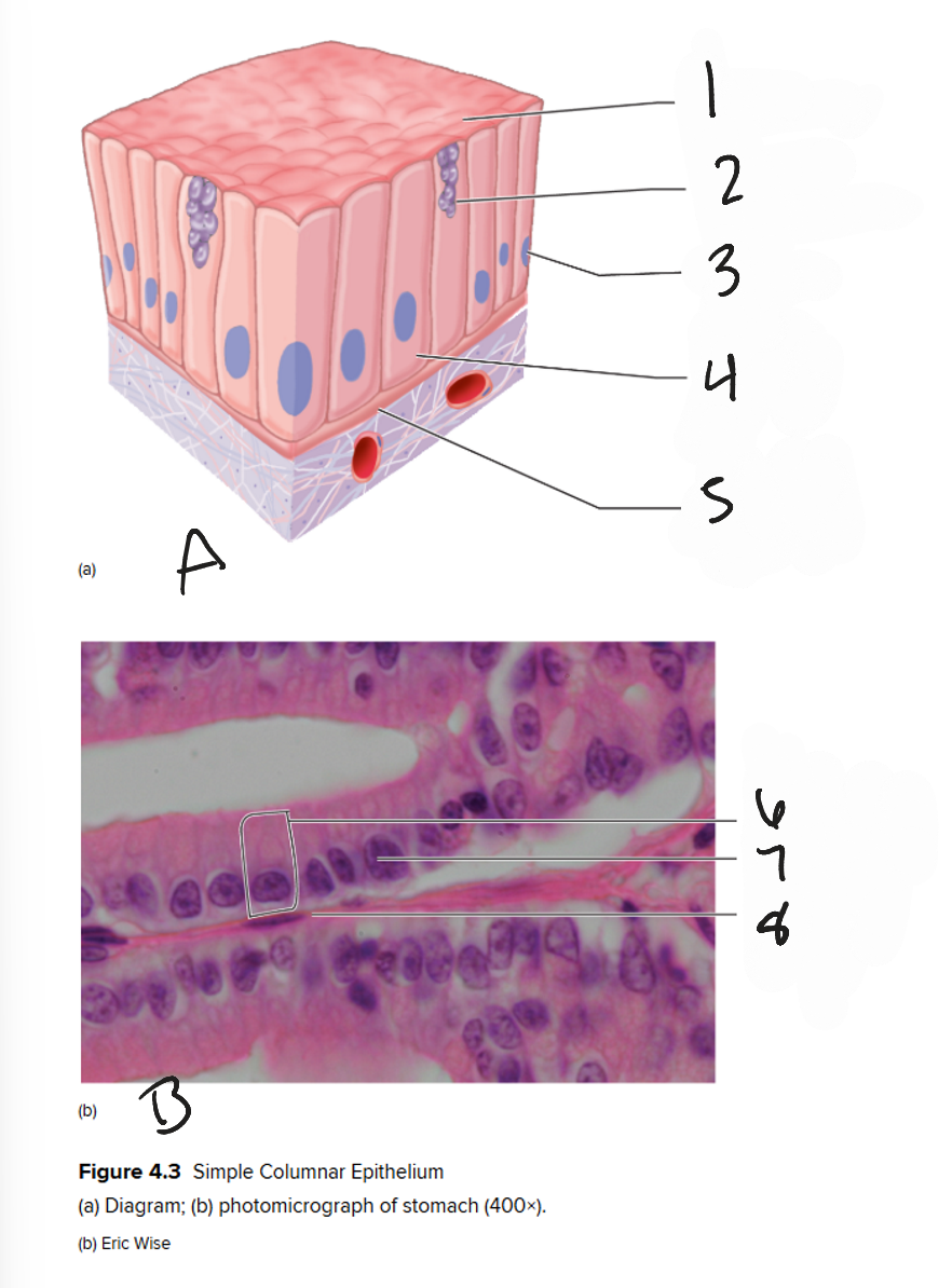
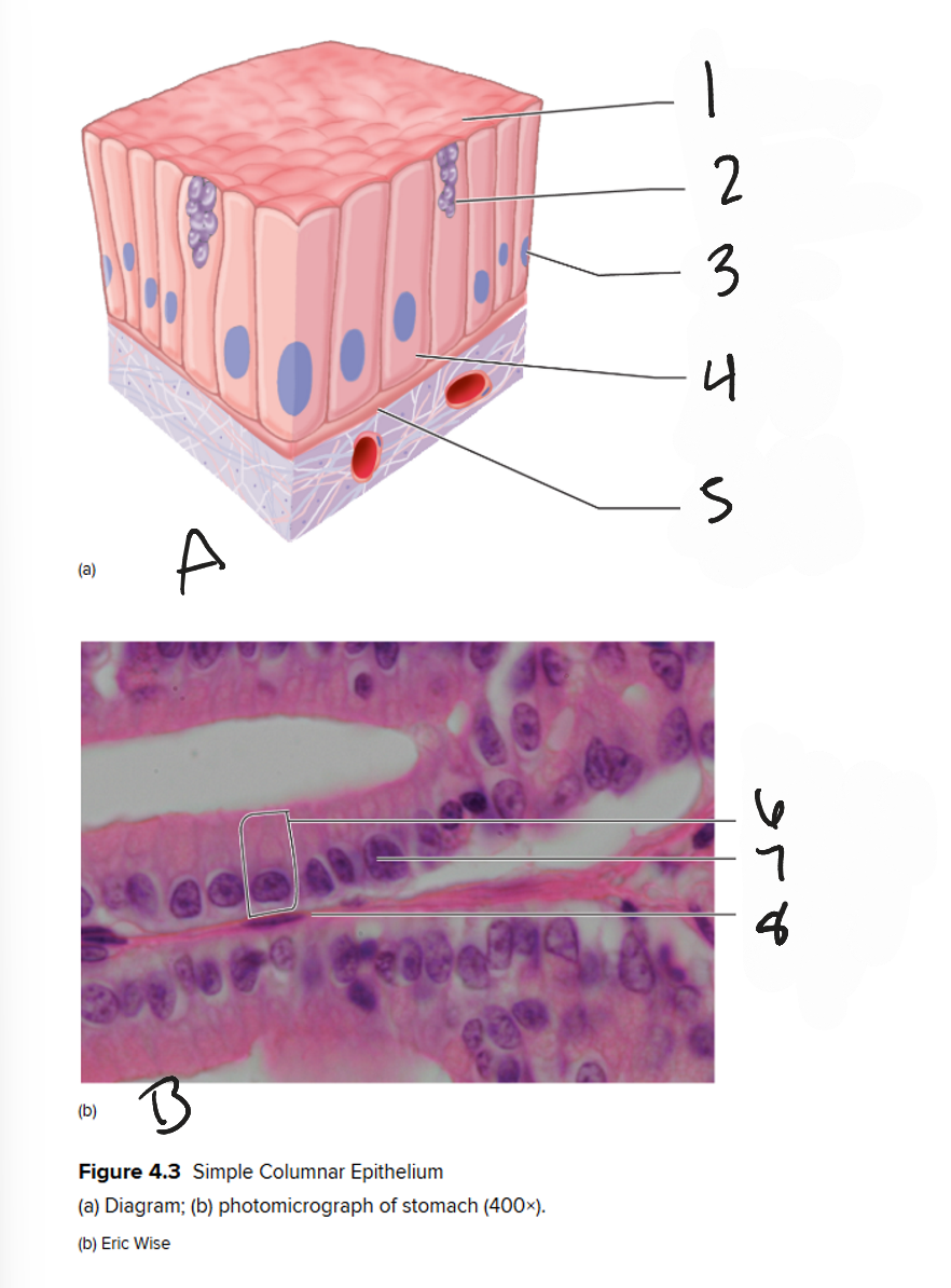
A
diagram of simple columnar epithelium

1
free surface
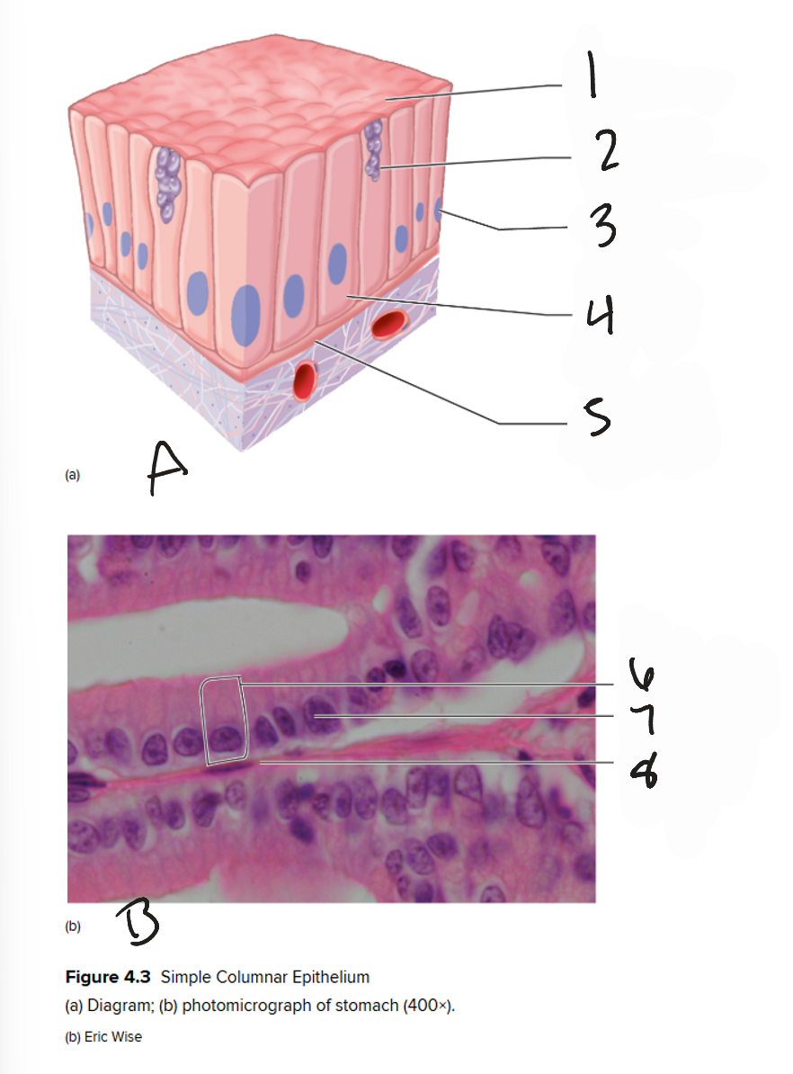
2
goblet cell containing mucus
3
nucleus
4
simple columnar epithelial cell
5
basement membrane
B
photomicrograph of stomach (400x)

6
cell membrane

7
nucleus

8
basement membrane
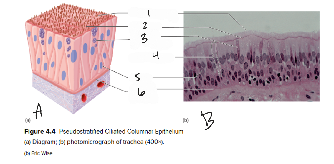
A
diagram of pseudostratified ciliated columnar epithelium
1
cilia on free surface
2
free surface
3
goblet cell containing mucus
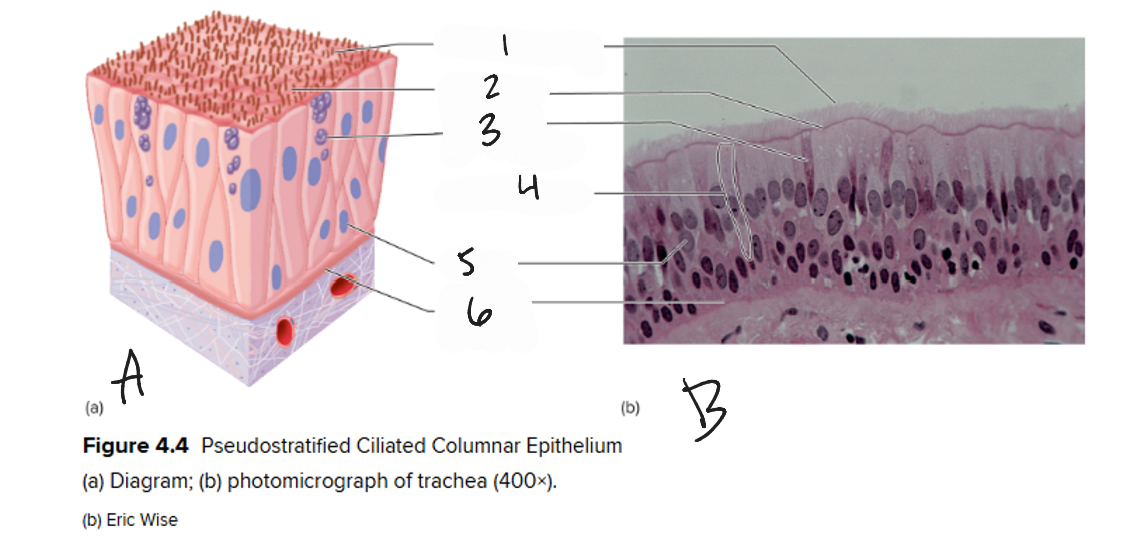
4
plasma membrane

5
nucleus

6
basement membrane
B
photomicrograph of trachea (400x)

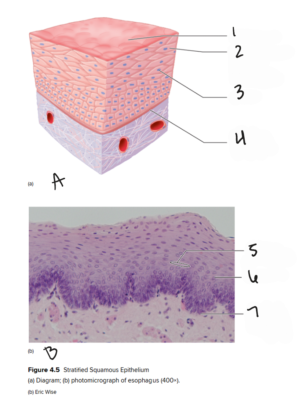
A
diagram of stratified squamous epithelium
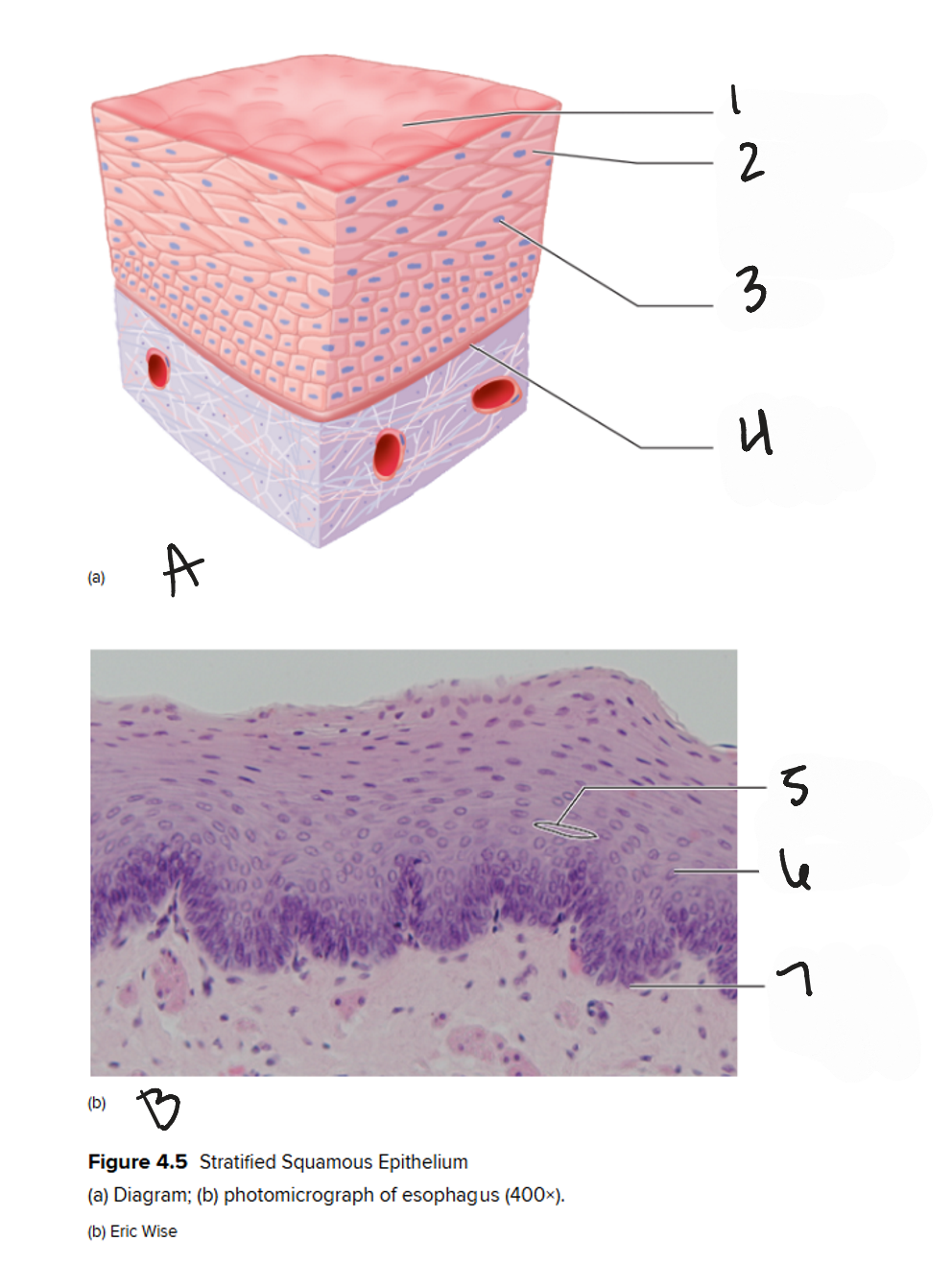
1
free surface
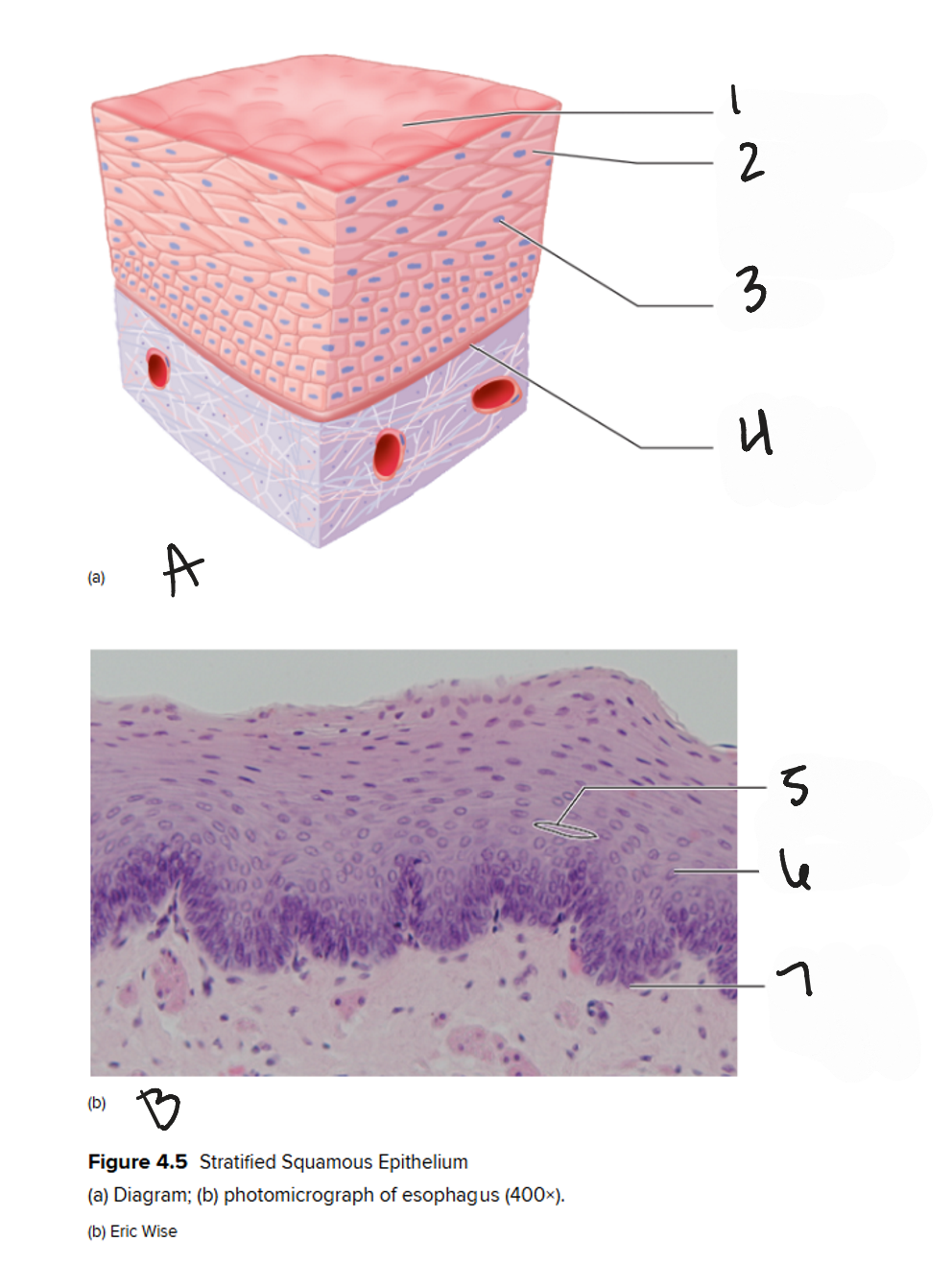
2
nonkeratinized stratified squamous epithelial cell
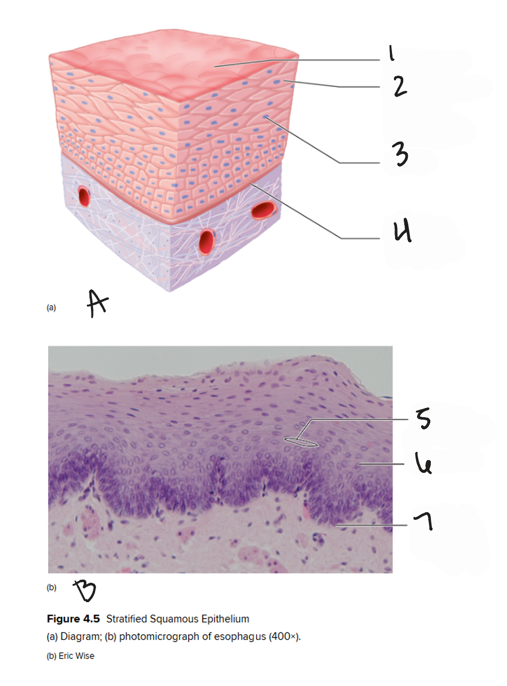
3
nuclei

4
basement membrane
B
photomicrograph of esophagus (400x)
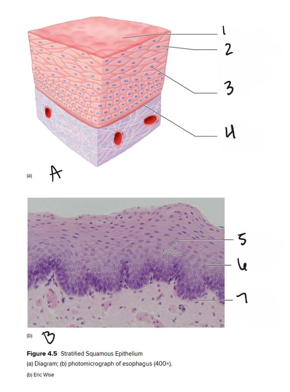
5
plasma membrane

6
nucleus

7
basement membrane
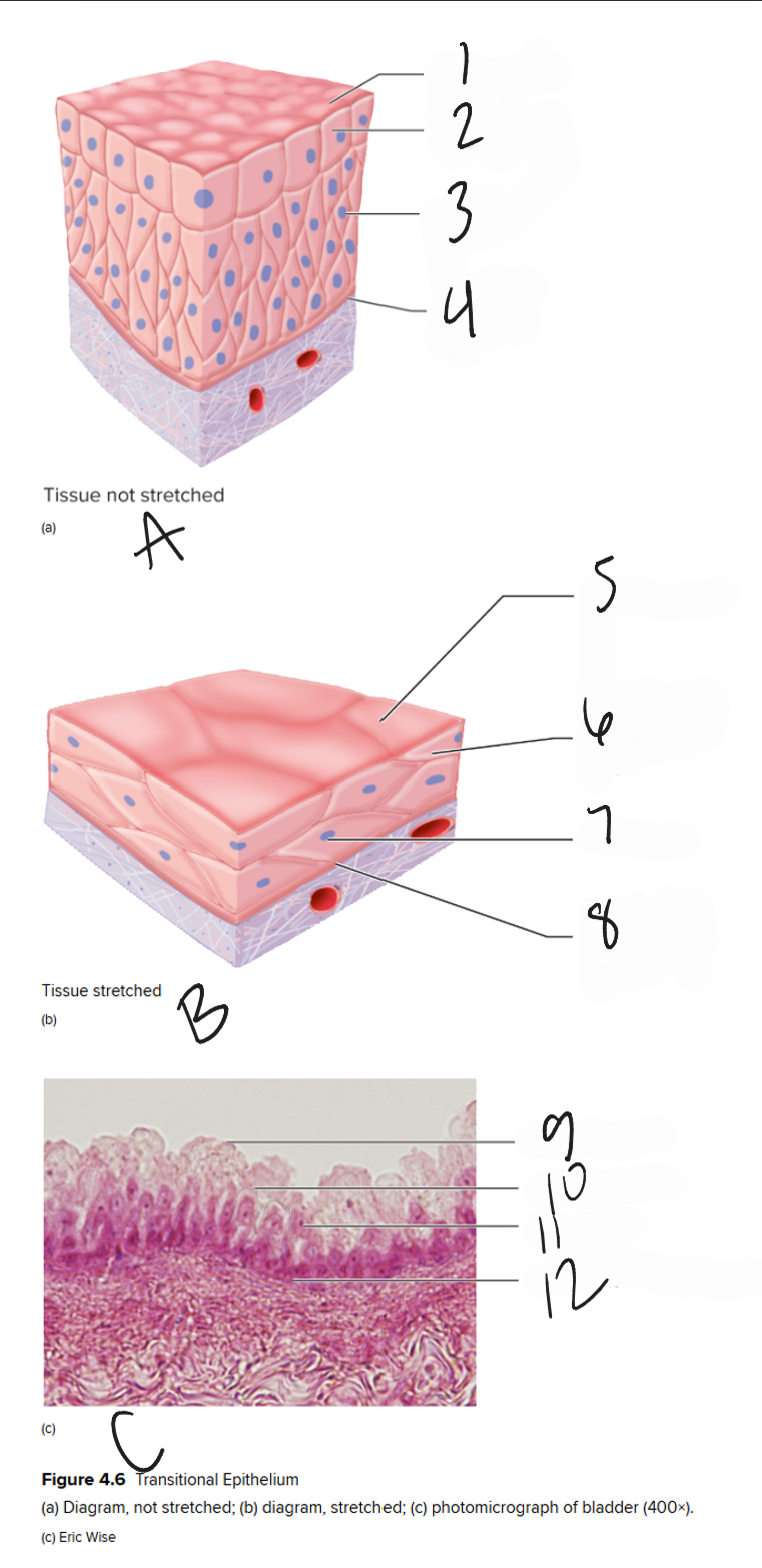
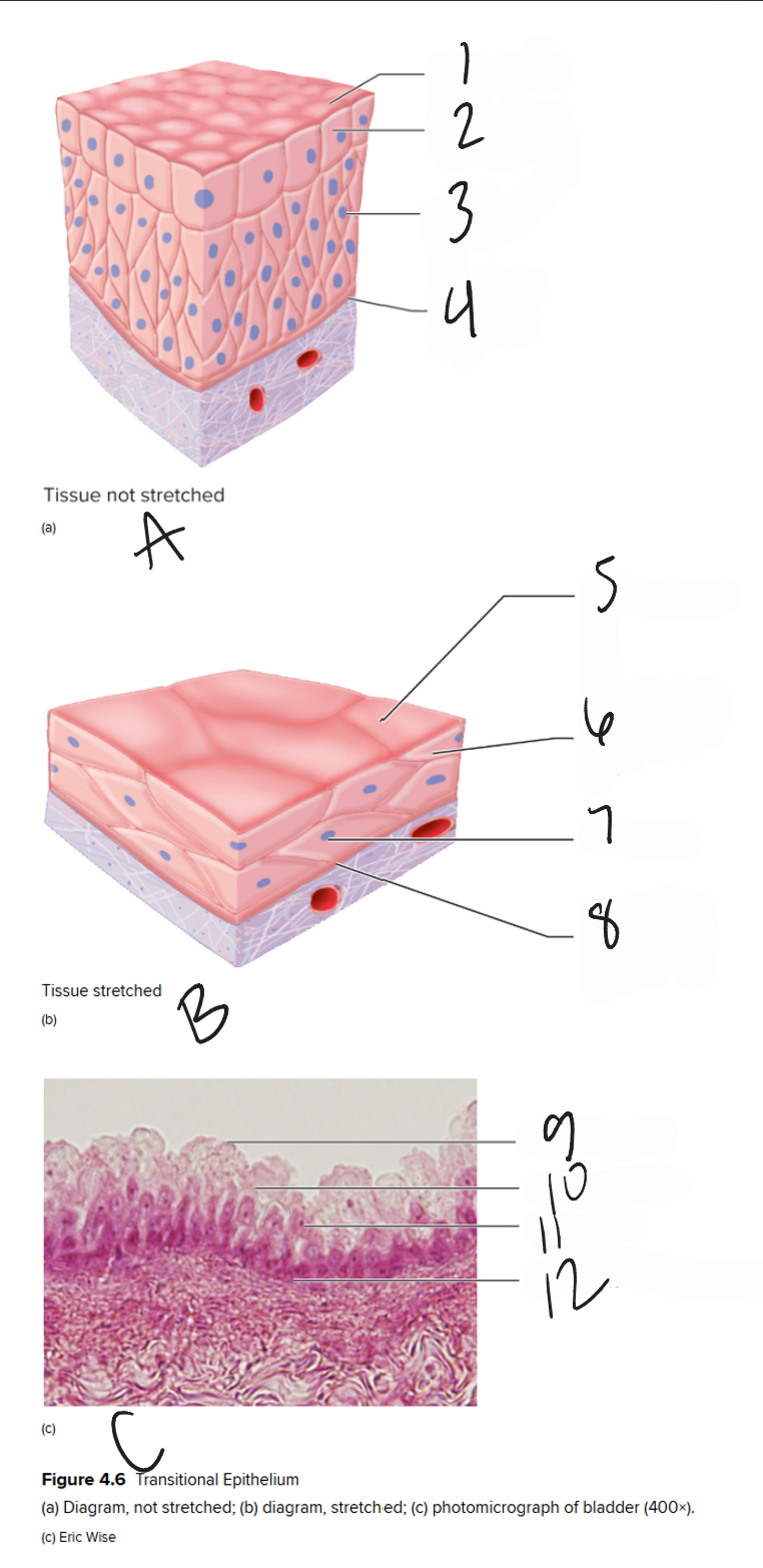
A
diagram of unstretched transitional epithelium tissue
1
free surface
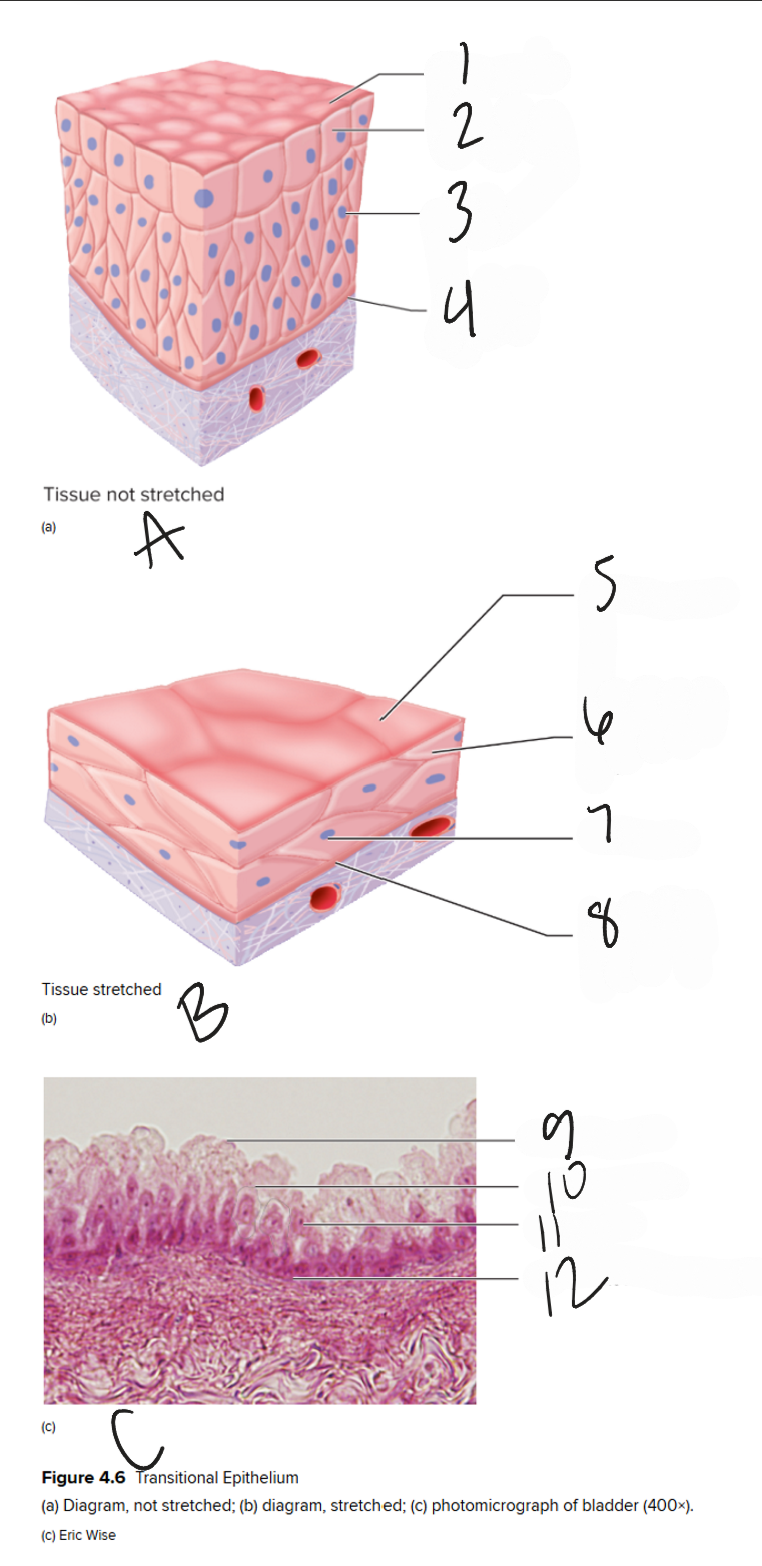
2
transitional epithelial cell
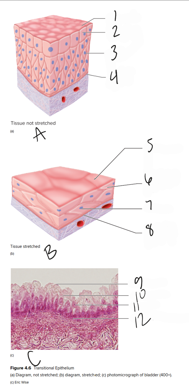
3
nucleus
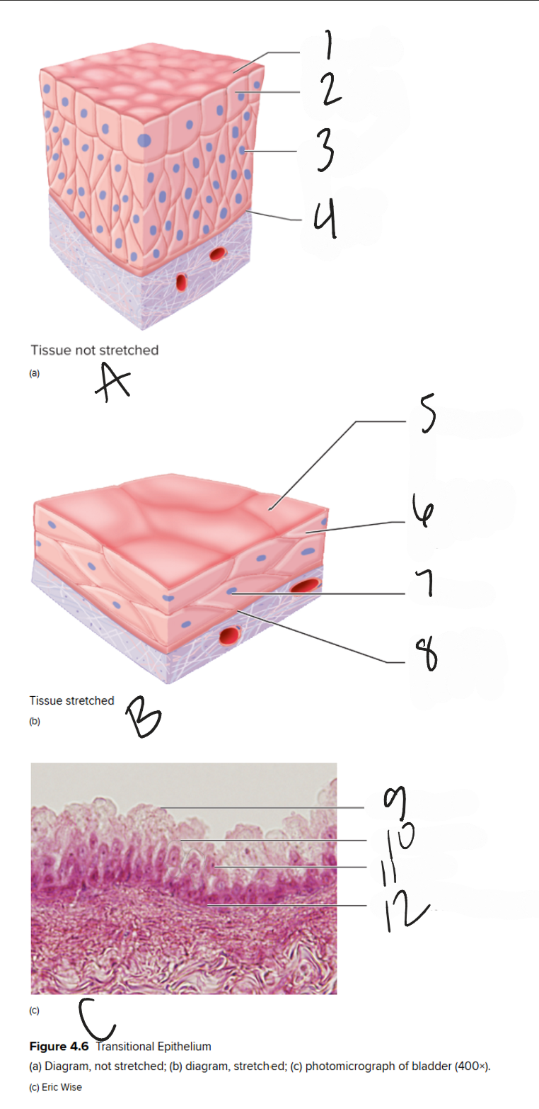
4
basement membrane

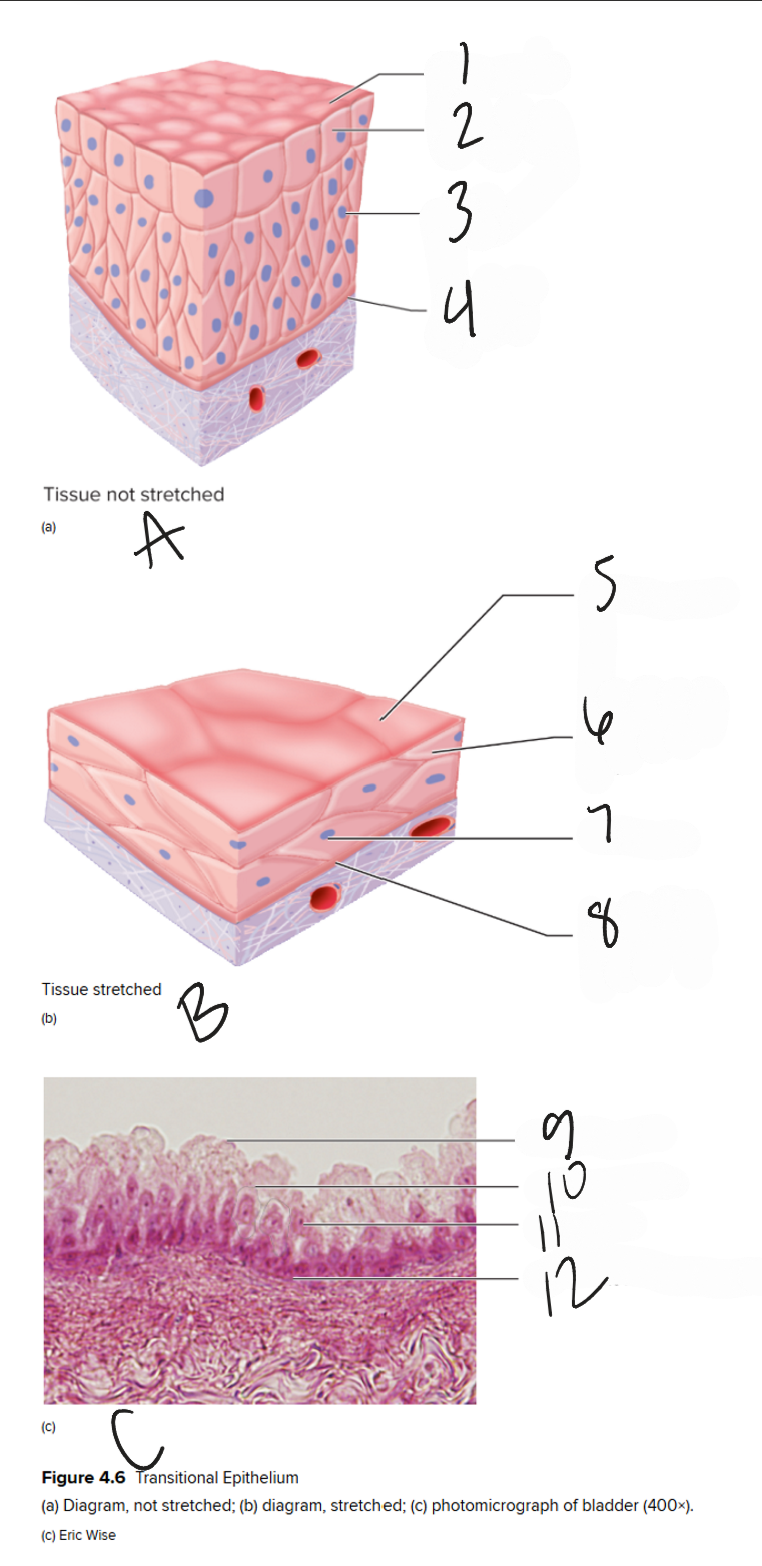
B
diagram of stretched transitional epithelial tissue
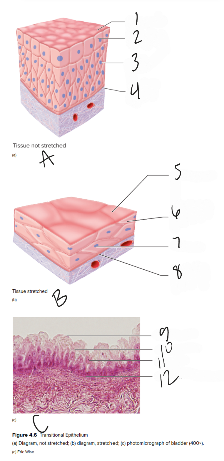
5
free surface

6
transitional epithelial cell

7
nucleus
8
basement membrane
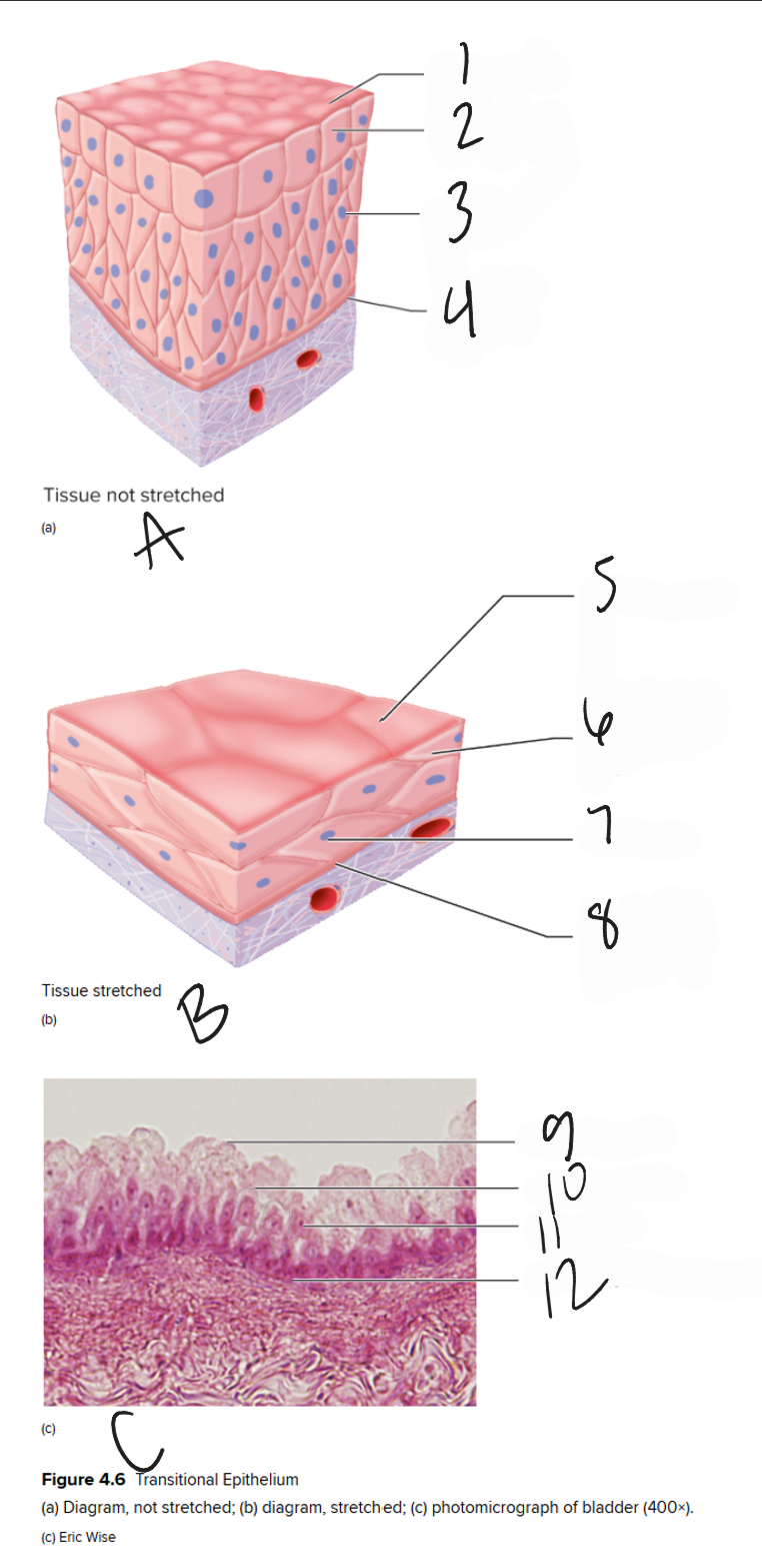
C
pictograph of bladder (400x)

9
apical surface
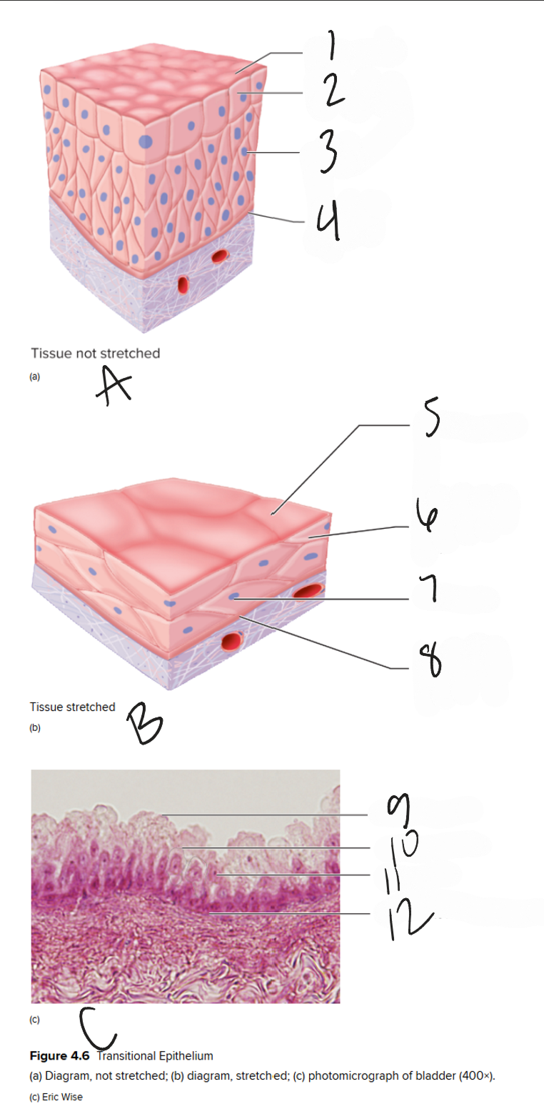
10
cell membrane
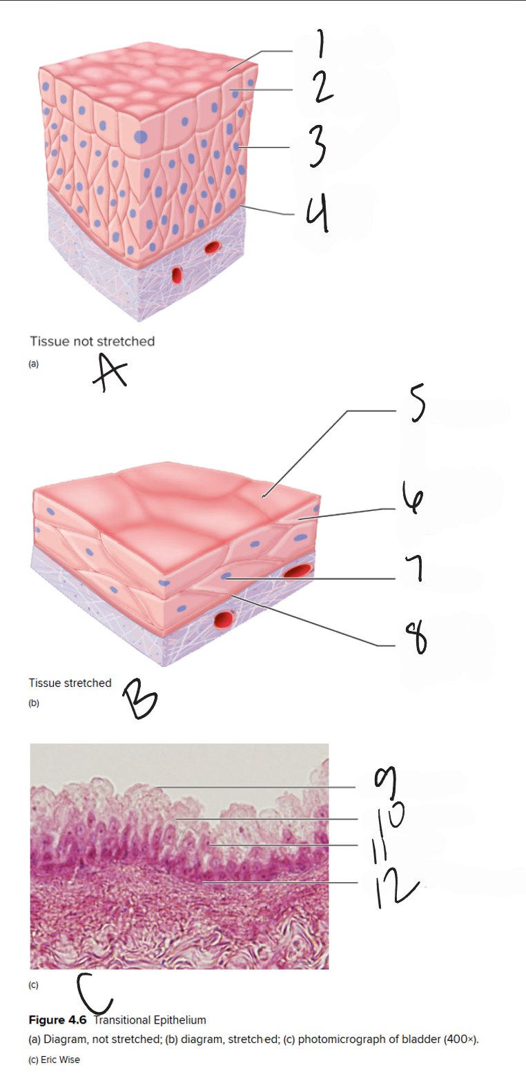
11
nucleus
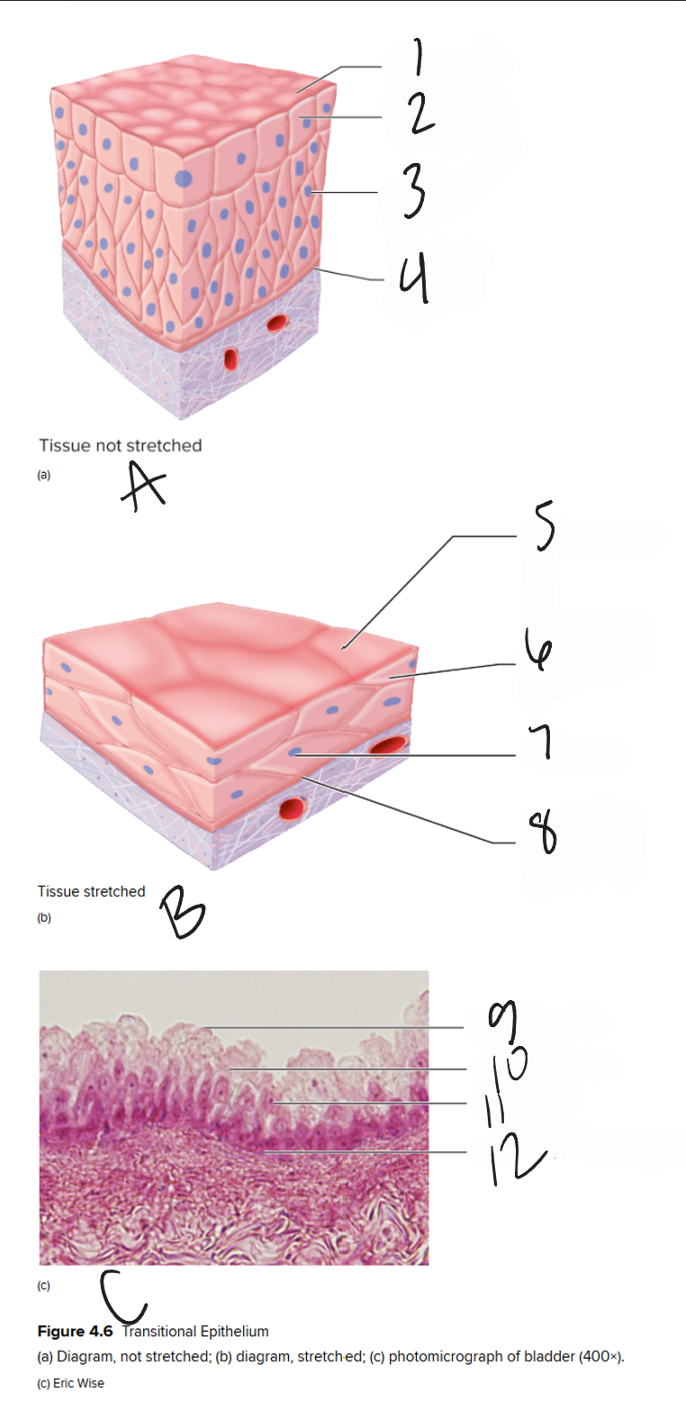
12
basement membrane
Epithelial Classification
Layer: SIMPLE
Cell Shapes (4):
Simple squamous epithelium
Simple cuboidal epithelium
Simple columnar epithelium
Pseudostratified columnar epithelium
Epithelial Classification
Layer: STRATIFIED
Cell Shapes (2):
Stratified squamous epithelium
Transitional epithelium
epithelial tissues
The tissues that cover body surfaces, line cavities, and form glands, characterized by closely packed cells and minimal extracellular matrix. They are classified based on the number of layers and cell shapes.
Basement membrane
A thin, noncellular adhesive layer that separates epithelial tissues from underlying connective tissues, providing support and anchoring the epithelium.
Cell Shape: Squamous
flattened
Cell shape: cuboidal
cube-like
Cell shape: columnar
tall and column-like
Cell shape: pseudostratified
a single layer of cells with varying heights, appearing stratified
Simple
cells in a single layer
Stratified
cells stacked in more than 1 layer
epithelial tissue is most commonly found on these surfaces
Free surfaces
simple squamous epithelium characteristics
It consists of a single layer of flat cells
smooth/slippery surface surface: diffusion and filtration
lines air sacs of lungs
lines blood vessels (endothelium)
surface layer of many membranes in various body cavities (mesothelium)
simple cuboidal epithelium characteristics
It consists of a single layer of cube-shaped cells
functions in secretion and reabsorption
lines kidney tubules
form many major glands and glandular organs (major tissue of the kidneys)
simple columnar epithelium characteristics
It consists of a single layer of column-shaped cells
nuclei are frequently in a row
can be ciliated on apical surface or have microvilli
cells with microvilli line inner portion of d. tract (absorption)
mucus-secreting goblet cells are often present
ciliated cells: line the uterine tubes