Option A- neurology and behaviour
1/79
Earn XP
Description and Tags
Name | Mastery | Learn | Test | Matching | Spaced |
|---|
No study sessions yet.
80 Terms
neural tube
hollow structure from which the brain and spinal cord form- runs the length of the dorsal side of the body
which processes are necessary for the development of the neural tube
cell division and differentiation
why is the genus Xenopus used in neuroscience studies? include why using animal models is useful in research
less ethical concerns
nervous system less complex
The eggs are large and easily manipulated
external development of the embryo is easier to observe
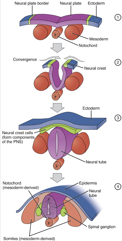
stages of neurulation
ectoderm tissue differentiates to form the neural plate and neural plate border. notocord is derived from the mesoderm tissue
neural plate folds inwards and downwards. notocords is ‘pushed’ downwards by the folding. eventually neural plate borders meet to form the neural crest
closure of the neural tube separates the neural crest from the ectoderm. neural crest cells will develop to form the majority of the PNS e.g. ganglions
mesoderm differentiates to become somites, which will eventually give rise to parts of the skeleton and muscle systems, including the vertebrae. the neural tube will eventually form the brain and spinal cord. notocord degenerates to eventually become the invertebral discs
from where is the spinal cord formed
from a region of the embryonic neural tube
what causes spina bifida
A gap in the neural tube: gap arises if infolding of part of the neural plate, to form the neural tube during neurulation, is incomplete
Incomplete closure of the neural tube leaving the spinal cord nerves exposed and prone to damage
how can spina bifida be treated
often corrected by surgery
most cases can be prevented if the mother gets enough folic acid during pregnancy
how does neural migration occur
by contractile actin filaments moving the cell along glial cells and its organelles in a given direction
why is the migration of neurons particularly important in brain development
Immature neurons must migrate in order to adopt precise final positions that allow for the formation of neural circuitries
This migration process is critical for the development of brain and spinal architecture
embryogenesis
development of a fully-formed organism from a fertilised egg
what embryonic tissues do all tissues derive from
All tissues are derived from three initial germ layers (ectoderm, mesoderm, endoderm) formed via gastrulation
how are neurons produced
Neurons are produced by progenitor neuroblasts via a process known as neurogenesis
how are nerve cells formed
The neural tube contains multipotent neuronal stem cells which can differentiate to form the different types of nerve cells
process of neural migration
Immature neurons must migrate in order to adopt precise final positions that allow for the formation of neural circuitries
This migration process is critical for the development of brain and spinal architecture
Neural migration may occur via one of two distinct processes – glial guidance or somal translocation
Glial cells may provide a scaffolding network along which an immature neuron can be directed to its final location
Alternatively, the neuron may form an extension at the cell’s perimeter and then translocate its soma along this length
what are axons
long narrow outgrowths from the cell body which carry the impulse from neuron to neuron
how do axons develop
An immature neuron consists of a cell body (soma) containing a nucleus and cytoplasm
Axons and dendrites will grow from each immature neuron out of the neural tube in response to chemical signals from surrounding cells
Some axons may be quite short (within the CNS) but others may extend to other parts of the body (within the PNS)
what is a synapse
A synapse is a junction at which a neuron transmits a signal to another cell (relay neuron or effector)
what happens to synapses which are used more and less frequently
As an organism matures, some synapses are used more frequently and these connections are consequently strengthened
Other synapses are not used as often and these connections are weakened and do not persist
This strengthening and weakening of certain neural pathways is central to the concept of how organisms learn
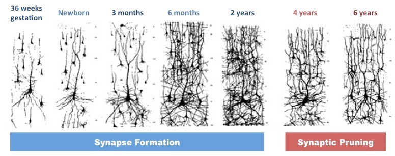
what is neural pruning
Neural pruning involves the loss of unused neurons (by removing excess axons and eliminating their synaptic connections) through apopstosis/ programmed cell death
The purpose of neural pruning seems to be to reinforce complex wiring patterns associated with learned behaviour
Pruning is influenced by environmental factors and is mediated by the release of chemical signals from glial cells
comparion of neurons and synaptic connection between brains of infants and adults
Infant and adult brains typically have the same total number of neurons (roughly 100 billion neurons in total)
However infant brains form vastly more synaptic connections (approximately twice the number found in adult brains)
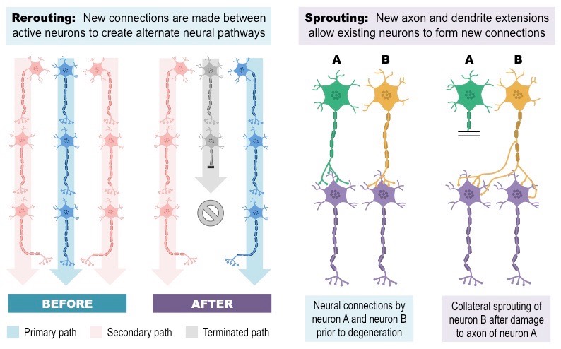
define neuroplasticity
Neuroplasticity describes the capacity for the nervous system to change and rewire its synaptic connections
2 primary mechanisms for neuroplasticity
Rerouting involves creating re-establishing an existing nervous connection via an alternative neural pathway
Sprouting involves the growth of new axon or dendrite fibres to enable new neural connections to be formed
define stroke
A stroke is the sudden death of brain cells in a localised area due to inadequate blood flow- This results in the improper functioning of the brain, due to the loss of neural connections in the affected area
2 types of strokes
Ischemic and Hemorrhagic
Ischemic strokes result from a clot within the blood restricting oxygenation to an associated region of the brain
Hemorrhagic strokes result from a ruptured blood vessel causing bleeding within a section of the brain
what can happen to the brain following a stroke? can it repair itself?
Strokes symptoms may be temporary if the brain is able to reorganise its neural architecture to restore function
Following a stroke, healthy areas of the brain may adopt the functionality of damaged regions
This capacity for the restoration of normal function is made possible due to the neuroplasticity of the brain

what is the process of embryo development
During embryonic development, the neural tube will enlarge and develop into different components of the nervous system:
The anterior (front) part of the neural tube will expand to form the brain during cephalisation (development of the head)
The remainder of the neural tube will develop into the spinal cord
Cells that comprised the neural crest will differentiate to form most of the peripheral nervous system
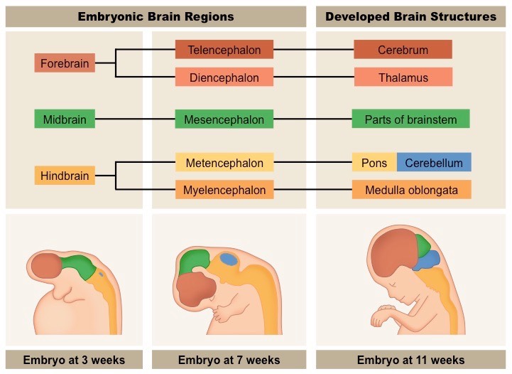
what are the primary vesicles and what do they eventually develop into
distinct bulges developed from the anterior end of the neural tube- eventually develop and form the fore, mid, and hind sections of the brain
what are the 3 primary structures of the brain and what will they eventually develop into?
The embryonic brain will initially be composed of three primary structures – the forebrain, midbrain and hindbrain- will eventually give rise to the identifiable components of the developed brain
forebrain → cerebrum + thalamus
midbrain → parts of brainstem
hindbrain → pons + cerebellum + medulla oblongata
what is the process of the anterior end of the neural tube developing into the brain called
neurulation
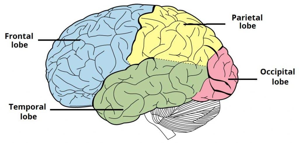
parts of the cerebral cortex and their functions
The cerebral cortex is the outer layer of neural tissue found in the cerebrum
parts: frontal lobe, parietal lobe, temporal lobe, occipital lobe
frontal: controls motor activity and tasks associated with the dopamine system (memory, attention, etc.)
parietal: responsible for touch sensation (tactility) as well as spatial navigation (proprioception)
temporal: involved in auditory processing and language comprehension
occipital: is the visual processing centre of the brain and is responsible for sight perception

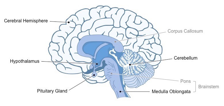
what is the cerebellum
The cerebellum appears as a separate structure at the base of the brain, underneath the cerebral hemispheres
both hemispheres transmit info through the corpus collosum
It is responsible for coordinating unconscious/rote motor functions – such as balance and movement coordination
what is the brainstem
The brainstem is the posterior (back) part of the brain that connects to the spinal cord (which relays signals to and from the body)
The brainstem includes the pons, medulla oblongata (often referred to as the medulla) and the midbrain
The brainstem (via the medulla) controls automatic and involuntary activities (breathing, swallowing, heart rate, etc.)
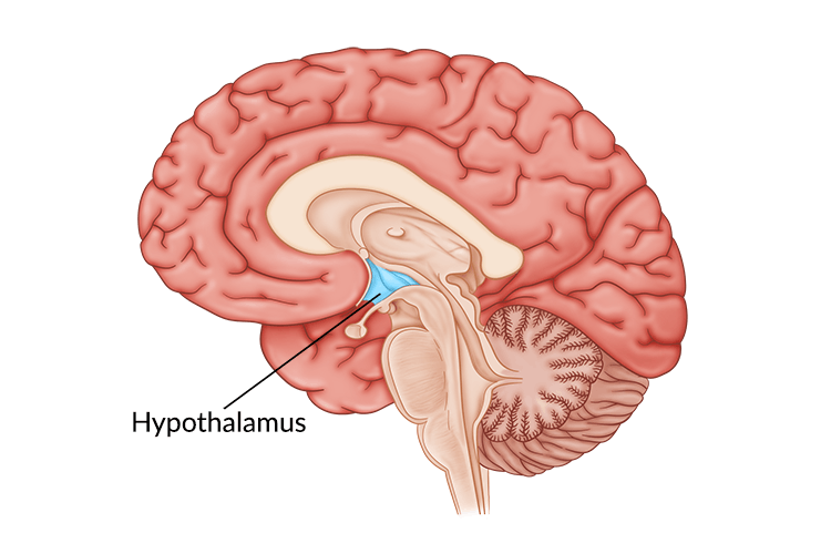
what is the hypothalamus
is the region of the brain that functions as the interface with the pituitary gland
As such, the hypothalamus functions to maintain homeostasis via the coordination of the nervous and endocrine systems
The hypothalamus also produces some hormones directly, which are secreted via the posterior pituitary (neurohypophysis)
what is the pituitary gland
considered the ‘master’ gland – it produces hormones that regulate other glands and target organs
anterior lobe secretes hormones such as FSH, LH, growth hormone and prolactin
posterior lobe secretes hormones such as ADH and oxytocin
diagram of the brain with each part labelled

what is the visual cortex
Located within the occipital lobe of the cerebrum and receives neural impulses from light-sensitive cells in the eyes
is the region of the brain responsible for visual perception (sight)
what is broca’s area
Located within the frontal lobe of the left cerebral hemisphere (not present in the right hemisphere)
Is responsible for speech production (if damaged, the individual cannot produce meaningful speech despite intending to)
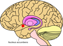
what is the nucleus accumbens
is involved in the pleasure reward pathway and is found within each cerebral hemisphere
It secretes neurotransmitters responsible for feelings of pleasure (dopamine) and satiety / satisfaction (serotonin)
It communicates with other centres involved in the mechanisms of pleasure

how are animal experiments used to help identify brain functions
can be used to identify function by stimulating regions with electrodes or removing via lobotomy
Experimentation on animals involves less ethical restrictions than human studies
BUT they are limited by the differences between animal and human brains, making valid comparisons difficult
how do lesions help to identify brain functions
lesions: abnormal areas of brain tissue which can indicate the effect of the loss of a brain area
can be identified via post-mortem analysis (autopsy) or via scans of the brain (CT scans or MRI)
effects of lesions can be difficult to identify, as many functions may involve multiple brain areas
AND the brain has the capacity to re-learn certain skills by re-routing instructions to other areas (plasticity)
how are autopsies used to help identify brain functions
autopsy: a post-mortem examination of a corpse via dissection in order to evaluate causes of death
Comparisons can be made between the brains of healthy and diseased corpses to identify affected brain areas
Include example- e.g. damage to Broca’s area affects speech
how are fmri scans used to help identify brain functions
fmri records changes in blood flow within the brain to identify activated areas
Oxygenated haemoglobin responds differently to a magnetic field than deoxygenated haemoglobin
These differences in oxygenation can be represented visually and reflect differences in the level of brain activity
non-invasive and can be used to identify multiple brain regions involved in complex, integrated brain activities
how has the cerebral cortex of humans changed through the process of evolution
the cerebral hemispheres are responsible for higher order functions
Through evolution, the human cranium has increased in size and the folding has become more extensive
foldings permits high surface area to volume ratio
increased surface/volume permits more synapses
both changes have allowed an increase in the number of neurons present in the cerebral cortex
folding decreases distances therefore faster communication
what functions are each of the brain’s hemispheres responsible for
Function
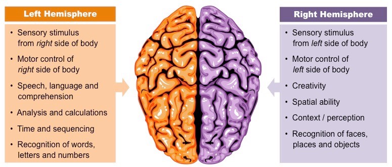
each hemisphere of the brain processes what kind of information
contralateral processing
brain is cross wired- the left cerebral hemisphere is responsible for processing sensory information from the right side of the body and vice versa
nerves from each eye will meet at the chiasma
information from inner fields cross at the chiasma
tactile (movement) information is transferred in the spinal cord or brainstem
visual information is transferred at the optic chiasma from the optic nerve to form an image in the visual cortex
muscular contractions are coordinated by the motor cortex
what is the autonomic nervous system
The autonomic nervous system controls involuntary processes in the body using centres located mostly within the brainstem
has 2 antagonistic branches
Sympathetic nerves release noradrenaline (adrenergic) to mobilise body systems
Parasympathetic nerves release acetylcholine (cholinergic) to relax body systems and conserve energy (‘rest and digest’)
what is the medulla oblongata
The medulla oblongata is a part of the brainstem responsible for coordinating many autonomic (involuntary) activities
This includes the regulation of body activities such as swallowing, breathing and heart rate
describe the roles of sympathetic and parasympathetic nervous system in the regulation of the rate of heartbeat
sympathetic
stimulus- blood ph decreases as co2 concentration increases
receptor: breathing and heart rate centers in the medulla oblongata contain chemoreceptors in their blood vessels
responses: intercostal muscles and diaphragm relax and move up to increase the rate and size of contractions- increasing the rate and depth of ventilation (sinoatrial node) to increase the heart rate
parasympathetic
stimulus- blood ph increases as co2 concentration decreases
receptor: breathing and heart rate centers in the medulla oblongata contain chemoreceptors in their blood vessels
responses: intercostal muscles and diaphragm contract and move down to decrease the rate and size of contractions- decreasing the rate and depth of ventilation (heart’s pacemaker) to decrease the heart rate
what happens during a pupil reflex
The pupil reflex is an involuntary response originating at the brainstem and under the control of the autonomic nervous system
Pupils constrict in bright light (to prevent overstimulation of photoreceptors) and dilate in dim light (to maximise light exposure)
In bright light, parasympathetic nerves trigger circular muscles to contract and cause the pupils to constrict
In dim light, sympathetic nerves trigger radial muscles to contract and cause the pupils to dilate
explain how the pupil reflex test will show whether an unconscious victim is ‘brain dead’ or severely brain damaged
Brain death is defined as the permanent absence of measurable activity in both the cerebrum and brainstem
Hence, individuals with a non-functioning cerebrum but a functioning brainstem may be kept alive in a vegetative state
failure of pupil reflex indicates damage to the medulla oblongata (brainstem)
if brain stem fails then the organism can no longer function and it is unlikely that higher order brain functions can persist
different responses from each eye could also indicate brain damage
more testing is needed to determine the area/ extent of brain damage
how much energy is used up by the brain and what processed is it used to sustain/carry out
The human brain consumes ~20% of the body’s energy levels- rate of energy consumption varies little, regardless of the level of physical exertion by the body
processes:
Energy is needed to maintain a resting potential when neurons are not firing (Na+/K+ pump uses ATP)
Energy is used to synthesise large numbers of neurotransmitters to facilitate neuronal communication
explain how the visual stimuli from the right/left eye reaches the visual cortex of the brain
a. right eye receives information/stimuli/light from both left and right visual field
b. light from the left visual field goes to the right side of the retina
OR vice versa
c. impulses from retina carried along the optic nerve
d. optic nerves cross at optic chiasma
e. impulses from the left side of the retina goes to the left side of the brain
OR vice versa
what is sensitivity
Sensitivity describes the ability of an organism to detect external and internal changes and respond accordingly
Receptors detect these changes as stimuli, and generate nerve impulses which are relayed to the brain and effector organs
main types of receptors in humans and their functions, stimuli, and location
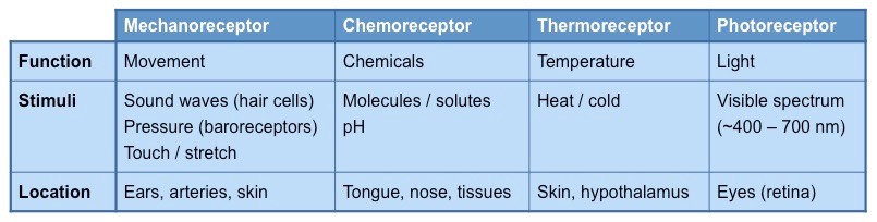
diagram showing labels of different parts of the eye
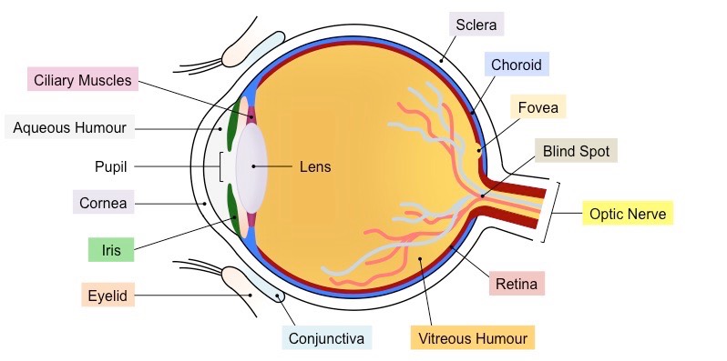
what is the retina
light-sensitive layer of tissue that forms the innermost coat of the internal surface of the eye
contains receptor cells (rods and cones) that are sensitive to light
what is the vitreous humor
is full of transparent fluid which helps to hold the eye in shape
what is the cornea
thin transparent layer covering the front of the eye which is lubricated by conjunctiva
refracts the light rays as they enter the eye
cannot change shape
what is the lens
responsible for fine adjustments to focusing, can change shape
what are the suspensory ligaments
strong fibres that hold lens in place
what are the ciliary muscles
the lens is attached to ciliary muscles
a ring of muscles that contract or relax to change the shape of the lens
what is the iris
constricts and dilates to control how much light enters the eye via the pupil
what is the sclera
a tough outer covering around the eye
what is the choroid
a dark layer behind the retina, which absorbs light so that it does not bounce around inside the eye
what is the optic nerve
carries impulses to the brain
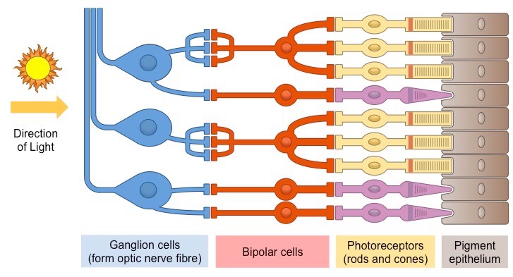
diagram showing how light moves through the retina to the brain
difference between rod cells and cone cells
rods
dominant in dim light conditions
1 type
distributed throughout the retina
responsive to all visible wavelengths
high receptor-ganglion ratio: many rods connect to one ganglion
poor visual acuity
cone
dominant in bright light conditions
3 types
concentrated around fovea
sensitive to short wavelengths- violets, medium- greens, long- reds
very low receptor-ganglion ratio
high visual acuity
explain how light is transmitted to the brain
Photoreceptors (rods and cones) convert light stimuli into electrical nerve impulses (action potential).
Neural information is relayed to the brain through bipolar cells and ganglion cells.
Bipolar cells transmit nerve impulses from photoreceptors to ganglion cells.
Rod cells may synapse with multiple bipolar cells, resulting in low resolution (poor acuity).
Cone cells typically synapse with a single bipolar cell, resulting in high resolution (high acuity).
Ganglion cells transmit nerve impulses via long axonal fibers composing the optic nerve.
Signals from ganglion cells may go to the visual cortex for sight or other brain regions for eye movements or circadian rhythms.
The region where ganglion axon fibers feed into the optic nerve lacks photoreceptors, termed the "blind spot".
The brain fills in details from surrounding regions, preventing individuals from perceiving a visual blind spot.
explain how contralateral processing occurs
Information from the right half of the visual field is detected by the left half of the retina in both eyes and is processed by the left hemisphere (and vice versa for the left half of the visual field)
Information from each eye may swap at the optic chiasma, so that the right or left visual field is processed together
The optic nerves that swap sides are moving contralaterally, while those that stay on the same side remain ipsilateral
Impulses are conducted by the optic nerve to the thalamus, before being transmitted to the visual cortex (occipital lobe)
Thalamic structures (e.g. lateral geniculate nuclei) are involved in coordinating eye movements and circadian rhythms
explain how sound is perceived by the human ear to the brain
eardrum/tympanic membrane is moved by sound waves
eardrum causes movement of the bones of the middle ear
bones of the middle ear amplify sound (by 20x)
bones of the middle ear on the round window move
causing movement of fluid within the cochlea
different hair cells (mechanoreceptors) respond to different wavelengths/ pitch of sound
hair cells release a chemical neurotransmitter when stimulated
sounds/vibrations are transformed into nerve impulse/action potentials (in the cochlea)
carried by auditory nerve to brain
round window releases pressure/ dissipates sound
this allows fluid in cochlea to vibrate
how is the middle ear separated from the outer ear
The middle ear is separated from the outer ear by the eardrum and the inner ear by the round window
It is an air-filled chamber that houses three small bones (collectively called the ossicles)
The bones of the middle ear are individually called the malleus (hammer), incus (anvil) and stapes (stirrup)
The malleus is in contact with the eardrum and the stapes contacts the oval window (while the incus connects the two)
explain how sound is transmitted and amplified by structures in the middle ear
The function of the ossicles is to amplify the sound vibrations by acting like levers to reduce the force distribution
Sound travelling through air is mostly reflected when in contact with a liquid medium (due to the incompressibility of fluids)
The amplification of sound by the ossicles (bones of the middle ear) allows the vibrational pressure to pass to the cochlear fluid with very little loss
The oval window is smaller than the ear drum, which also assists in amplifying the sound energy
what is the cochlea
The cochlea is a fluid-filled spiral tube within the inner ear that converts sound vibrations into nerve impulses
explain how hair cells in the semicircular canals detect movement of the head
Within the semicircular canals are gelatinous caps called cupula, which are embedded with numerous hair cells
When the head moves, the fluid in the semicircular canals follows the direction of movement
This fluid movement exerts pressure on the hair cells embedded in the cupula, triggering nerve impulses
There are three semicircular canals at 90º angles to one another, allowing head movement to be detected in all three planes
The brain integrates information from the semicircular canals in each ear in order to identify head position and movement
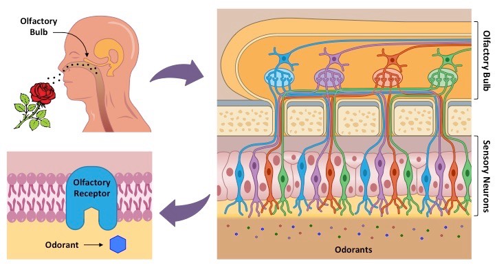
what is olfaction and where does it occur
Olfaction is the ability to detect airborne chemicals (odorants) as scents or smells; occurs inside the upper part of the nose
explain how olfactory receptors detect chemicals in the air
At the back of the nasal cavity is a patch of tissue called the olfactory epithelium, which is embedded with chemoreceptors
The olfactory epithelium is lined with mucus, in which odorant molecules will dissolve before binding to the chemoreceptors
Binding of an odorant molecule will trigger a nerve impulse, which is transferred via the olfactory bulb to the brain
The combination of olfactory receptors activated determines the specific scent perceived by the brain
BASICALLY
odor/odorant molecules dissolved by mucus in olfactory epithelium → odorant molecules bind to chemoreceptors in the epithelium → triggers a nerve impulse → nerve impulse transferred by the olfactory bulb to the brain
what are the names of the genes responsible for the production of photoreceptive pigments in cone cells
OPN1MW and OPN1LW
explain what happens in the eye when an individual has red-green color blindness
There are three different types of cone cells, each of which absorbs different wavelengths (trichromatic: red, green, blue)
The genes responsible for producing red or green photoreceptors are located on the X chromosome (sex-linked)
If either of the genes responsible for production of pigments are mutated, the pigments are not produced properly and the eye cannot distinguish between green wavelengths and red wavelengths in the visible spectrum
As these genes are recessive and located on the X chromosome, red-green colour-blindness is more common in males
explain the use of cochlear implants in deaf patients
Cochlear implants may be used to stimulate the auditory centres of the brain in patients with non-functioning hair cells
Standard hearing aids are ineffective in deaf patients as they amplify sounds but do not bypass defective hearing structures
Cochlear implants consist of two parts – an external part (microphone / transmitter) and an internal part (receiver / stimulator)
The external components detect sounds, filter out extraneous frequencies and then transmit the signals to the internal parts
The internal components receive the transmissions and produce electrical signals via electrodes embedded in the cochlea
The electrical signals are then transferred via the auditory nerve to be processed by the brain
explain how sounds of different wavelengths are distinguished by the ear
only write 3 out of 4 of the points below
movement of eardrum and ossicles causes vibration of cochlear fluid
different hair /cilia vibrate at different wavelengths
different hair cells send different nerve signals in the auditory nerve