Abnormal ocular conditions- L13 and L14 part 3 Benign Peripheral Retinal Disorders
1/21
There's no tags or description
Looks like no tags are added yet.
Name | Mastery | Learn | Test | Matching | Spaced |
|---|
No study sessions yet.
22 Terms
Peripheral retinal conditions that are unlikely to affect vision
White without pressure
• CHRPE
• Chorioretinal scar
• RPE window defect
• Choroidal naevus
• Pavingstone degeneration
• Reticular degeneration
• Cystoid degeneration
• Equatorial drusen
• Pars Plana cyst
White without pressure : aetiology
Asymptomatic
•White-grey appearance of the equator and/or peripheral retina
•Well demarcated scalloped borders
•Seen without scleral indentation (CF: white with pressure)
•Occurs in about 30% of all patients
• More common in patients with dark skin
•Usually bilateral
•Benign
White without pressure: differential diagnosis
-Subclinical retinal detachment
• Retinoschisis
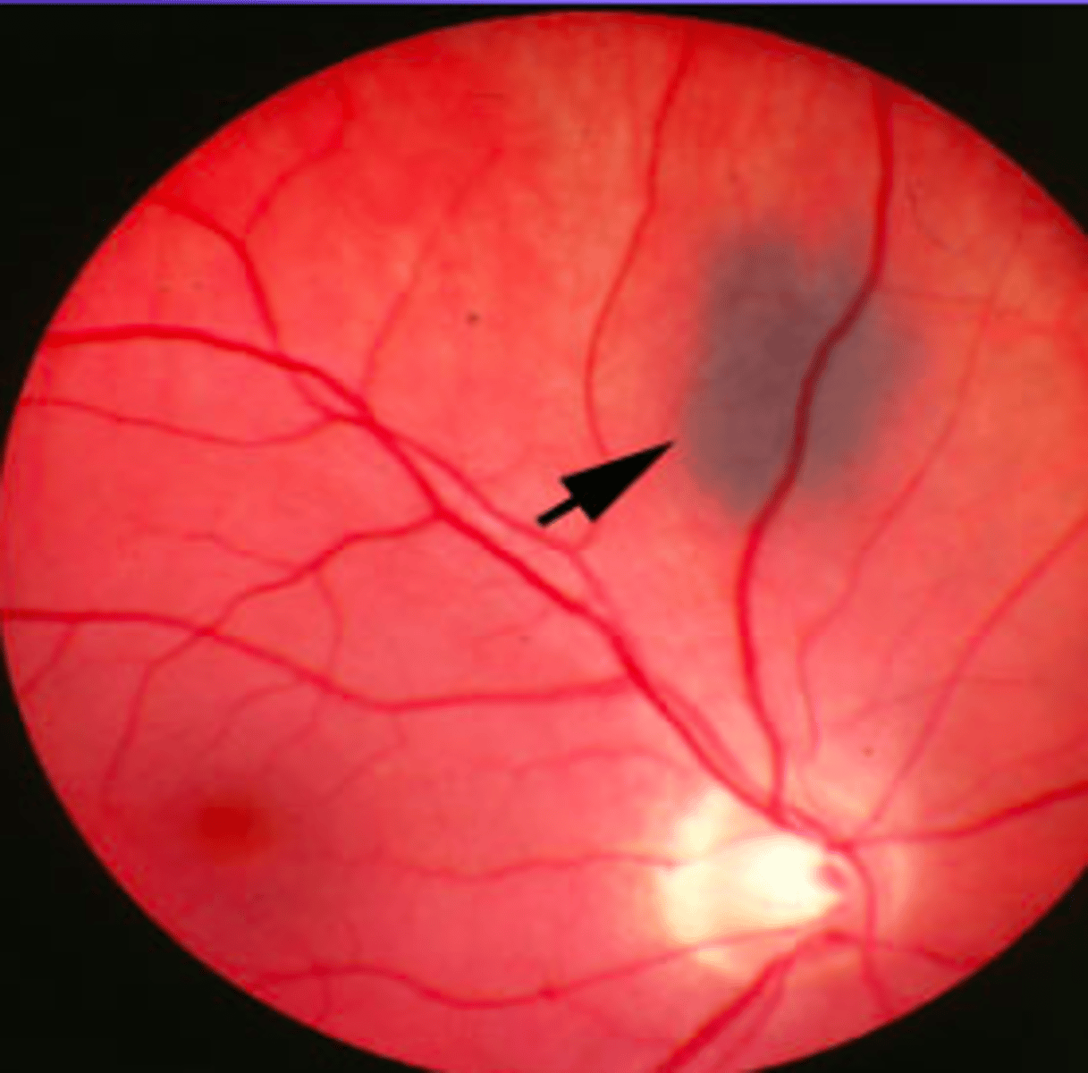
Congenital hypertrophy of the retinal pigment epithelium (CHRPE)
bear tracks (cat's paws)
• Congenital thickening of the RPE
- prevalence: 1.2 %
(CHRPE): three variants
Solitary (unifocal)
• Grouped (multifocal)
• Atypical (bilateral and multifocal)
(CHRPE): risk factors
Typically benign
• No associated preceding event
• Atypical associated colonic polyps and carcinoma
(CHRPE): symptoms
asymptomatic
(CHRPE): signs
Typically round, flat, hyperpigmented lesions
• Dark black to light grey
• Distinct margins
• May have areas of chorioretinal atrophy within lesion (lacunae)
• May have a depigmented halo around lesion
• Normal overlying retina and vasculature, no vitreous traction
• Generally located equatorially
• May cause a scotoma
(CHRPE) : Differential Diagnosis
Choroidal Melanoma
• Choroidal Naevus
• Melanocytoma
• Focal pigmentation (injury, inflammation or drug toxicity)
(CHRPE): prognosis
Generally benign
• Relationship to colonic polyps/carcinoma
• Multiple lesions (4 or more) or bilateral lesions
- or a family history of colon cancer
should undergo further testing
(CHRPE): management
No active intervention usually indicated A typical CHRPE
RPE Window defect aetiology
Relatively common
•Results from the absence of melanin in the RPE (RPE is present)
•Round, well circumscribed, white/yellow area
•No surrounding RPE hyperplasia (unlike in chorioretinal scar)
•Differentiate from retinal holes which appear red/brown with oedema
and/or hyperplasia
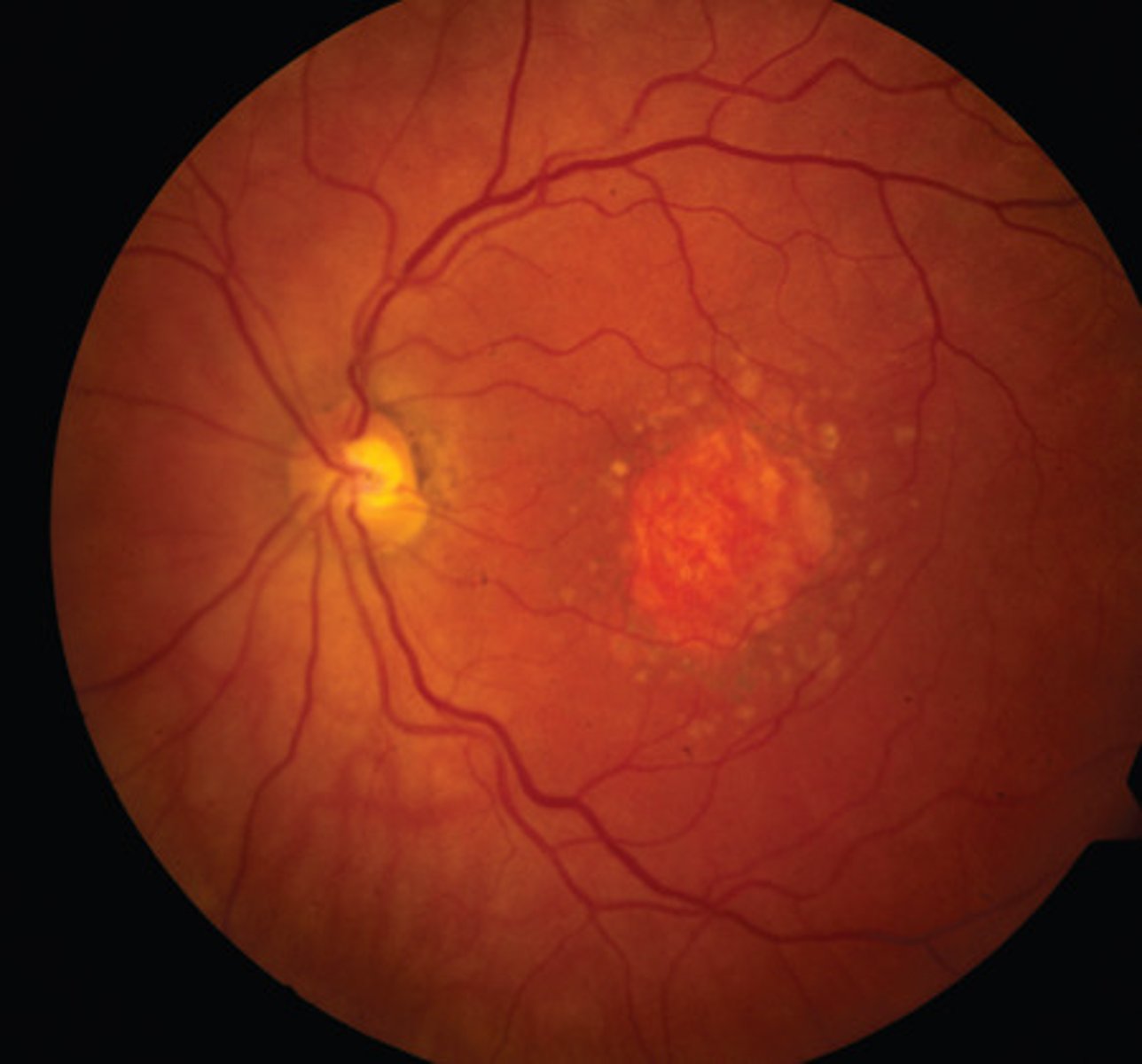
Choroidal naevus : Aetiology
Area of increased choroidal pigmentation
•Benign - low malignancy potential
•Common - prevalence of 2-7%
•Prevalence increases with age
Choroidal naevus signs
Typically small, flat, grey lesions
• May be mottled (drusen and RPE variations)
• Variable shape - round to oval to irregular indistinct margins
• Usually no chorioretinal atrophy, no haloes around the lesion
Choroidal naevus- differential diagnosis
Choroidal melanoma
Choroidal Naevi: Management
Baseline colour photography (or careful drawing)
• OCT and autofluorescence to look for sub-retinal fluid and lipofuscin
• Urgent (within 2 weeks) referral:
• Any one of – thickness > 2mm (or LBD > 7mm), collar stud configuration,
documented growth
• Any two of – thickness > 1.5mm (or LBD > 6mm), orange pigment, serous
detachment, symptoms
• Otherwise routine exam with patient education
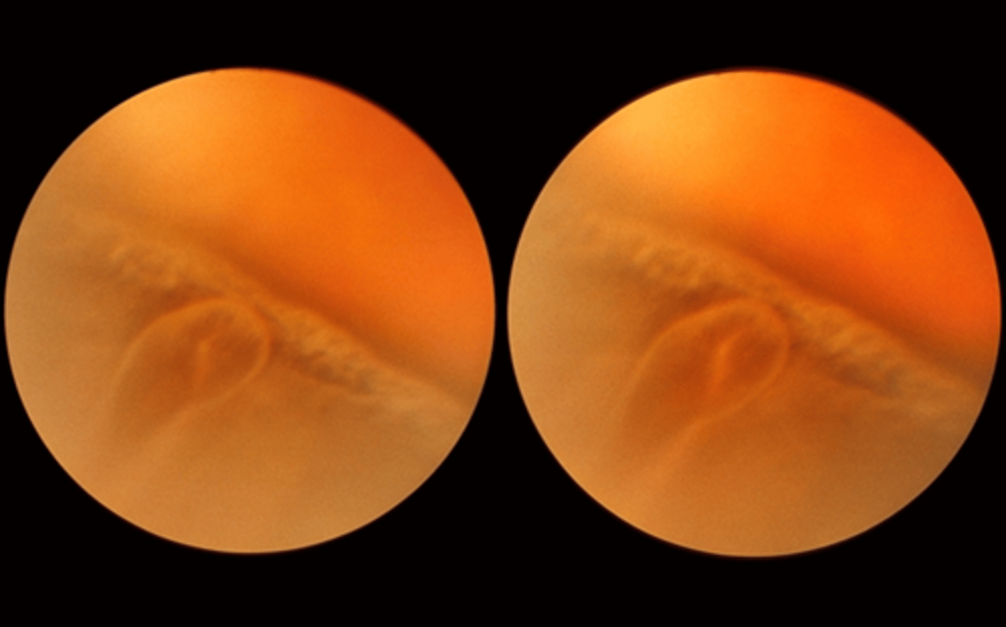
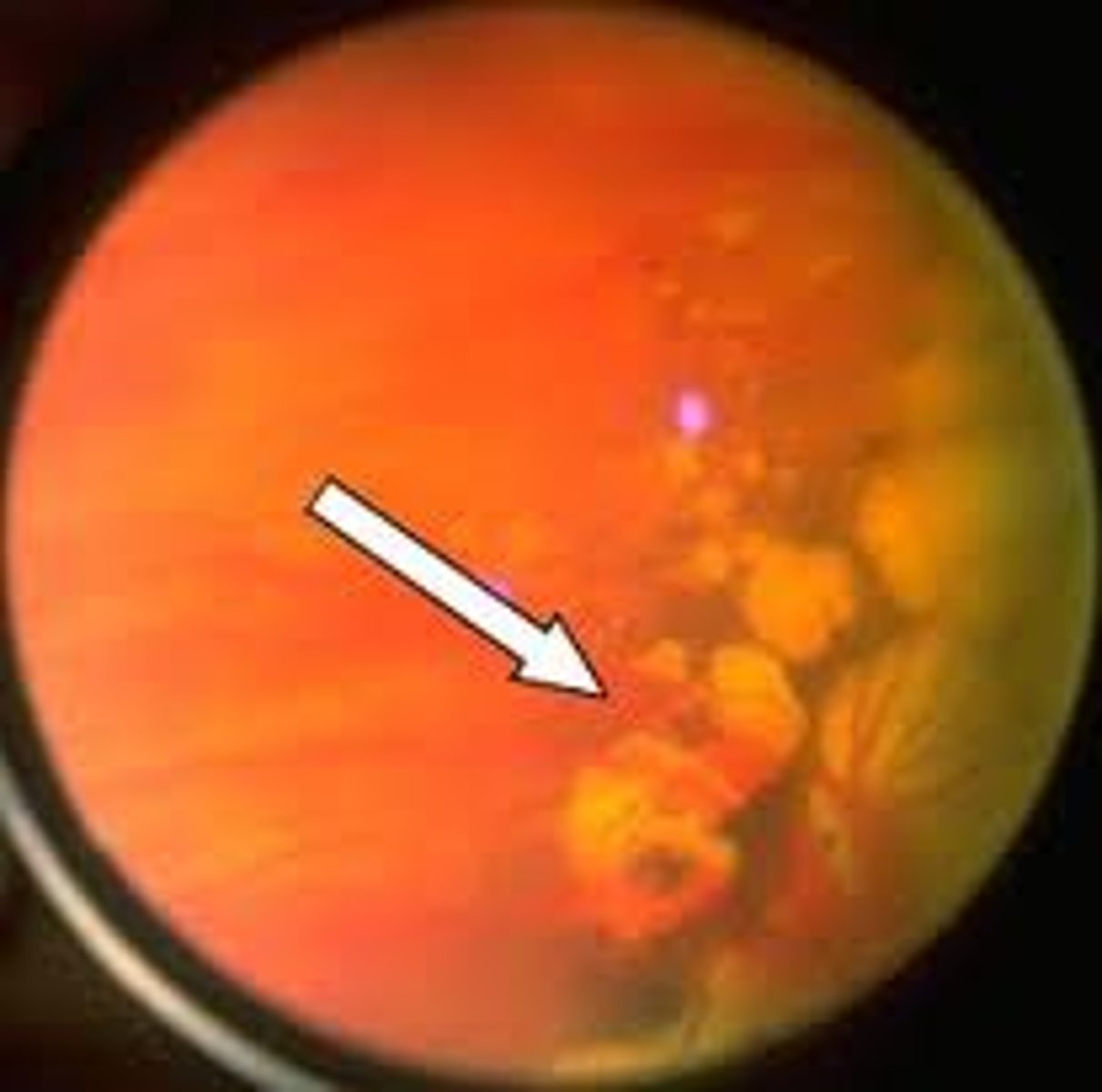
Pavingstone degeneration: aetiology
Occurs in about 30% of all patients
• Peripheral retinal degeneration
• Small, pale, non-elevated, depigmented areas
• Usually bilateral, mostly seen inferiorly
• Choroidal vessels seen due to RPE and outer retina loss
• May increase with age
• Benign
• Differentiate from chorioretinal scars, retinal holes, lattice degeneration
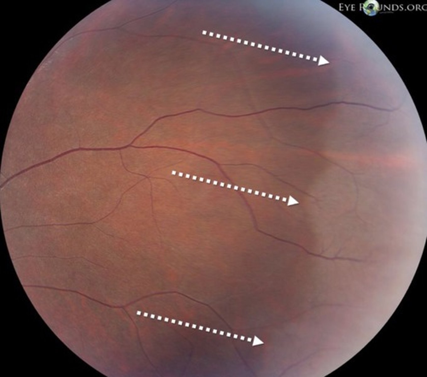
Peripheral pigmentary (reticular) degeneration: aetiology
• Polygonal, honeycomb pattern of granular pigmentation
• Retinal periphery
• Occurs in 20% over age 40 and increases with age
• Benign
Peripheral pigmentary (reticular) degeneration: differential diagnosis
Retinitis pigmentosa
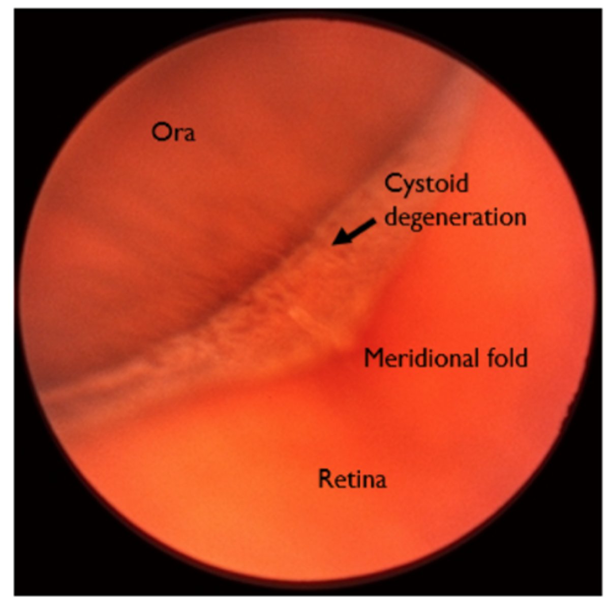
Cystoid degeneration aetiology
Present in ~100% of eyes over 80 years of age (increases with age)
• Tiny vesicles with indistinct boundaries on a greyish-white background
• Usually close to the ora serrata
• May resemble lattice degeneration -cystoid is elevated while lattice is
depressed
• Benign (although possible link with degenerative retinoschisis)
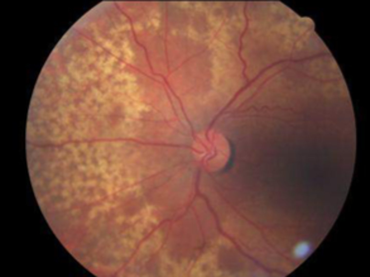
Equatorial drusen aetiology
Peripheral drusen
•Deposits of material on Bruch's membrane
•Found in over 70% of patients over 50 years of age
•Histologically similar to macula drusen
•Benign (as long as not approaching macula) and not associated with
any other conditions
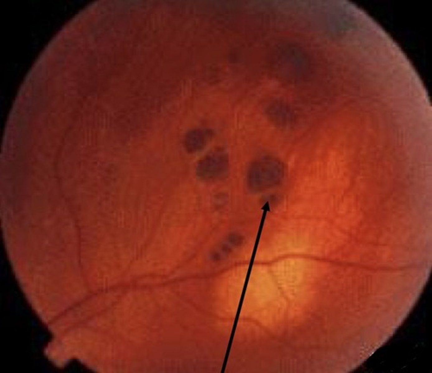
Pars Plana Cyst: aetiology
Clear bullous elevation of the non-pigmented ciliary epithelium of pars plana
•Seen in 5-10%
•At the vitreous base
•Harmless