IOS neuro
 Call Kai
 Learn
Learn Practice Test
 Spaced Repetition
 Match
Match Flashcards
 Knowt Play
1/111

There's no tags or description
Looks like no tags are added yet.
Name | Mastery | Learn | Test | Matching | Spaced | Call with Kai | Chat |
|---|
No analytics yet
Send a link to your students to track their progress
112 Terms
the cranium is divided into the:
viscerocranium, and neurocranium
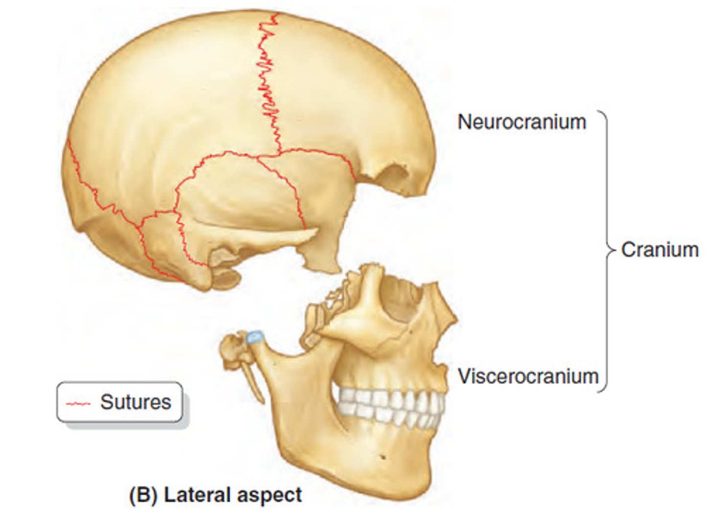
what is the viscerocranium
facial skeleton (bones of the face)
where is the viscerocranium located
beneath the line between the eye's orbit and external acoustic meatus; ANTEROINFERIOR REGION OF THE CRANIUM
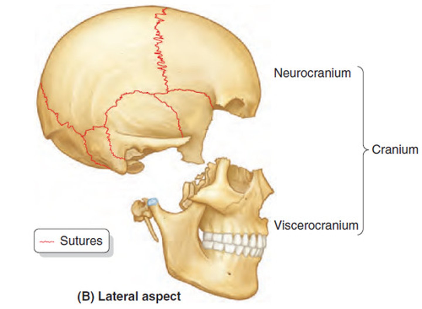
what is the neurocranium
the bones that encase the brain
where is the neurocranium located
above the line between the eye's orbit and external acoustic meatus; POSTEROSUPERIOR REGION OF THE CRANIUM
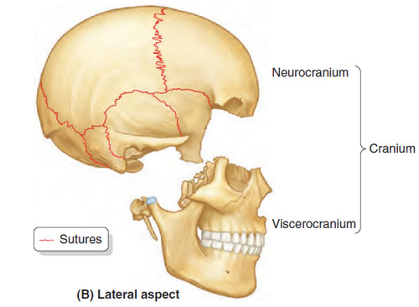
What bones do the viscerocranium not include
the mandible
How common are craniofacial disorders at birth?
They account for about 1/3 of all congenital defects.
When does craniofacial development mainly occur?
second month of embryonic life; weeks 4–8
What structures give rise to the face and parts of the neck?
The pharyngeal arches.
What are pharyngeal arches (PAs)?
Transient embryonic structures that form in the head and neck region.
When do pharyngeal arches appear?
During the 4th week of embryonic development.
What animal structure are pharyngeal arches evolutionarily related to?
The branchial (gill) arches in fish.
What is the function of pharyngeal arches in humans?
To organize the development of facial and neck structures.
How many pharyngeal arches form in human embryos?
Five: arches 1, 2, 3, 4, and 6.
What happens to the 5th pharyngeal arch?
It does not form or regresses rapidly.
What happens to pharyngeal arches 4 and 6?
They fuse and are often referred to together as arch 4-6.
What are the three layers of each pharyngeal arch?
Mesodermal core, ectodermal outer layer, and endodermal inner lining.
What is the origin of the outer layer of pharyngeal arches?
Ectoderm.
What is the origin of the inner lining of pharyngeal arches?
Endoderm from the pharynx.
Around what day do the 1st and 2nd arches appear?
Around day 26.
What major neural event occurs around day 26?
Closure of the cranial neuropore.
Which cells invade the pharyngeal arches?
Cranial neural crest cells (CNCCs).
What is the origin of cranial neural crest cells?
The dorsal margins of the neural tube.
CNCCs from the forebrain migrate to which region?
The frontonasal region.
CNCCs from the midbrain and hindbrain migrate to...?
The pharyngeal arches.
What do CNCCs differentiate into within the arches?
Cartilage, nerves, and vascular components.
What does CNCC proliferation and migration give rise to in the face?
Facial prominences.
What are facial prominences?
Buds of tissue that develop into facial structures.
What does the frontonasal prominence become?
The forehead, bridge of the nose, and midline upper face.
What structures come from CNCCs migrating into the 1st pharyngeal arch?
The maxillary and mandibular prominences.
What does the maxillary prominence form?
The upper jaw (maxilla).
What does the mandibular prominence form?
The lower jaw (mandible).
Which embryonic structures give rise to the bones and cartilages of the face and neck?
The pharyngeal arches.
What is the origin of the mesenchyme in the pharyngeal arches?
Cranial neural crest cells.
Which arches give rise to facial bones?
The 1st and 2nd pharyngeal arches.
Which arches give rise to laryngeal cartilages?
The 3rd, 4th, and 6th pharyngeal arches.
What are the two processes of the 1st pharyngeal arch?
Maxillary process and mandibular process.
What bones does the maxillary process form?
Zygomatic bone, maxilla, and squamous part of the temporal bone.
What does the mandibular process form?
The mandible.
What cartilage is found in the mandibular process of the 1st PA?
Meckel's cartilage.
What bones come from the 1st pharyngeal arch?
Maxilla, mandible, zygomatic bone, squamous temporal bone, incus.
What middle ear bone comes from the 1st PA?
Incus.
What is the 2nd pharyngeal arch also called?
The hyoid arch.
What bones/structures come from the 2nd PA?
Stapes, styloid process, stylohyoid ligament, upper part of hyoid.
What middle ear bone comes from the 2nd PA?
Stapes.
What does the 3rd pharyngeal arch form?
The lower part of the hyoid bone.
Which arches contribute to the hyoid bone?
The 2nd and 3rd pharyngeal arches.
What does the 4th pharyngeal arch form?
Part of the thyroid cartilage (laryngeal).
What does the 6th pharyngeal arch form?
The cricoid cartilage (laryngeal).
Which pharyngeal arches fuse together?
The 4th and 6th arches.
What type of ossification occurs in bones from the 1st pharyngeal arch?
Membranous ossification.
What type of ossification occurs in bones from the 2nd to 6th pharyngeal arches?
Endochondral ossification.
What is membranous ossification?
Direct formation of bone from mesenchyme, without a cartilage stage.
What is endochondral ossification?
Bone formation through a cartilage model that is later replaced by bone.
Which bones undergo membranous ossification?
Flat bones of the face and skull from the 1st PA (e.g., maxilla, mandible, zygomatic bone).
Which bones undergo endochondral ossification?
Bones/cartilages from PAs 2-6 (e.g., stapes, hyoid, thyroid cartilage, cricoid).
In the histological images, what does alizarin red stain?
Bone.
In the histological images, what does alcian blue stain?
Cartilage.
At what fetal age are the ossification patterns shown in the images?
12 weeks.
What type of anatomical sections are shown in the images?
Midsagittal and parasagittal sections.
What are the components of the axial skeleton?
Skull, vertebral column, ribs, and sternum.
What embryological tissue forms the axial skeleton?
Paraxial mesoderm and cranial neural crest cells.
What part of the skull comes from cranial neural crest cells?
The face and anterior skull base.
What are the components of the appendicular skeleton?
Limbs and their girdles (pelvic and shoulder).
What is the embryological origin of the appendicular skeleton?
Parietal layer of the lateral plate mesoderm.
What is the embryological origin of the vertebrae and ribs?
Paraxial mesoderm (sclerotome of somites).
What bones/cartilages come from Pharyngeal Arch 1?
Maxilla, mandible, malleus, incus.
What bones/cartilages come from Pharyngeal Arch 2?
Stapes, part of the hyoid bone.
What bones/cartilages come from Pharyngeal Arch 3?
Lower part of the hyoid bone, some tracheal cartilage.
What cartilages come from Pharyngeal Arches 4 and 6?
Tracheal and laryngeal cartilages.
💪 Muscle Derivatives
What muscles come from Pharyngeal Arch 1?
Muscles of mastication and tensor tympani.
What muscles come from Pharyngeal Arch 2?
Muscles of facial expression and stapedius.
What muscle comes from Pharyngeal Arch 3?
Stylopharyngeus.
What muscles come from Pharyngeal Arches 4 and 6?
Pharyngeal and laryngeal muscles.
What muscles come from occipital myotomes?
Tongue muscles.
Cranial Nerve Associations
Which cranial nerve is associated with Pharyngeal Arch 1?
CN V (Trigeminal nerve - mandibular branch).
Which cranial nerve is associated with Pharyngeal Arch 2?
CN VII (Facial nerve).
Which cranial nerve is associated with Pharyngeal Arch 3?
CN IX (Glossopharyngeal nerve).
Which cranial nerve is associated with Pharyngeal Arches 4 and 6?
CN X (Vagus nerve).
Which cranial nerve is associated with tongue muscles from occipital myotomes?
CN XII (Hypoglossal nerve).
What pharyngeal arch gives rise to the anterior 2/3 of the tongue?
The 1st pharyngeal arch.
What forms the anterior 2/3 of the tongue?
Two lateral lingual swellings and one median lingual swelling.
Where do the lateral and median lingual swellings fuse?
At the midline.
What gives rise to the posterior 1/3 of the tongue?
The 3rd and 4th pharyngeal arches.
What embryological structure overgrows the 2nd pharyngeal arch?
The hypopharyngeal eminence (from the 3rd arch).
What structure in the embryo forms the epiglottis?
The 4th pharyngeal arch.
Does the 2nd pharyngeal arch contribute to the adult tongue structure?
No, it is overgrown, but it contributes taste innervation to the anterior 2/3.
💪 Motor Innervation
What innervates all tongue muscles (except palatoglossus)?
CN XII (Hypoglossal nerve).
What is the embryological origin of tongue muscles?
Occipital somites (myotomes).
Somatic Sensory Innervation (Touch, Pain, Temperature)
What nerve provides general sensory innervation to the anterior 2/3 of the tongue?
CN V3 (mandibular branch of trigeminal).
What nerve provides general sensory innervation to the posterior 1/3 of the tongue?
CN IX (glossopharyngeal).
What nerve provides sensory innervation to the epiglottis?
CN X (vagus).
👅 Taste Innervation
What nerve carries taste from the anterior 2/3 of the tongue?
CN VII (facial nerve, via chorda tympani).
What nerve carries taste from the posterior 1/3 of the tongue?
CN IX (glossopharyngeal).
What nerve carries taste from the epiglottis?
CN X (vagus).
Summary Facts
Which arch is responsible for the surface and vasculature of the anterior 2/3 of the tongue?
The 1st pharyngeal arch.
Which arches form the posterior 1/3 of the tongue and epiglottis?
The 3rd and 4th pharyngeal arches, respectively.
What does the 1st pharyngeal groove form?
The external auditory canal (ear canal).
What does the distal part of the 1st pharyngeal pouch form?
The tympanic cavity (middle ear cavity).
What does the proximal part of the 1st pharyngeal pouch form?
The Eustachian (auditory) tube.