Exam 1 Combined
 Call Kai
 Learn
Learn Practice Test
 Spaced Repetition
 Match
Match Flashcards
 Knowt Play
1/3002

There's no tags or description
Looks like no tags are added yet.
Name | Mastery | Learn | Test | Matching | Spaced | Call with Kai | Chat |
|---|
No analytics yet
Send a link to your students to track their progress
3003 Terms
Extends from the ileocecal junction to the upper part of the anal canal
Where is the large intestine located?
Water, electrolytes, and vitamin K
What is absorbed in the large intestine?
Cecum
defined as large pouch forming the beginning of the large intestine, connected to it is small blind tube or diverticulum called appendix
Colon
divided into 4 distinct segments;ascending, transverse, descending, and sigmoid
Rectum
the pelvic part of the large intestine
Anal canal
the last subdivision of the large intestine
T10 umbilical region
referred pain from the appendix can be felt in what region?
RLQ
the get a positive Blumberg's sign, you must palpate at the McBurney's located in the :
ileocecal junction
where do the large intestines begin?
Cecum
identify the structure
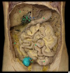
Ileocecal junction
identify the structure
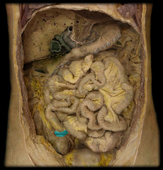
appendix
identify the structure
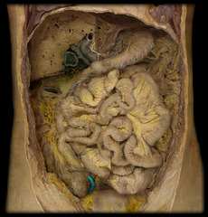
ascending colon
identify the structure
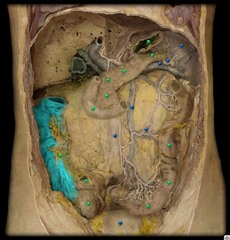
right colic (hepatic) flexure
identify the structure
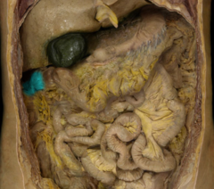
transverse colon
identify the structure
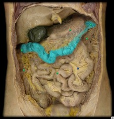
left colic flexure
identify the structure
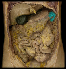
descending colon
identify the structure
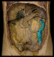
sigmoid colon
identify the structure
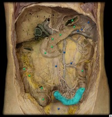
Mesentery of small intestine
identify the structure
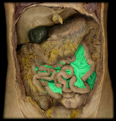
Ileum
identify the structure
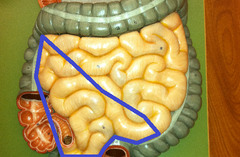
Lymphoid tissue, naturalizes pathogens
What does the appendix contain?
Appendicitis
What is the most common cause of surgery in the abdomen?
epiploic appendages
identify the structure
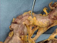
small, fat filled pouches
What are epiploic appendanges?
teniae coli
identify the structure
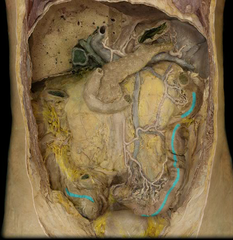
Teniae coli
3 longitudinal smooth muscular bands. Not present in
rectum or anal canal
haustra
identify the structure
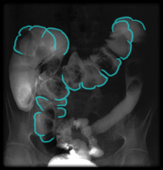
Haustra
sacculations of the colon from the tightening of the teniae coli
Mass movements (peristaltic contractions): The circular muscles contract simultaneously with the teniae coli of the colon wall to move the colon contents a considerable distance toward the anus
What are the mass movements (peristaltic contractions) of the large intestine?
superior mesenteric a. (SMA)
inferior mesenteric a. (IMA)
what main abdominal arteries supply the large intestine?
ileocolic a.
right colic a.
middle colic a.
name the 3 arteries that branch from the SMA to supply the large intestine:
left colic a.
sigmoid aa.
superior rectal a.
name the 3 arteries that branch from the IMA to supply the large intestine:
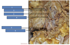
Right colic artery
Identify the structure at #1
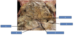
Ileocolic artery
Identify the structure at #2
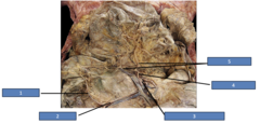
Superior mesenteric artery
Identify the structure at #3
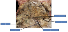
Middle colic artery
Identify the structure at #4
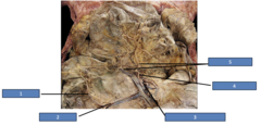
Marginal artery
Identify the structure at #5
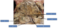
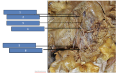
Inferior mesenteric artery
Identify the structure at #1
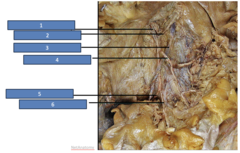
Left colic artery
Identify the structure at #2
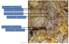
Inferior mesenteric artery
Identify the structure at #3
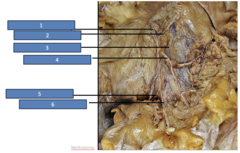
Sigmoid artery
Identify the structure at #4
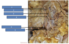
Superior rectal artery
Identify the structure at #5
sigmoid artery
Identify the structure at #6
rectal folds (superior, middle, inferior)
these structures help maintain the fecal content inside the rectum
marginal a.
what artery anastamoses and connects the arterial supply from the SMA and IMA?
superior mesenteric v.
venous drainage for the midgut portion of the large intestine:
inferior mesenteric v.
venous drainage for the hindgut portion of the large intestine:
hepatic portal v.
to reach the portal venous system, the superior mesenteric v. will drain into the:
splenic v.
to reach the portal venous system, the inferior mesenteric v. will drain into the:
Splenic v.
Identify the structure
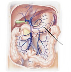
Portal vein
Rule of thumb: All blood from the gut should go to the liver through the...
cisterna chyli
lymph nodes surrounding the celiac trunk, SMA and IMA will drain into the:
thoracic duct
the cisterna chyli drains lymph received from abdominal nodes into the:
Celiac --> foregut
Superior mesenteric --> midgut
Inferior mesenteric --> hindgut
All of the lymph nodes report to major lymph nodes:
Celiac -->
Superior mesenteric -->
Inferior mesenteric -->
Celiac lymph nodes
What lymph do the stomach, liver, spleen, and pancreas drain into?
Superior and inferior mesenteric
What lymph do the intestines drain into?
Upper
Where are the anal columns located in the anal canal?
pectinate line
the inferior border of the anal columns demarcate the:
pectinate line
what line separates upper anal canal from lower anal canal?
anorectal junction
the superior border of the anal columns demarcate the:
involuntary (somatic)
the internal anal sphincter is under what muscular control?
voluntary
the external anal sphincter is under what muscular control?
endoderm
the upper anal canal originates from what embryonic layer?
Internal anal sphincter
Under involuntary control, modified extension of the circular muscles of rectum. Relaxes to allow defecation, constricts to maintain fecal continence
External anal sphincter
Under voluntary control, relaxation allows defecation, constricts to maintain fecal continence
ectoderm
the lower anal canal originates from what embryonic layer?
Endoderm
The upper anal canal originates from what embryonic layer?
pectinate line
the embryological anal membrane will turn into the:
superior rectal a.
the upper anal canal receives blood from the:
inferior mesenteric a.
the superior rectal a. is a branch of the:
haemorrhoids
In their normal state, they are cushions that help with stool control. They become a disease when swollen or prolapsed
middle rectal a.
inferior rectal a.
the lower anal canal receives blood from the:
superior rectal vein to the inferior mesenteric v.
the upper anal canal venous drainage is done by:
middle rectal v.
inferior rectal v.
the lower anal canal venous drainage is done by:
internal iliac v.
the middle rectal v. and inferior rectal v. drain into the:
internal iliac a.
the middle rectal a. and inferior rectal a. branch from the:
Left gastric vein
Which of the following veins is involved in the formation of esophageal varices as part of a portacaval anastomosis?
Superior rectal vein and inferior rectal vein
Anorectal varices result from increased pressure in the portal system. Which veins are primarily involved in this anastomosis?
internal iliac nodes
inferior mesenteric nodes
lymph drainage for above the pectinate line travels to:
superficial inguinal nodes
lymph drainage for below the pectinate line travels to:
abdominal lymph nodes, such as the inferior mesenteric and internal iliac nodes.
Pathology above the pectinate line (upper anal canal) will tend to spread to the...
picked up by the superficial inguinal lymph nodes
Pathology below the pectinate line (lower anal canal) will be...
pudendal n. (S2-S4)
somatic motor to external anal sphincter and somatic sensory to skin around the anal region is done by:
Autonomic and visceral sensory innervation
What type of innervation is found above the pectinate line in the anal canal?
intestinal mucosa
false diverticula involve swelling of only:
entire intestinal wall
true diverticula involve:
Sigmoid colon
What is the most common location for diverticula to develop in the colon?
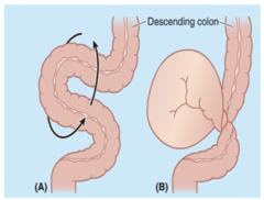
volvulus of Sigmoid Colon
Rotations and twisting of the mobile loop of the sigmoid colon:
family history, IBD, old age, African American race, low-fiber diet, alcohol, smoking, obesity
risk factors for colorectal cancer
colonoscopy
diagnosis for colorectal cancer:
Surgery, chemotherapy, radiation
Three primary treatment options for colorectal cancer:
Crohn's Disease
patient presents to your office with •Cobblestonning of buccal mucosa and gingiva, Aphtous ulcers and Mucosal tags. Patient has a history of abdominal pain and sudden weight loss. What might be the diagnosis?
Diffuse labial, gingival or mucosal swelling Cobblestonning of buccal mucosa and gingiva
Aphtous ulcers
Mucosal tags
Pyostomatitis Vegetans
Angular cheilitis
Oral manifestations from Crohn's disease:
B) It is foregut structure
Which of the following statement is correct regarding the marked structure:
A) Received its blood supply from the superior mesenteric artery
B) It is foregut structure
C) Receives blood supply from both celiac trunk and SMA
D) It is retroperitoneal
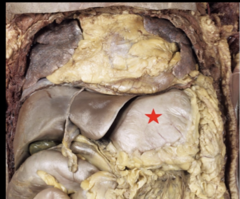
A) Esophageal varices- Systemic hypertension
They are typically associated with portal hypertension
Which one of the following pairings is INCORRECT
A) Esophageal varices- Systemic hypertension
B) Duodenum-Retroperitoneal space
C) Ampulla of Vater- Head of pancreas
D H. Pylori. Peptic ulcer disease
False
T/F: One of these structures are mislabeled
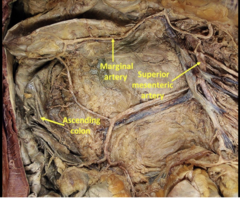
D) Rectum-Tenia Coli
Which one of the following pairs don't fit together
A) Transverse colon-Mesocolon
B) Crohn's disease-Ileocecal junction
C) positive Blumberg's sign-Appendicitis
D) Rectum-Tenia Coli
B) SMA
C) Marginal artery
5) This part of the Gl tract receives blood supply from (check all that applies):
A) Gastroduodenal artery
B) SMA
C) Marginal artery
D) Superior Pancreaticoduodenal
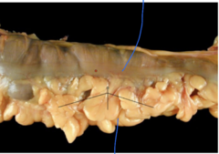
A) Internal anal sphincter-puborectalis muscle (internal anal sphincter is smooth muscle controlled by ANS and puborectalis is skeletal muscle)
D) Anal canal below pectinate line- Portal vein (should be inferior rectal vein)
Which of the following pairings is incorrect (check all that applies)
A) Internal anal sphincter-puborectalis muscle
B) Upper part of anal canal- Hindgut
C Rectum-Superior rectal artery
D) Anal canal below pectinate line- Portal vein
Pudenal nerves
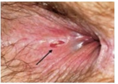
The picture depicts an anal fissure, a painful tear in the lining of the anus: what nerve supply somatic sensory fibers to this