B3192 - Musculoskeletal: Bone Disorders Pt.1
 Call Kai
 Learn
Learn Practice Test
 Spaced Repetition
 Match
Match Flashcards
 Knowt Play
1/148

There's no tags or description
Looks like no tags are added yet.
Name | Mastery | Learn | Test | Matching | Spaced | Call with Kai | Chat |
|---|
No analytics yet
Send a link to your students to track their progress
149 Terms
What are the three factors involved in controlling osteoblast and osteoclast activity?
RANK
RANKL
Osteoprogeterin (OPG)
Function of RANKL
RANKL: A protein produced by osteoblasts that bind to RANK receptors on osteoclasts. Once it's bound, it promotes and activates osteoclast activity while inhibiting osteoblasts

Function of RANK?
A receptor on the osteoclasts; activates osteoclasts when RANKL binds to RANK
Function of OPG?
A "decoy" receptor for RANKL and is produced by osteoblasts. It essentially soaks up RANKL and prevents it from binding onto RANK, inhibiting osteoclasts while promoting osteoblast activity

T/F: Does OPG compete with RANKL to bind to RANK receptors?
F: OPG does not compete with RANKL; it competes with RANK receptors by soaking up RANKL, preventing RANKL from binding onto RANK and activating osteoclast activity
RANK ___ osteoclast activity whereas OPG ___osteoclast activity
promotes
inhibits
Significance of Wnt signalling?
It plays a key role in bone formation by inhibiting osteoclast activity and promoting osteoblast activity
Sclerostin (SOST)
a Wnt inhibitor = promotes osteoclast activity and inhibits osteoblasts, results in decreased bone formation

Effects of Sclerostin on osteoclast, osteoblast, RANKL, and OPG
Osteoclast = increased
Osteoblast = decreased
OPG = decreased
RANKL = increased
These overall results in decreased bone formation and increased turnover
Results of sclerostin inhibitor
Inhibits the effects of SOST and promotes bone formation by increasing osteoblast and OPG activity while inhibiting osteoclast and RANKL
What nutrients are required for bone health? How are they important? (7)
Calcium and Phosphate (minerals): aids in the formation of the inorganic matrix of the bone; required for bone formation and growth
Vitamin D: enhances the absorption of calcium in the GI tract
Vitamin C: required for collagen synthesis, which forms the organic components of the bone
Vitamin A: stimulates osteoblast activity during bone remodelling
Vitmain K and B12: synthesis of bone proteins
Hormonal factors required for bone health
GH and Insulin-like GH factors: promote osteoblast activity and protein synthesis
Thyroid hormones: stimulate osteoblast
sex hormones:
- Slows bone resorption by osteoclasts
- promotes bone deposition by osteoblasts
- sex hormones cause osteoblasts to produce more OPG, inhibit osteoclasts, and induce apoptosis of osteoclasts
Calcitonin: inhibits osteoclasts
PTH:
- promotes osteoclast activity
- formation of calcitriol in the kidneys
-calcium reabsorption from urine
Bone fracture
Break in the continuity of a bone that occurs when stress > force bone can absorb
How fractures are classified
Pattern along fracture line
Degree of break in continuity
Communication with external environment
Character of fracture pieces
Three major causes of fractures
Acute injury
Fatigue or stress
Pathologic (weakened bones)
Patterns along fracture lines
-Transverse
-Longitudinal
-Spiral
-Oblique
-Greenstick

Degree of breaks in continuity
-Complete
-Incomplete/partial

Complete fracture
Parts of the bone are completely separated
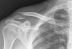
Incomplete/partial fracture
Break is incomplete, ex. greenstick fracture

Communication with external environment
-Open (compound)
-Closed (simple
Open fracture
Broken bones protrude through skin

Closed fracture
Broken bones do not break skin

character of fracture pieces
-Comminuted
-Compression
-Impacted
-Stress
-Avulsion
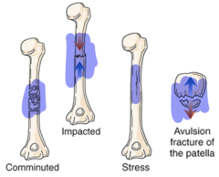
Comminuted fracture
Bone in broken into two or more pieces

Compression fracture
Bones are crushed or squeezed together

Impacted fracture
One end of fractured bone is driven into interior of the other end

Stress fracture
Failure of one external surface of the bone

Avulsion fracture
Separation of small bone piece at site of ligament or tendon

Pathogenesis of bone fractures
Broken bones cause damage to the surrounding tissues, periosteum, and the blood vessels in the cortex and bone marrow
- Broken blood vessels lead to hematoma formation
The damage to the bone tissue triggers an inflammatory response
Cause of fracture manifestations
Inflammatory response
This is because a fracture causes DAMAGE to the surrounding tissue, periosteum, and blood vessels in the cortex and bone marrow
Manifestations of fractures
-Pain
-Unnatural alignment
-Swelling
-Loss of function
-Blood loss
-Impaired sensation
Diagnosis of fractures
-X-ray
For fractures that are difficult to detect (occult fractures):
-CT
-MRI
What type of fracture may not be initially visible using X-rays? And why is it good to have more than two angles when imagining a fracture?
Stress fracture
Having two angles when imaging a fracture allows for a complete picture of the fracture, which can help differentiate oblique and spiral fractures
Tx of fractures?
Reduction: Align the bones close to their anatomical position - helps regain normal function
Immobilization: Maintain proper alignment during the healing process (casts, splints, traction, etc.)
Closed manipulation: NON-SURGICAL, bones are manipulated back into place (Pushing ends of bones into their correct position)
Open manipulation: SURGICAL, surgical realignment of bone (screws, nails, plates, wires, glue, cement, etc.)
RICO treats fractures
Bone readily regenerates via _____
processes involved in embryogenesis
Four steps of fracture repair
Hematoma formation
Fibrocartilaginous callus formation
Bony callus formation
Remodeling
Hematoma formation (fracture repair)
Torn blood vessels bleed into and around bone to form a hematoma, facilitating formation of fibrin meshwork

Fibrin meshwork
Aids in fracture repair by sealing off site and providing a framework for healing processes
Fibrocartilaginous callus formation (fracture repair)
Fibroblasts proliferate, creating granulation tissue forms to connect the bone fragments
Procallus
Granulation tissue
Bony callus formation
Callus ossifies and osteoprogenitors develop into osteoblasts to produce spongy bone
Ossification
Conversion of soft cartilage to bony callus
Bone remodeling (fracture repair)
Compact bone replaces spongy bone and osteoclasts remove dead portions of bone
Complications of fracture healing (9)
-Delayed union
-Malunion
-Nonunion
-Osteonecrosis
-Infection
-Compartment syndrome
-Fat emboli
-DVT/PE
-Osteoarthritis
Delayed union - why does this happen? (Complications of fracture healing)
Failure of the fracture to heal within the predicted time
Due to infection, smoking, malnutrition, corticosteroid use, and decreased vascularization; common underlying lesson is due to IMPAIRED IMMUNE FUNCTION
Adverse effects of corticosteroids in bone remodelling?
Corticosteroids can cause an increase in RANKL and a decrease OPG, which leads to an increase in bone resorption
Malunion - why does this happen? (Complications of fracture healing)
Deformity at the fracture site
Could potentially be due to misalignment or immobilization
Nonunion - why does this happen? (Complications of fracture healing)
Failure of the bone to heal
Due to a lack of nutrients, osteoblast dysfunction, or hormonal imbalance
Osteonecrosis - why does this happen? (Complications of fracture healing)
Death of bone tissue due to ischemia
- Fractures can damage or block blood vessels; The bones are not being actively perfused
Compartment syndrome - why does this happen? (Complications of fracture healing)
Increased pressure in the muscle comparment leading to poor blood flow and damage to the muscle/nerves
muscles are enclosed in fascia, when bones and muscles are damaged = inflammation = which puts more pressure and more damage to tissues and compresses blood vessles = ischemia, necrosis, and nerve damage
Fat emboli - why does this happen? (Complications of fracture healing)
Yellow bone marrow contains a lot of fat, which, when released into the bloodstream due to a fracture, can cause an occlusion and impair perfusion.
DVT and PE - why does this happen? (Complications of fracture healing)
Inflammation increases the risk of clotting (hypercoagulability), which is a crucial factor in the development of DVT and PE.
What to watch out for
PE:
-sudden shortness of breath
-sharp chest pain that worsens with breathing
- rapid or irregular heartbeat
-coughing that may produce bloody mucus.
DVT:
- swelling in the affected limb
- pain or tenderness
- warmth to the touch
- skin discoloration (redness or blueness)
- The majority may be asymptomatic due to the formation of collateral blood vessels
Osteoarthritis - why does this happen? (Complications of fracture healing)
Wear and tear of joints, damage to the joint.
Fractures can cause osteoarthritis if they do not heal properly. Fractures alter the joint's weight distribution and increase pressure on the joint, leading to damage and inflammation that can result in osteoarthritis.
Dislocation (mechanical alteration in bones)
Temporary displacement of two bones with complete loss of contact between articular cartilage
Subluxation (mechanical alteration in bones)
Contact between joint surfaces is only partially lost
Osteomyelitis
Pyogenic inflammation of bone and bone marrow due to infection
Hallmark: bone infection is PUS-PRODUCING
Manifestations of osteomyelitis
Inflammation
Fever
Pain
Necrotic bone

Infection origins of osteomyelitis
-Spread from open wound
-Hematogenous
-Spread from skin infections
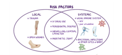
Common causative agents of osteomyelitis
S. aureus
S. pneumonia
E. coli (in immunocompromised patients)
Pathogenesis of osteomyelitis
Pathogens enter bone and cause inflammation. Thrombosis of local blood vessels causes ischemic necrosis and fibrosis

Complications of osteomyelitis
Septicemia
Arthritis
Pathologic fractures
Sequestra
Sequestra
Necrotic bone that is a medium for continued bacterial proliferation
Diagnosis of osteomyelitis
- Xrays or bone scans
- Bone biopsy and culture

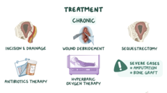
Tx of osteomyelitis
- IV abx - FIRST LINE OF TX
- Debridement
- Amputation
- Hyperbaric oxygen therapy (enhances bone and soft tissue healing)
Skeletal tuberculosis? How does it spread?
Extrapulmonary form of TB, where pathogen spreads from the lungs to the bone
It spreads to the bone via the blood stream or lymphatic drainage
Common sites of skeletal TB infections
- Weight-bearing joints
- Vertebrae
Pott disease
TB of the spine
Result of TB in the vertebrae
- Neurological impairment
- paraplegia from vertebral necrosis and collapse
Tx of skeletal TB
- Anti-TB drugs
Origin cells of bone tumours
From ANY SKELETAL COMPONENT including bone cells, cartilage, fibroblasts, and bone marrow
Note: tumour cells are MONOCLONAL
T/F: Tumours are more likely to form in well-differentiated cells (e.g. osteocytes). Why?
False: Tumours are likely to occur in LESS-differentiated cells because these cells are either stem cells or progenitor cells that naturally divide more frequently, increasing the chance for accumulating genetic mutations that drive uncontrolled growth
Sarcoma
Malignant bone tumour
Osteoid osteoma? Location? S/S? Tx? Demographics affected?
Small BENIGN tumour found on the surface of long bones, flat bones, or the skull
- well localized, increasingly sever pain
- surgical excision, radiofrequency ablation
- Young adults
Composition of osteoid osteoma
Core contains osteoblasts, osteoclasts, and granulation tissue surrounded by sclerotic bone
Osteoblastoma
Larger, more lytic version of osteoid osteoma
Giant cell tumour (osteoclastoma)? Location? Tx? Demographics affected? Metastasis?
BENIGN Tumour of multinucleated giant osteoclasts is usually located at the junction of the metaphysis and epiphysis of long bones. 50% chance of metastasis after surgical disruption of og. tumour
- locally aggressive
- overexpresses RANK
Tx:
-curettage (scraping and scooping)
- bone grafts
Demographic:
- common in adults (20-40), esp. females
Effect of RANK overexpression in osteoclastoma
Hemorrhage from bone cortex thinning and rupturing
Osteosarcoma? Location? S/S? Tx? Demographics affected? Metastasis?
Highly MALIGNANT and most common primary bone cancer. Occurs around the knee - can metastasize to lung
S/s:
- painful, progressively enlargin mass, subject to sudden fracture
Tx:
- chemo
-radiation
- surgery
Demographics:
- young (10-25)
-elderly (>60)
Pathogenesis of osteosarcoma
Mutations in retinoblastoma or p53 gene lead to malignant osteoblasts
Chrondrosarcoma? Location? S/S? Tx? Demographics affected?
Primary MALIGNANT bone tumour of cartilage. Affects shoulder, pelvis, and ribs
S/s:
- painful, progressively enlarging mass (but slow growing)
Tx:
- chemo
- surgical excision
Demographics:
- males (40-70)
Pathogenesis of chrondrosarcoma
Cartilage expands and destroys bone, which stimulates osteoclasts to break down bone
Ewing sarcoma? Location? S/S? Tx? Demographics affected? Metastasis?
RARE and highly MALIGNANT tumour composed of undifferentiated cells that arise from long bones and pelvis
invades soft tissues and metastasizes widely
S/s:
- pain, fractures, and systemic signs of infection
Tx:
- chemo
- combined surgery
- radiation
Demographics:
- Children and young adults aged
Pathogenesis of Ewing sarcoma
Involves reciprocal translocation between chromosomes 11 and 22
invades soft tissues and metastasizes widely, leading
to pain, fractures, and systemic signs of infection
Fibrosarcoma? Location? Metastasis?
Fibrous mass of collagen, malignant fibroblasts, and osteoclast-like cells. Affects the metaphysis of femur or tibia
Metastasizes to the lungs
What is secondary bone cancer? Why is metastasis in bone common?
Secondary (metastatic) bone tumours - arise from a different organ and spread via lymph or blood
Bone is a common site for metastasis as its is highly vascularized - also why it is often MULTIFOCAL
Types of secondary bone tumours
Osteolytic
Bone-forming
Mixed
Osteolytic secondary bone tumour
Leads to pathological fractures from bone weakening
Bone-forming secondary bone tumour
Leads to overly dense bone foci
Osteogenesis imperfecta
Group of hereditary disorders caused by abnormal synthesis of type I collagen
Other name for osteogenesis imperfecta
Brittle bone disease
Manifestations of osteogenesis imperfecta (6)? Why?
- Basic morphological change: due to osteopenia, collagen is a key protein that provides strength and structural support to bones. This defective collagen makes bones abnormally weak, reducing bone density to a level below average (osteopenia)
- Fractures and dislocation: bone slack tensile strength due to osteopenia
- Blue sclera: lack of collagen, appears blue as it is thin
- Abnormal tooth development
- Deafness: an abnormality in the formation of bones in ars, causing irregularity in sound conduction
- Short stature
All of these are primarily due to defective collagen makeing bones abnormally weak, reducing bone density to a level below average (osteopenia)
Diagnosis of osteogenesis imperfecta
-Analysis of collagen production by fibroblasts
-Genetic testing
-Imaging
Tx of osteogenesis imperfecta
-Surgical correction of abnormalities
-Physiotherapy, orthotic supports, mobility devices
-Bisphosphonates
-Sclerostin inhibitors
Biphosphonates
Used in osteogenesis imperfecta to increase bone density by inhibiting osteoclast activity
Marfan syndrome
Autosomal dominant disorder of connective tissue - impaired crosslinking of collagen and elastic fibres
Pathogenesis of Marfan syndrome
Mutation of the FBN1 gene that results in impaired cross-linking of collagen and elastic fibers in the ECM
Skeletal manifestations of Marfan syndrome
-Excessive height
-Long extremities
-Arachnodactyly
-Laxity of joint ligaments
-Pectus excavatum/pectus carinatum
-Kyphosis and scoliosis
Ocular manifestations of Marfan syndrome
-Ectopia lentis
-Retinal detachment
-Severe myopia
The lenses are not directly held in place due to an abnormality in the crosslinking of collagen and elastic fibres (impaired structural integrity of connective tissue)
CVS manifestations of Marfan syndrome
-Mitral valve prolapse
-Dissection and rupture of aorta
Diagnosis of Marfan syndrome
-Genetic testing
-Imaging of bones and eyes
-ECG (for heart)