Microbiology - Section 1, Lesson 8
1/44
There's no tags or description
Looks like no tags are added yet.
Name | Mastery | Learn | Test | Matching | Spaced | Call with Kai |
|---|
No analytics yet
Send a link to your students to track their progress
45 Terms
Q: Who created the modern light microscope in 1830?
A: Joseph Jackson Lister.
Q: What does fluorescence microscopy use to visualize specimens?
A: Ultraviolet (UV) light.
Q: What does electron microscopy use for imaging?
A: Short-wavelength electron beams.
Q: What are the three main factors improved by modern microscopes?
A: Magnification, resolution, and contrast.
Q: What is a light microscope?
A: A microscope that uses light to visualize images.
Q: Name 3 types of light microscopes.
A: Brightfield, darkfield, fluorescence microscopes (others include phase-contrast, DIC, confocal, two-photon).
Q: What is the most commonly used type of light microscope?
A: Brightfield microscope.
Q: What kind of image does a brightfield microscope produce?
A: Dark image on a bright background.
Q: What is the total magnification formula in light microscopy?
A: Ocular magnification × Objective magnification.
Q: What is the typical ocular lens magnification?
A: 10×.
Q: What are common objective lens magnifications?
A: 4×, 10×, 40×, 100×.
Q: What is the platform where the slide is placed?
A: The stage.
Q: What do the x-y mechanical stage knobs do?
A: Move the slide side-to-side or forward-backward.
Q: When should the coarse focusing knob be used?
A: With 4× and 10× objective lenses (low magnification).
Q: When should the fine focusing knob be used?
A: With 40× or 100× lenses (high magnification).
Q: What provides light in a brightfield microscope?
A: The illuminator.
Q: What does the condenser lens do?
A: Focuses light onto the specimen.
Q: How do you adjust the light amount reaching the specimen?
A: Using the diaphragm or rheostat.
Q: What are chromophores?
A: Pigments that absorb and reflect specific wavelengths of light (used in staining).
Q: Why is staining useful in microscopy?
A: It increases contrast and resolution.
Q: Why is oil immersion used in microscopy?
A: To increase resolution by reducing light scattering.
Q: Why does oil work better than air between the lens and slide?
A: Its refractive index is similar to glass.
Q: What type of lens is designed for use with immersion oil?
A: Oil immersion lens (typically 100×).
Q: What limits the maximum resolution of a light microscope?
A: The wavelengths of visible light.
Q: What is the typical maximum magnification of most light microscopes?
A: 1,000⨯, or up to 1,500⨯ for some.
Q: What do electron microscopes use instead of light to achieve higher resolution?.
A: Short-wavelength electron beams
Q: What is the wavelength of an electron beam used in EM?
A: Approximately 0.005 nm.
Q: What is the maximum magnification of an electron microscope (EM)?
A: Up to 100,000⨯.
Q: Why can’t living material be viewed with an electron microscope?
A: Because specimen preparation requires dehydration, fixation, and vacuum conditions that kill the sample.
Q: What are the two basic types of electron microscopes?
A: Transmission electron microscope (TEM) and scanning electron microscope (SEM).
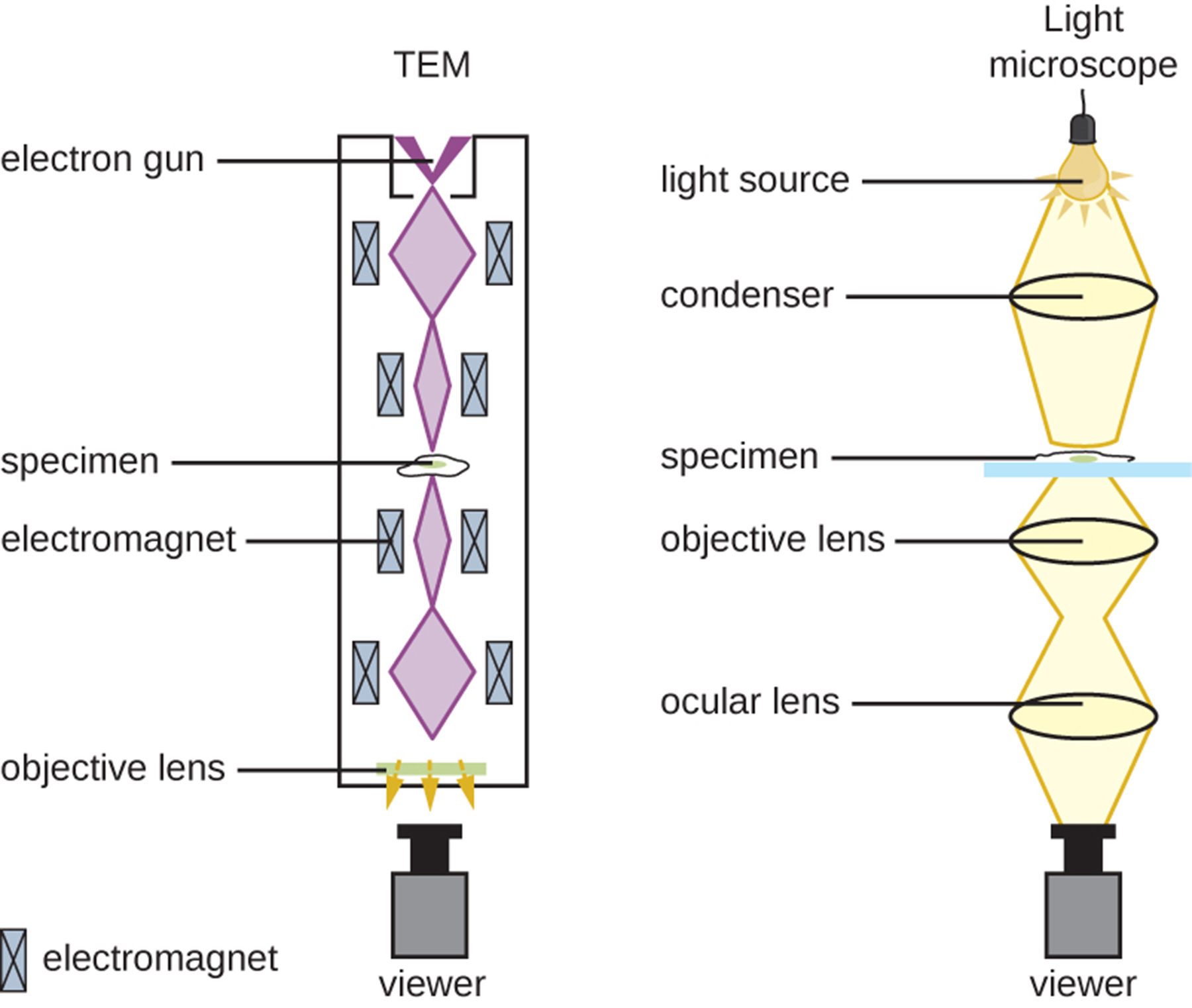
Q: How does a TEM work?
A: It uses an electron beam from above, focused by magnetic lenses, that passes through a thin specimen to form an image on a detector.
Q: How thick must a specimen be for TEM?
A: About 20–100 nm.
Q: What enhances contrast in TEM specimens?
A: Staining with electron-dense materials like heavy metals.
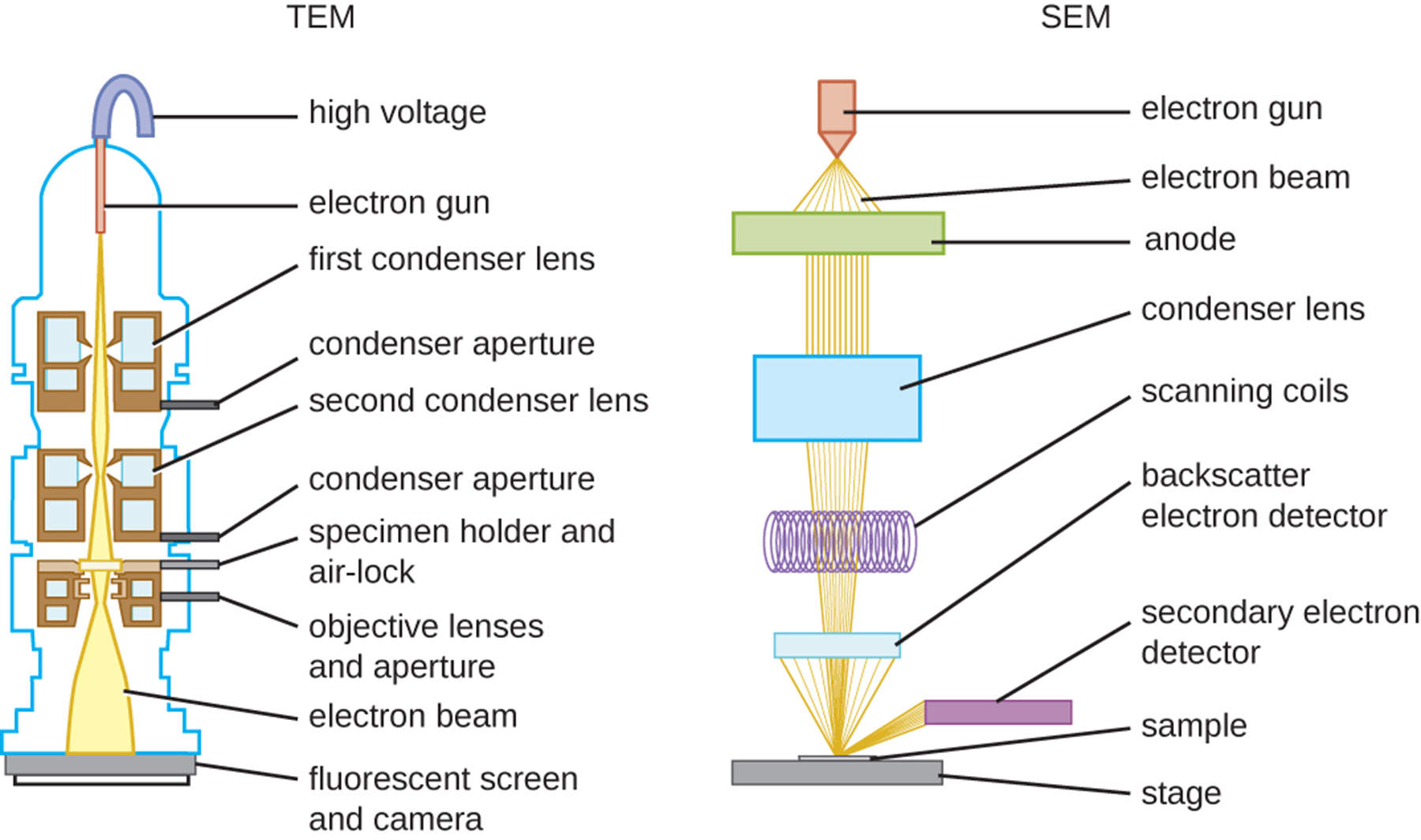
Q: How does an SEM work?
A: It detects electrons knocked off the surface of a specimen, producing a 3D image.
Q: What is done to SEM specimens before imaging?
A: They are dried, fixed, and coated with a thin layer of metal (e.g., gold).

Q: What type of structures does TEM best visualize?
A: Internal structures like organelles and membranes.
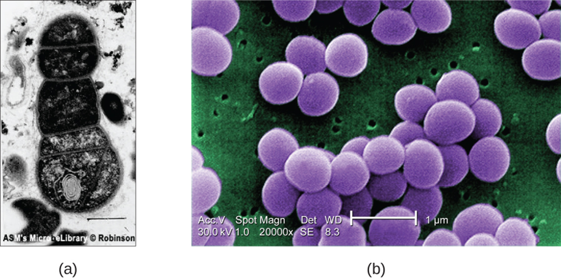
Q: What type of structures does SEM best visualize?
A: Surface details of specimens.
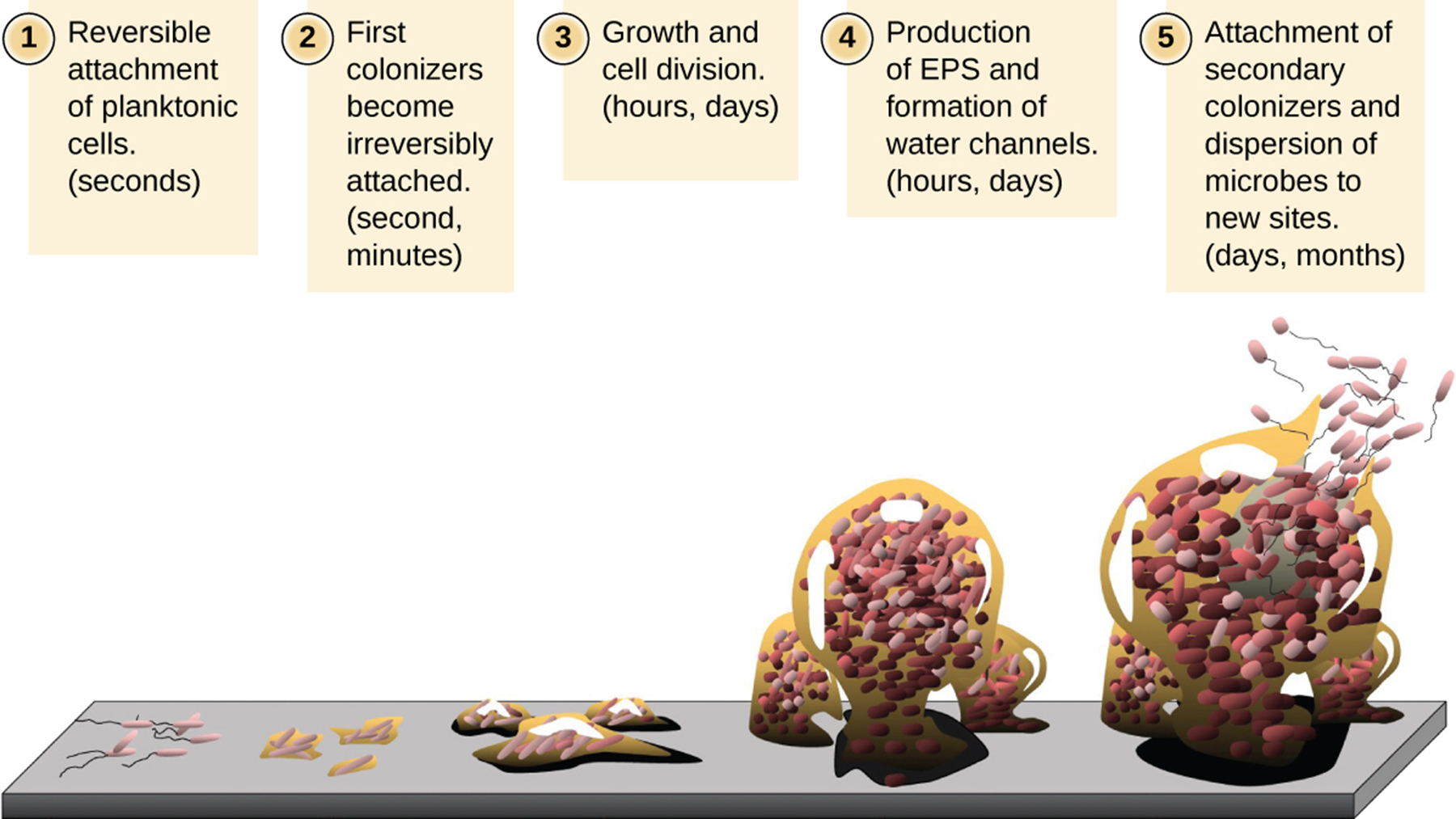
Q: What is a biofilm?
A: A complex microbial community embedded in an extrapolymeric substance (EPS) attached to a surface.
Q: Why are biofilms important in medicine?
A: They are resistant to immune responses and antimicrobial drugs.
Q: Why is light microscopy not ideal for observing biofilms?
A: Biofilms are thick and slicing them may disrupt the microbial community.
Q: How does confocal microscopy help in biofilm imaging?
A: It focuses on individual z-planes and produces 3D images of thick samples.
Q: What role do fluorescent dyes play in biofilm imaging?
A: They help identify cells within the EPS matrix.
Q: What is FISH (fluorescence in situ hybridization)?.
A: A technique using fluorescent probes to bind specific DNA sequences
Q: Why is electron microscopy limited in biofilm observation?
A: Dehydration needed for EM can distort thick biofilms.
Q: How can biofilm shape and water flow be studied?
A: Using video tracking of fluorescent beads moving through biofilm structures.