Balance Wk1 bootcamp LG pt.1
 Call Kai
 Learn
Learn Practice Test
 Spaced Repetition
 Match
Match Flashcards
 Knowt Play
1/19

Earn XP
Description and Tags
Introduction to the Kidney
Name | Mastery | Learn | Test | Matching | Spaced | Call with Kai | Chat |
|---|
No analytics yet
Send a link to your students to track their progress
20 Terms
Which structures are located in the renal cortex?
Answer: The glomerulus, proximal convoluted tubule, distal convoluted tubule, and cortical collecting duct.
Extra Information:
These are the cortical structures identified on the Bootcamp slide.
🧠 “Cortex Girls Prefer Drinking Coffee”
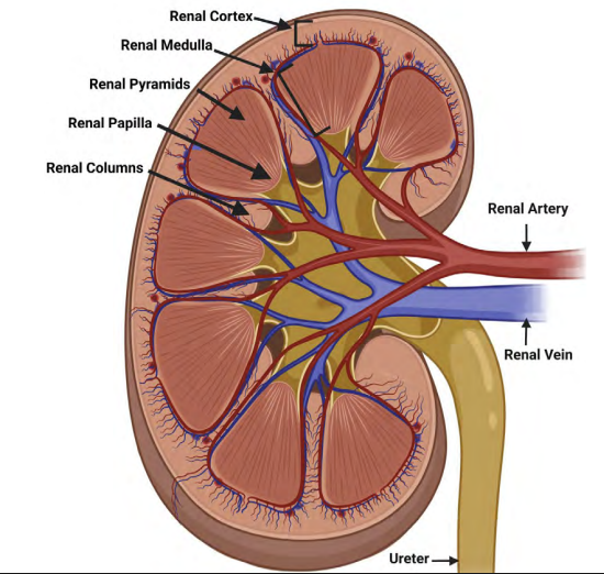
Which structures are located in the renal medulla?
Answer: Renal pyramids, renal columns, the Loop of Henle, and the medullary collecting duct.
Extra Information:
These are the medullary structures identified on the Bootcamp slide.
🧠 “Please Come Learn Medulla”

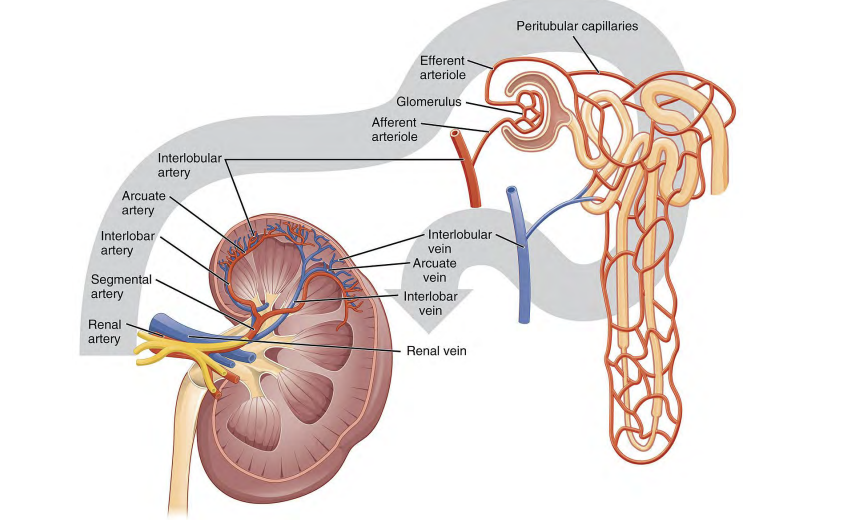
Trace the renal arterial supply from the renal artery to the glomerulus.
Answer: Renal artery → segmental artery → interlobar artery → arcuate artery → interlobular artery → afferent arteriole → glomerulus.
Extra Information:
This is the complete preglomerular arterial pathway shown on the slide.
Recall the sequence in order from the largest vessel to the glomerulus.
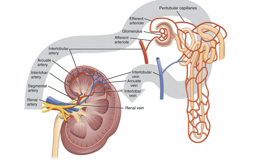
Which vessel immediately delivers blood into the glomerulus?
Answer: The afferent arteriole.
Extra Information:
The afferent arteriole comes after the interlobular artery in the arterial pathway.

Which vessel carries blood away from the glomerulus?
Answer: The efferent arteriole.
Extra Information:
Blood then enters the peritubular capillaries or vasa recta.

Trace blood flow from the glomerulus back toward the renal vein.
Answer: Glomerulus → efferent arteriole → peritubular capillaries or vasa recta → venous outflow pathway → renal vein.
Extra Information:
This is the postglomerular blood-flow pathway shown on the slide.
What structures make up the juxtaglomerular apparatus on this Bootcamp slide?
Answer: The macula densa and juxtaglomerular cells.
Extra Information:
The macula densa is associated with the distal convoluted tubule.
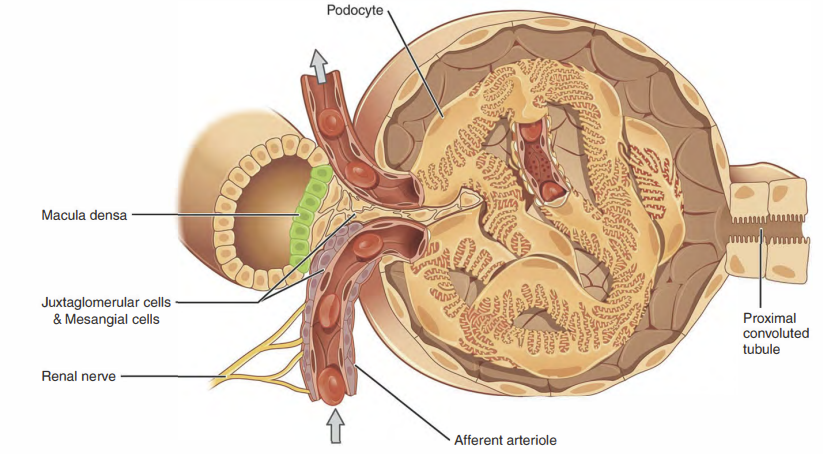
The macula densa is associated with the ______.
Answer: Distal convoluted tubule.
Extra Information:
The macula densa is identified as part of the juxtaglomerular apparatus.
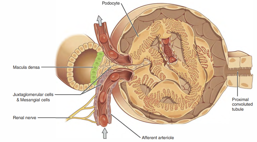
Juxtaglomerular cells are located in the ______ and secrete ______.
Answer: Afferent arteriole and renin.
Extra Information:
Juxtaglomerular cells are part of the juxtaglomerular apparatus.

What is the function of mesangial cells identified on this slide?
Answer: Mesangial cells remove debris.
Extra Information:
- This is the specific mesangial cell function emphasized on the Bootcamp slide.
How does the length of the left renal vein compare with the right renal vein?
Answer: The left renal vein is longer than the right renal vein.
Extra Information:
- This anatomical relationship is marked as high yield on the slide.

Which gonadal vein drains into the left renal vein?
Answer: The left gonadal vein.
Extra Information:
- The slide contrasts this relationship with the right gonadal vein.

What is nutcracker syndrome?
Answer: Compression of the left renal vein by the superior mesenteric artery.
Extra Information:
- This relationship is emphasized as a clinically important consequence of left renal vein anatomy.

What clinical presentation is associated with nutcracker syndrome?
Answer: Abdominal pain, hematuria (blood in urine), and varicocele.
Extra Information:
The underlying abnormality is compression of the left renal vein by the superior mesenteric artery.

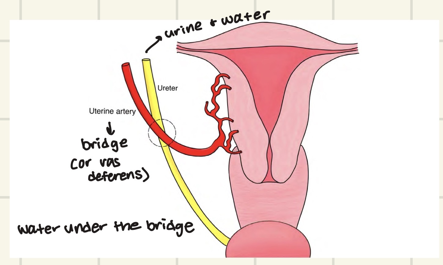
What does “water under the bridge” describe in ureter anatomy?
Answer: The ureter passes under the vas deferens or uterine artery.
Extra Information:
- “Water” represents the ureter.
- The slide uses this phrase to remember the anatomical relationship.
The ureter passes over the ______ artery.
Answer: Common iliac artery.
Extra Information:
- The slide emphasizes both the structures the ureter passes under and the vessel it passes over.

Why can the ureter be damaged during gynecologic procedures?
Answer: The ureter is in close proximity to the uterine artery.
Extra Information:
- The ureter passes under the uterine artery.
- This close anatomical relationship creates a risk of ureteral injury.

Where are the ureteral orifices located within the bladder?
Answer: The trigone.
Extra Information:
The ureteral orifice is one of the bladder structures highlighted on the slide.
The trigone of the bladder is a smooth, triangular area located at the base of the urinary bladder
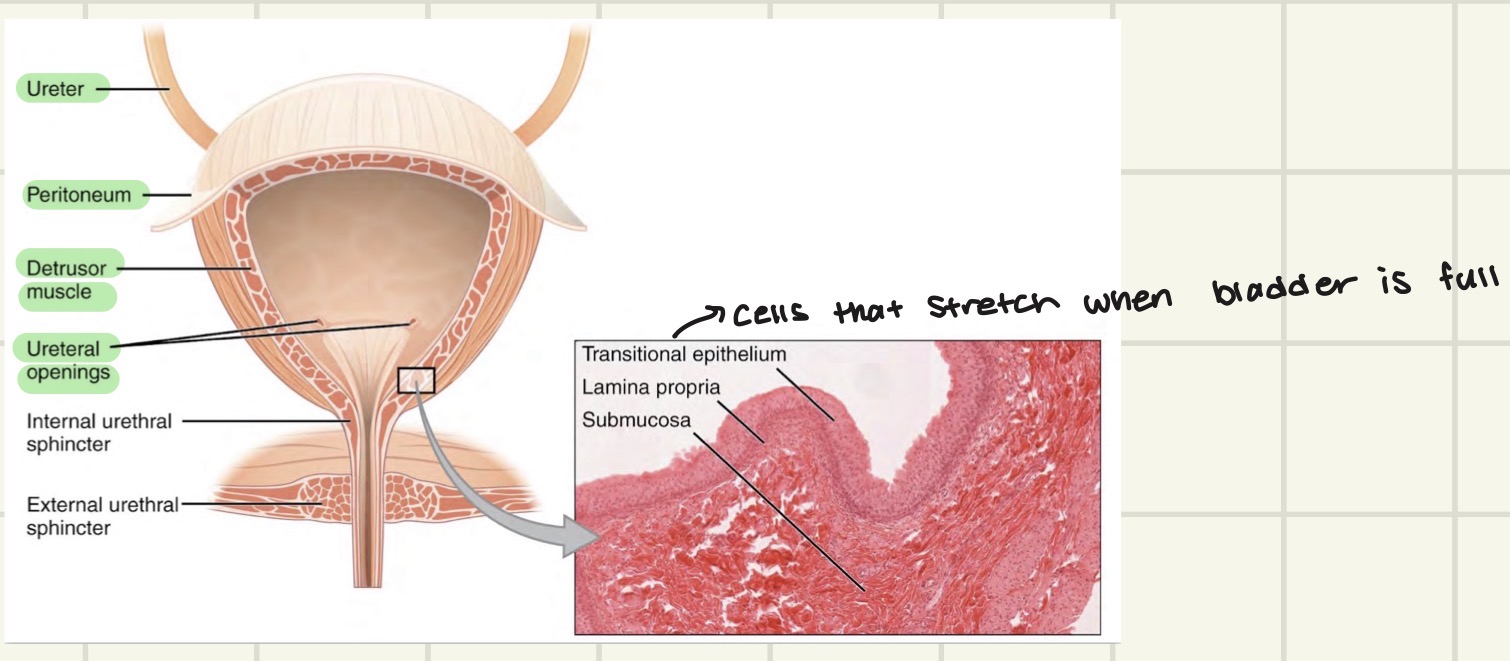
Which urethral sphincter is involuntary versus voluntary?
Answer: The internal urethral sphincter is involuntary, while the external urethral sphincter is voluntary.
Extra Information:
- Distinguishing the two sphincters by voluntary control is emphasized on the slide.

The detrusor muscle of the bladder is under ______ control.
Answer: Autonomic control.
Extra Information:
- The detrusor muscle is the bladder muscle specifically identified on the Bootcamp slide.
