ana300
 Call Kai
 Learn
Learn Practice Test
 Spaced Repetition
 Match
Match Flashcards
 Knowt Play
1/83

There's no tags or description
Looks like no tags are added yet.
Name | Mastery | Learn | Test | Matching | Spaced | Call with Kai | Chat |
|---|
No analytics yet
Send a link to your students to track their progress
84 Terms
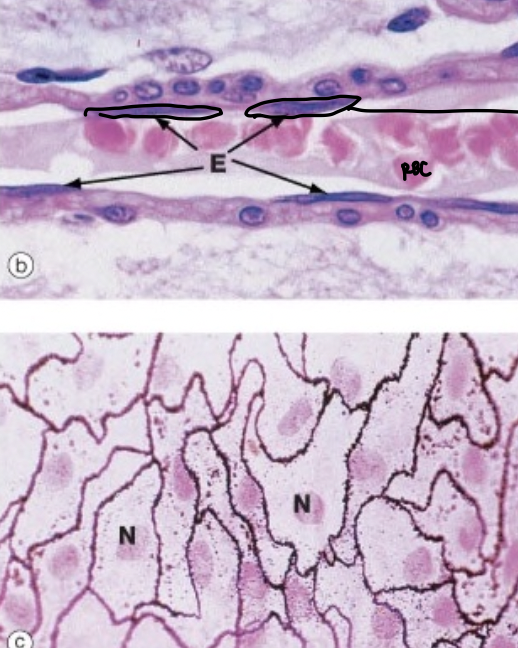
simple squamous (endothelium, mesothelium)
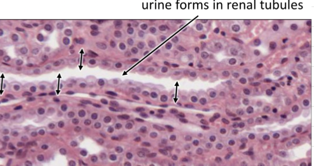
simple cuboidal, active secretion/absorption in renal tubes
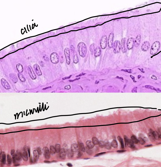
simple columnar (small intestine w/mv, gallbladder w/mv, uterine tubes w/cillia)
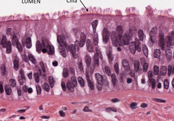
pseudostratified (resp tract w/goblet and cillia)
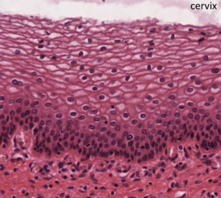
stratified squamous nonkeratinized (internal surfaces like cervix, mouth, throat, esophagus, rectum)
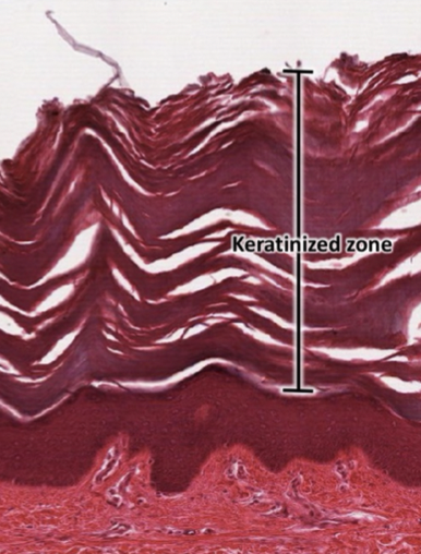
stratified squamous keratinized (external surface prevent dehydration)
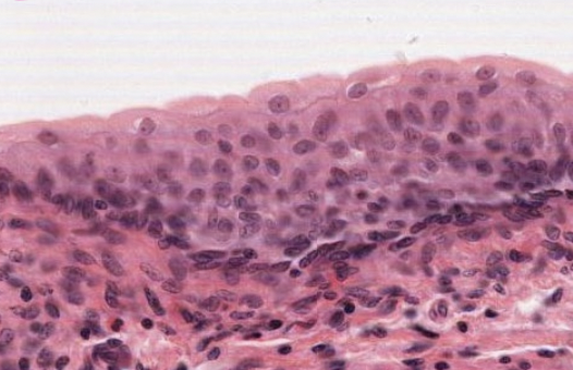
transitional (dome when empty), bladder
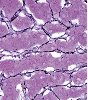
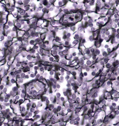
collagen type 3 reticular CT (stroma) in spleen, liver, lymph nodes
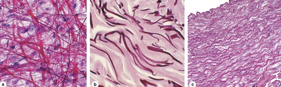
elastic CT (squiggly squiggly) resilience
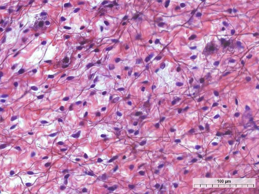
loose areolar CT (superficial fascia, btw muscles, around BV/N, around joint, lamina propria under mucous membrane GI)
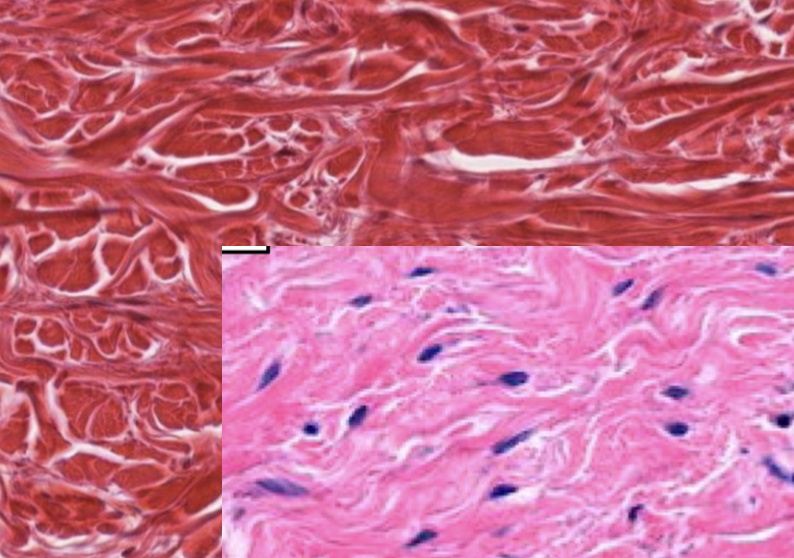
dense irregular CT (collagen type 1 in >1 direction, TENSILE strength)
(reticular layer of dermis (2), periosteum, perichondrium, organ/join capsule)
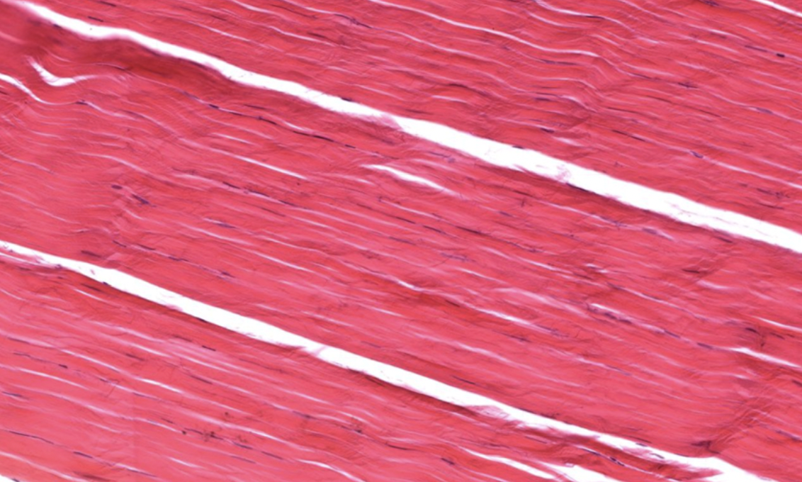
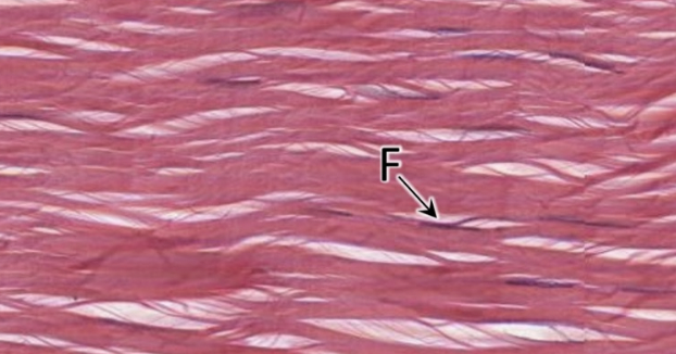
dense regular CT (collagen type 1 in 1 direction tensile strength)
(tendons, ligaments, some ligaments many elastic fibres (ligamentum flavum)
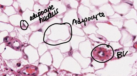
adipose tissue (very vascular, found with loose CT)
(1st dermis papillary and hypodermis, behind eyes around kidney)
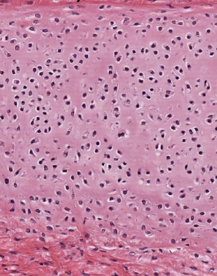
hyaline cartilage (collagen type 2 resist compression)
articular (NO PERICHONDRIUM), ribs, nose, trachea rings (yes perichondrium)
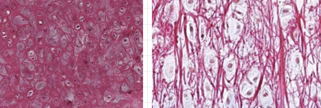
elastic cartilage CT2 and elastic (like hyaline but more elastic fibres for greater flexibility and resilience)
pinna, larynx epiglottis (yes perichondrium)
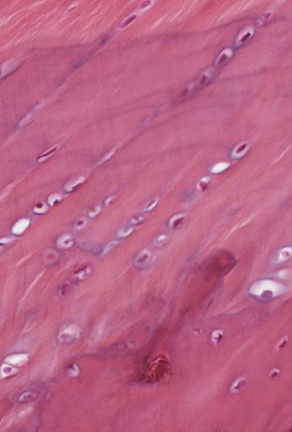
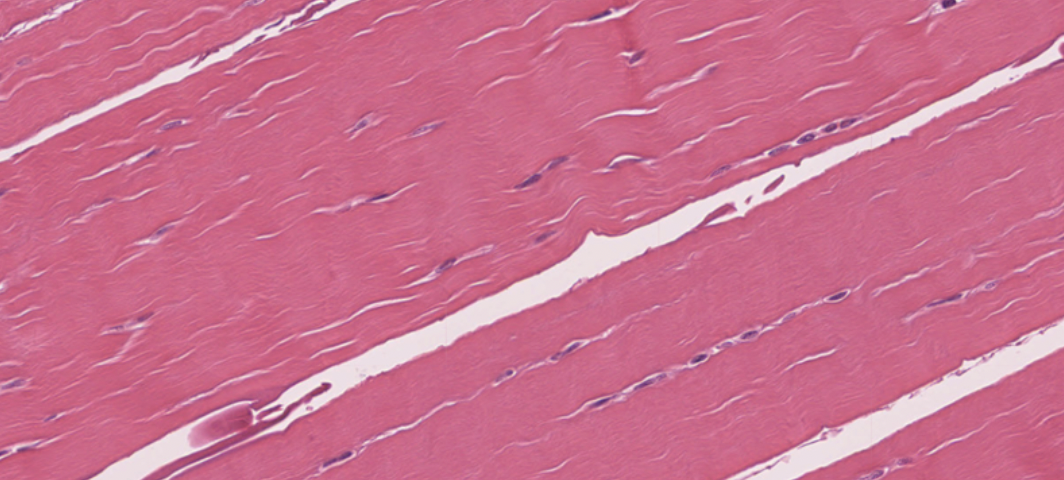
fibrocartilage (CT1&2) (like hyaline but more collagen type 1 Tensile force)
vertebral disc, knee meniscus (NO PERICHONDRIUM)
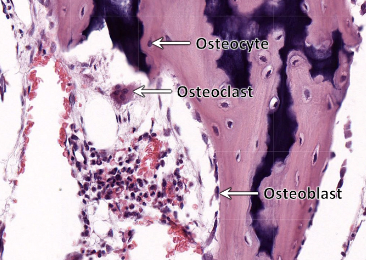
osteoblast (cuboidal)
osteocyte (in lacuna surrounded by matrix)
osteoclast from monocyte (look like big blob octopus, multinuclear)
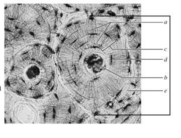
osteon
a. cannaliculi
b. central canal and perforating canal lined with endosteum
c. concentric lamellae
d. osteocyte
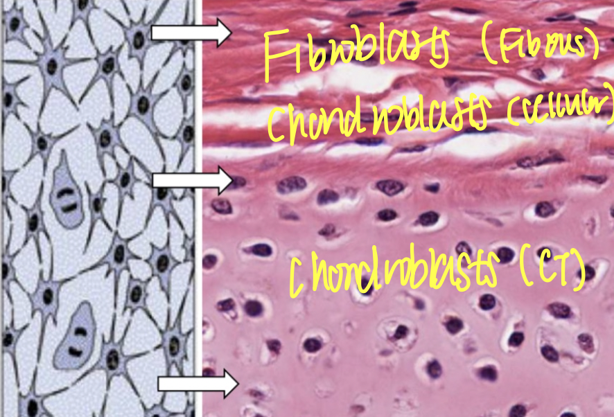
cartilage part (chondrocytes)
cellular layer of perichondrium (chondroblasts)
fibrous layer of perichondrium (fibroblasts)
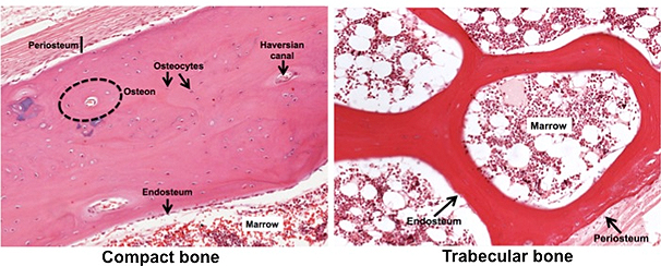
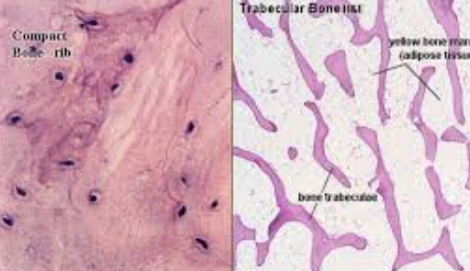
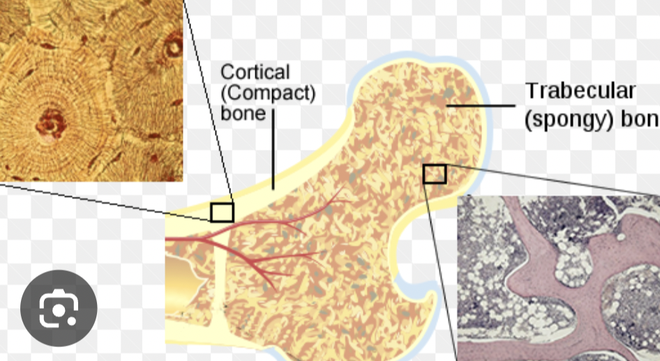
compact vs spongy bone
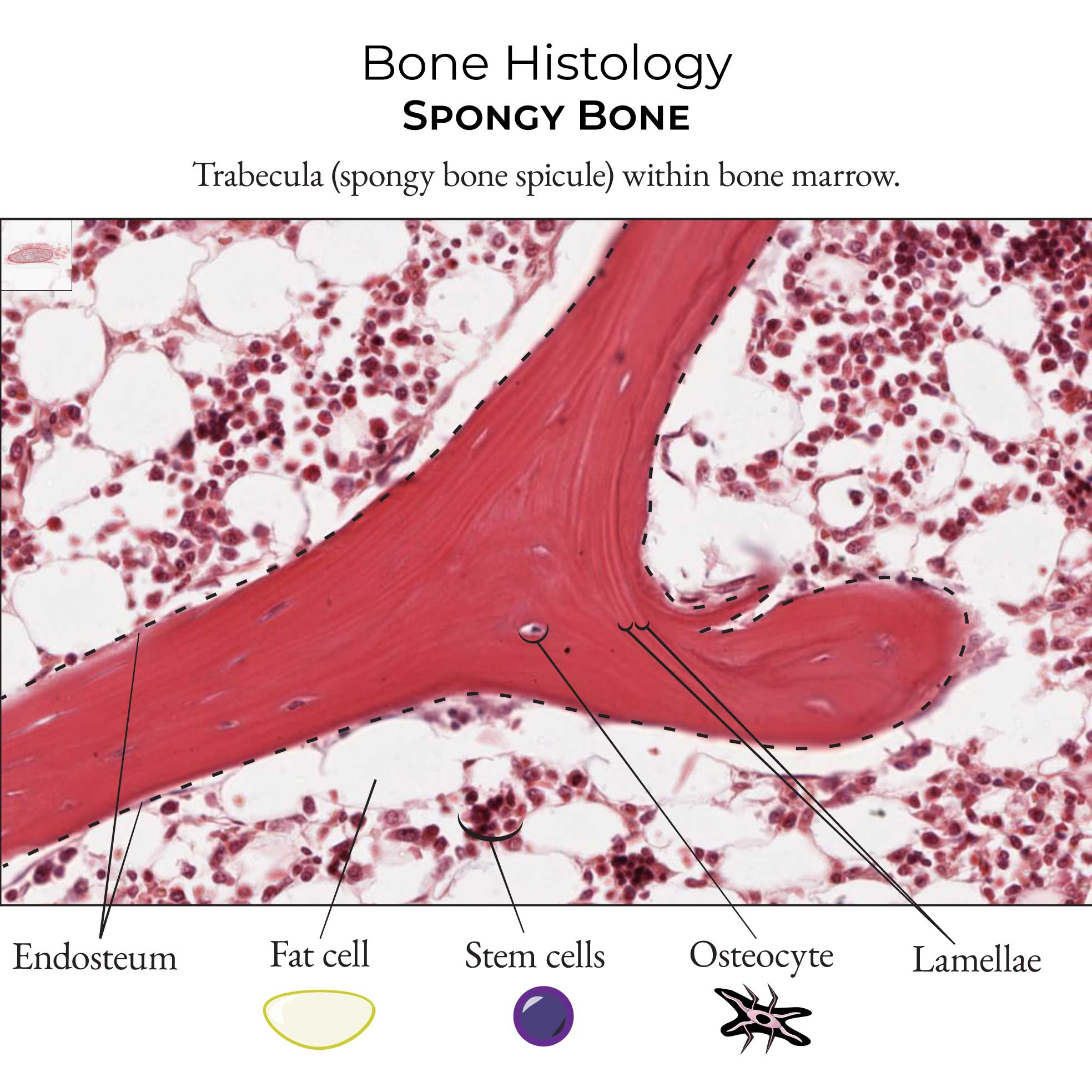
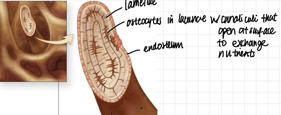
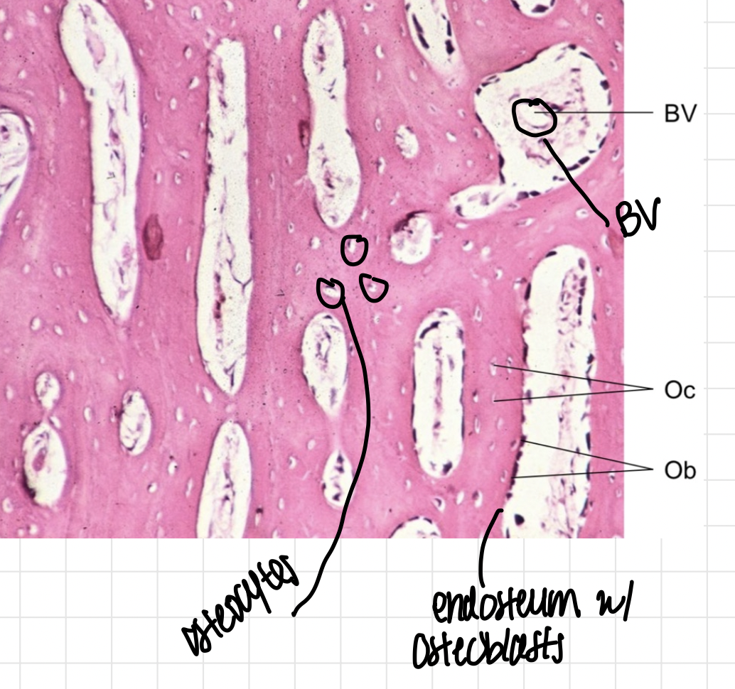
lamella
osteocyte in lacunae
surrounded by endosteum
red stuff in the trabeculae, the white stuff is yellow marrow!!!
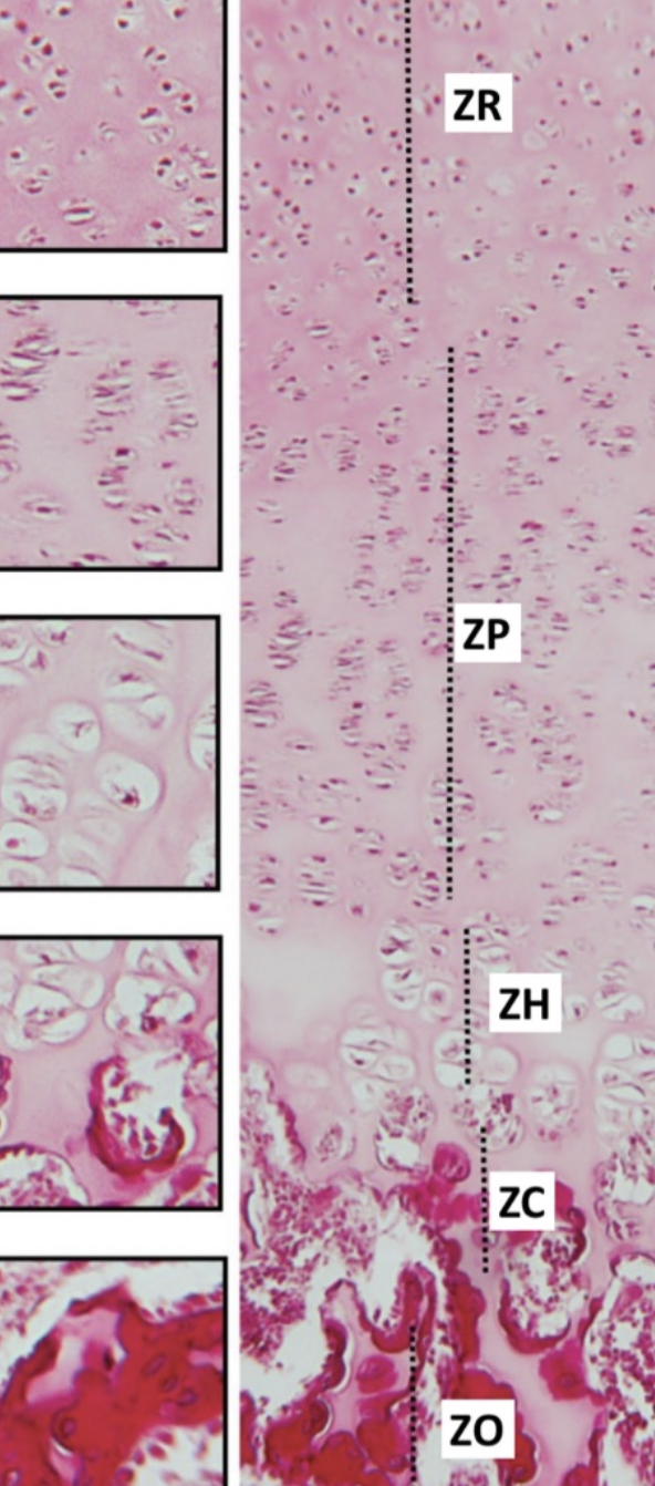
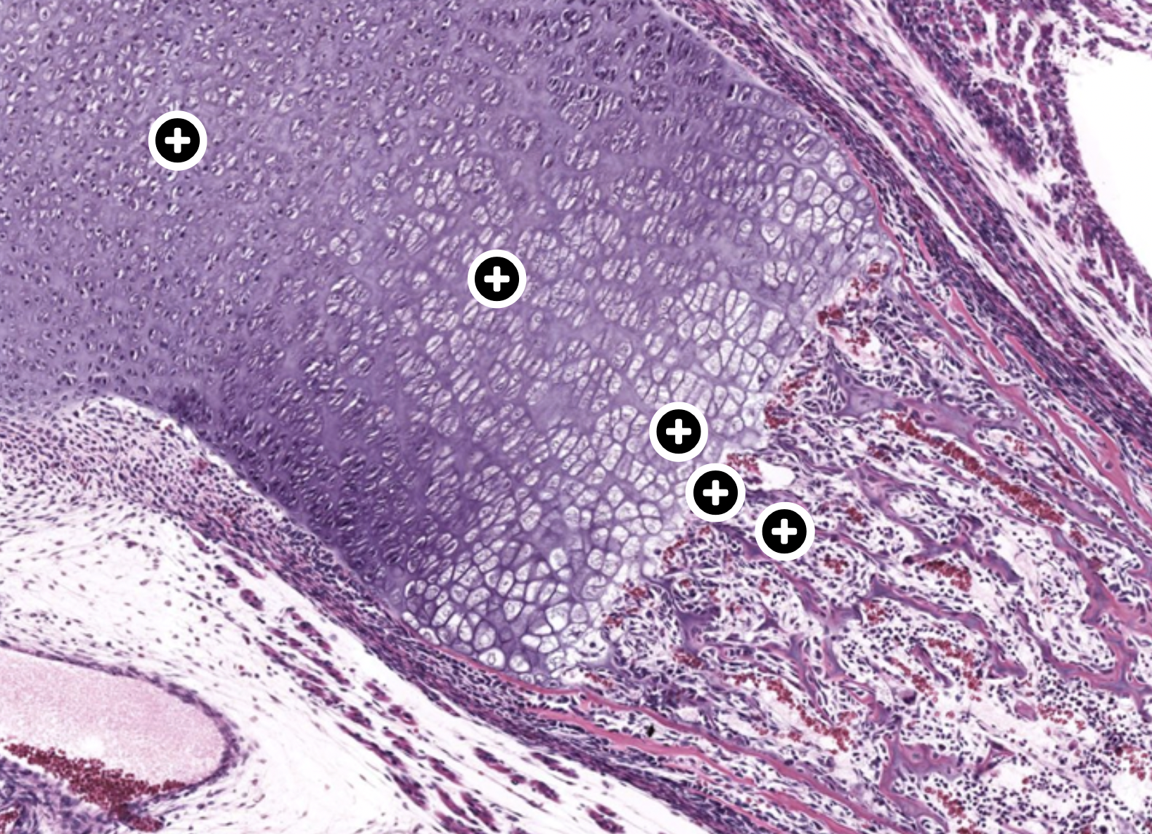
zone of reserve—chondrocytes quiescent
zone of proliferation— chondrocytes rapid cell cycle and growth, stacks of small dinner places
zone of hypertrophy— chondrocytes mature (very large lacunae stacks)
zone of calcifying cartilage— chondrocytes die!!! explosions
zone of ossification— endochondral bone formation
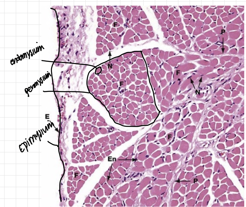
skeletal muscle connective tissue
skeletal muscle: epimysium (dense irregular CT)
muscle fascicle: perimysium (dense irregular CT)
muscle fibre: endomysium (loose CT)
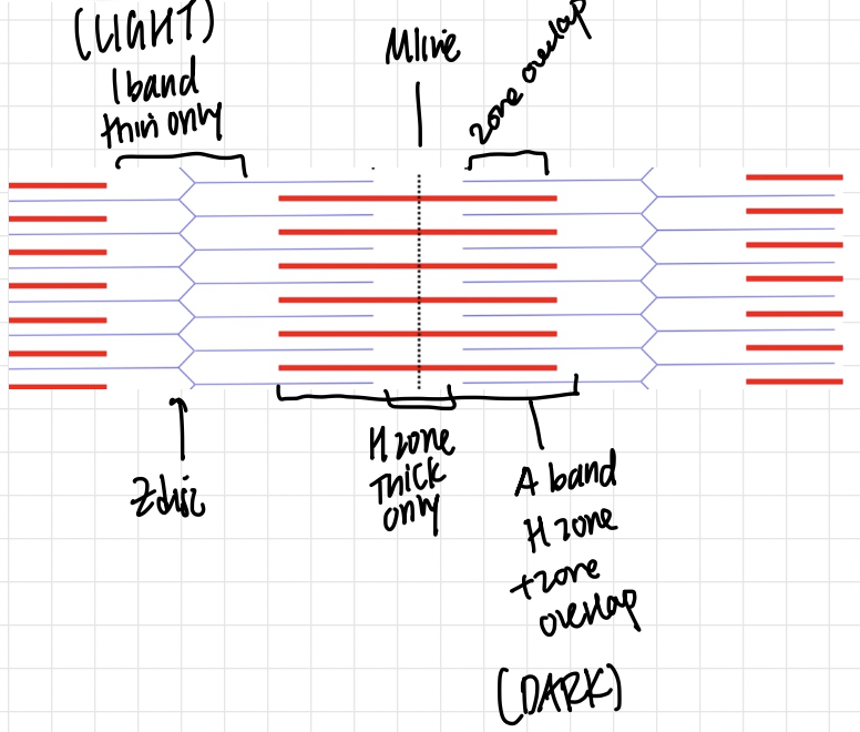
LIGHT: I BAND thin actin only
DARK: A BAND thick H zone only + overlap
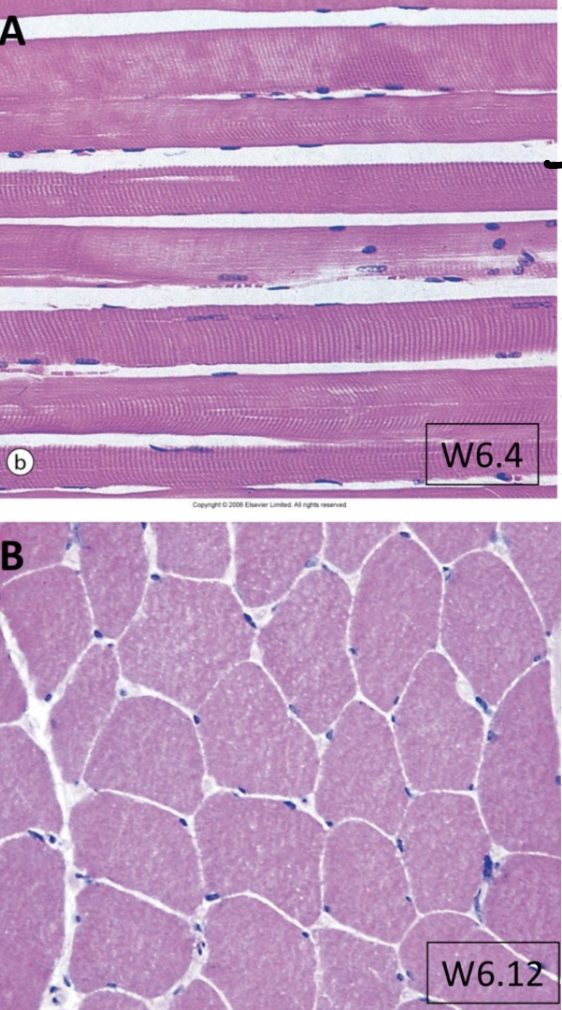
skeletal muscle fibre
Cross section: many nuclei PERIPHERAL! large fibres that are angular in shape
Myosatellite cells
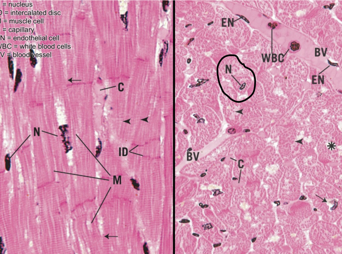
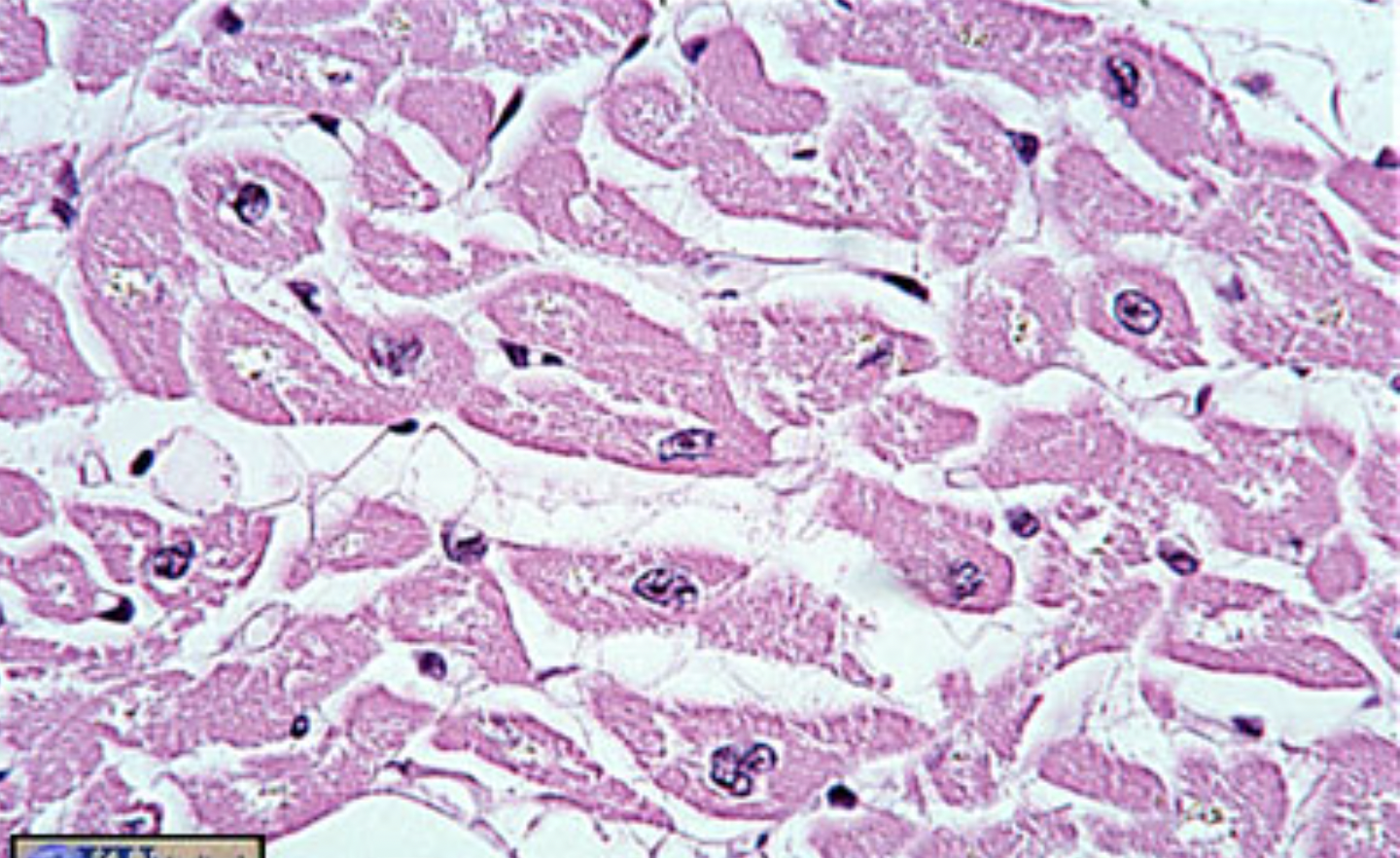
cardiac muscle
Cross section: CENTRAL nucleus sometimes 2, more endomysium than skeletal
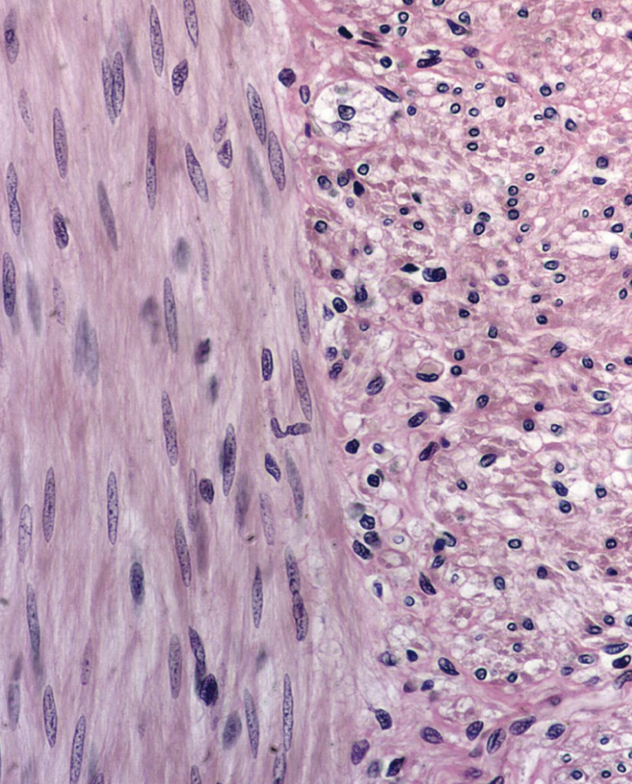
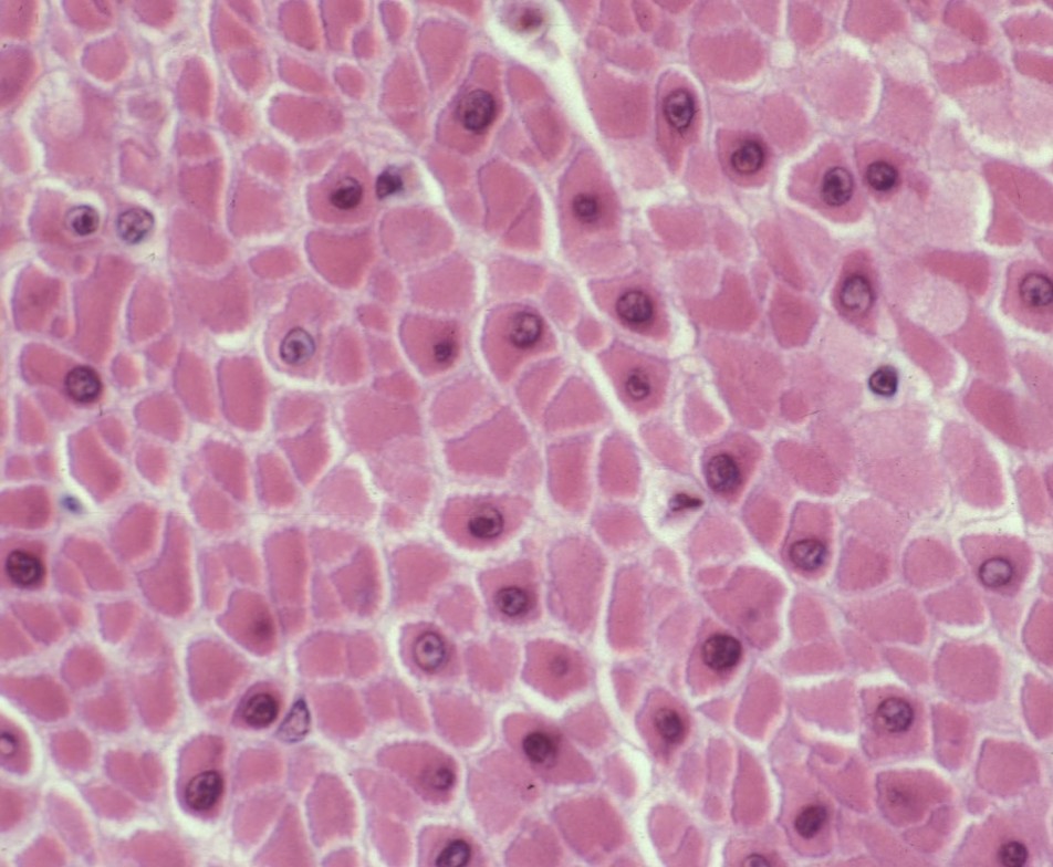
smooth muscle
Cross section: SINGLE central nucleus, some cells no nucleus, smallest cells
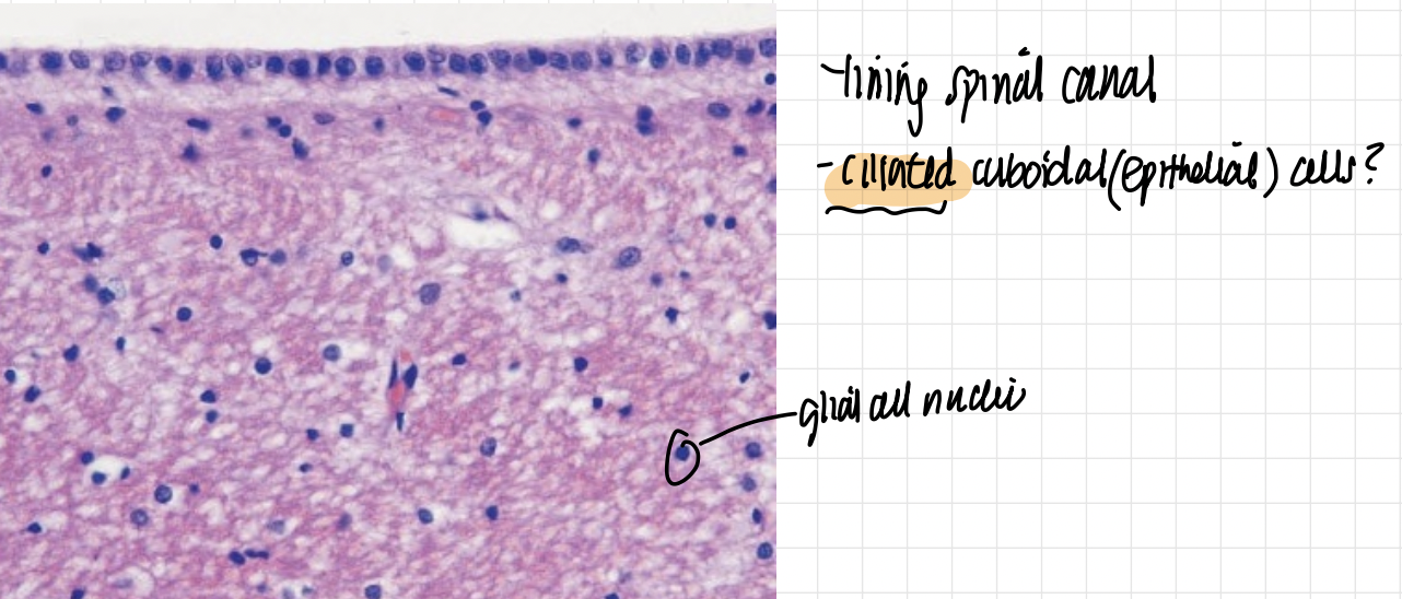
ependymal cells in CNS (glia) — ciliated cuboideal cells?
and glial cell nuclei
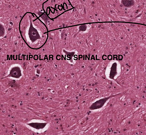
MULTIPOLAR CNS neuron (nissl substance RER very basophilic)
neuropil very dark pink background (glial cell nuclei)
no satellite cells
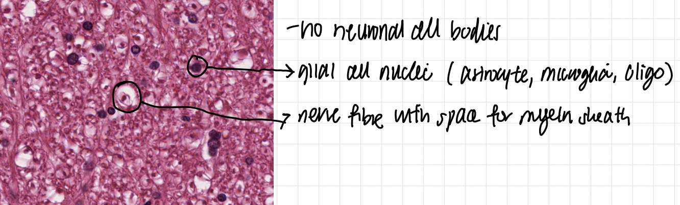
no neuronal cell bodies!!
dark things are glial nuclei (astrocyte, microglia, oligo)
also nerve fibre with space for myelin sheath by oligo
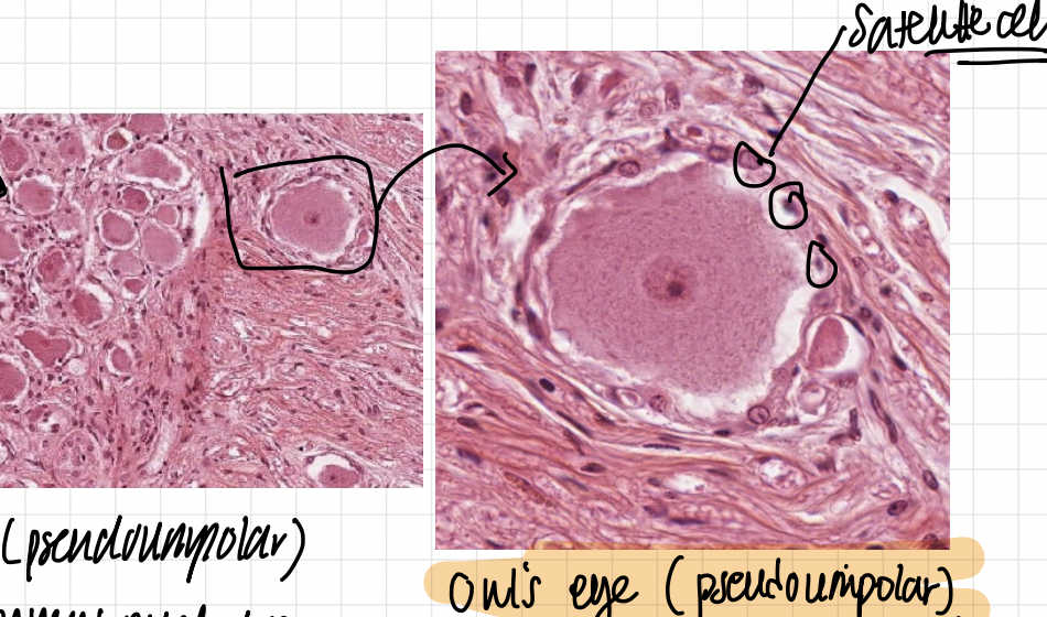
PNS SENSORY GANGLIA (GREY MATTER) PSEUDOUNIPOLAR
owls eye ROUND CELL BODY with many many satellite cells around in complete ring
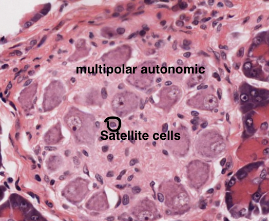
PNS autonomic ganglion (GREY MATTER) MULTIPOLAR
irregular cell bodies with less satellite cells
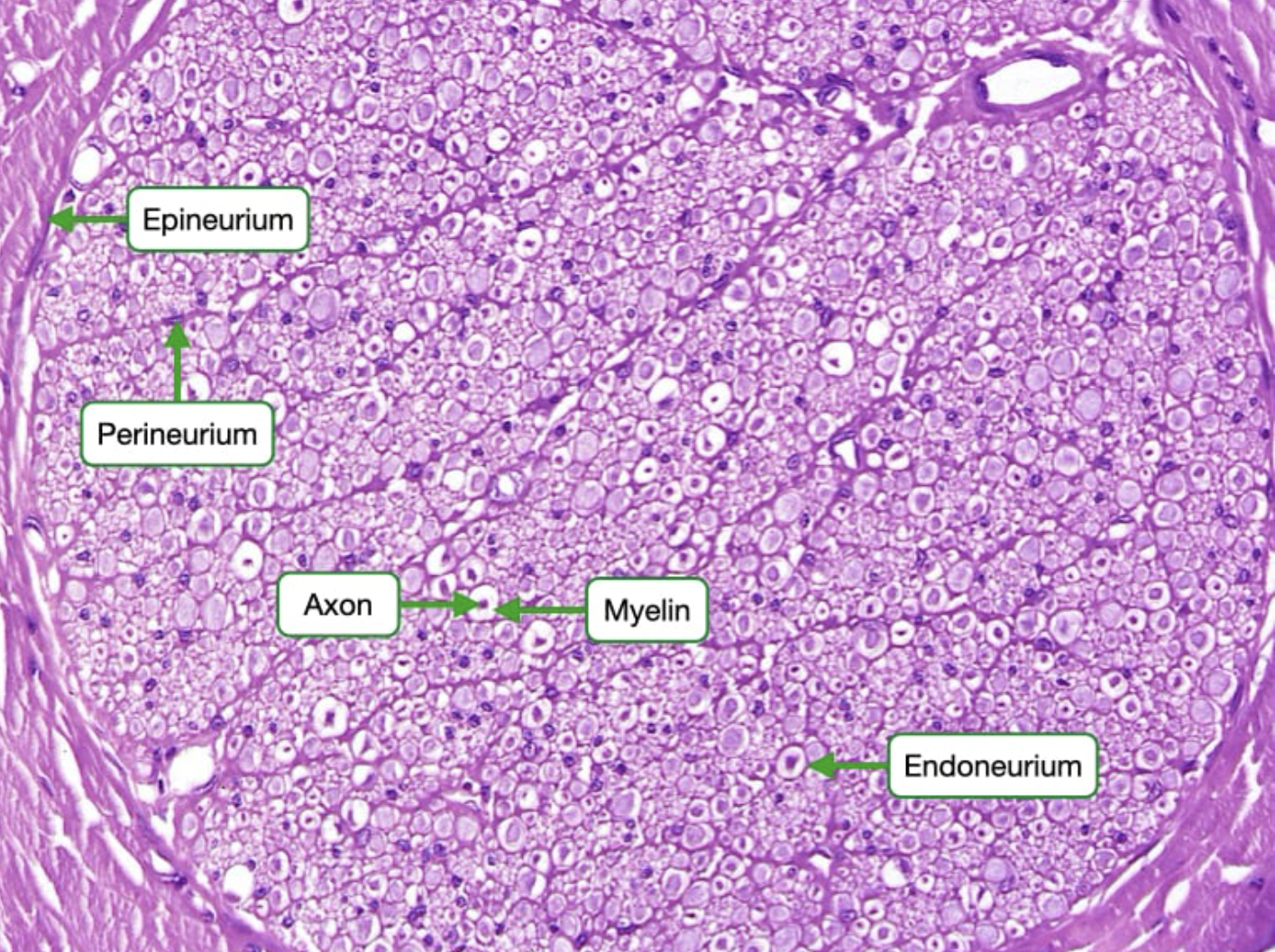
PNS white patter peripheral nerve
endoneurium, perineurium, epineurium
axons are the dark dots in the middle
the white spaces are myelin empty, the side black dots are schwann cell nuclei
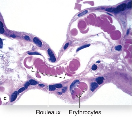
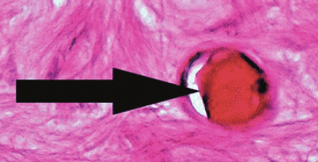
capillary longitudinal and cross section
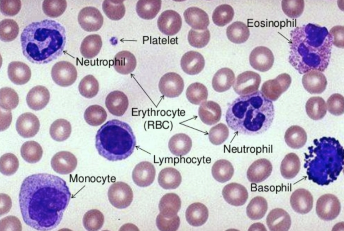
neutrophils (3-5 lobes, 50-70% WBC) phagocyte bacteria
eosinophils (bilobed, 2-4% WBC), parasites and allergy, AB-AG
basophils (S shape, <1% WBC), inflammation, allergy
monoctye (kidney shape)
lymphocyte: little cytoplasm, only WBC recirculate, 20-30% diff into b,t,nk
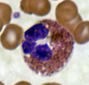
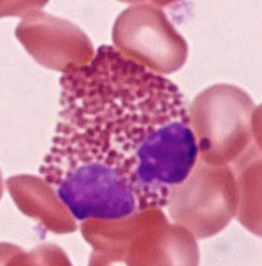
eosinophil (bilobed parasite, allergy)
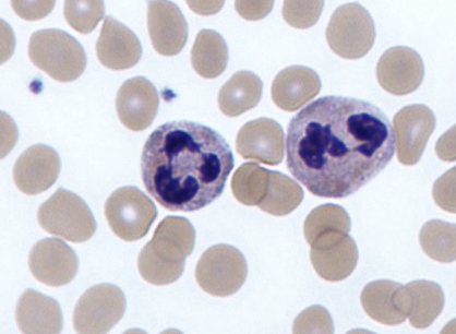
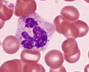
neutrophil (3 -5 lobes), bacteria phagocytose
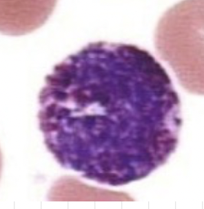
Basophil, s shape, inflammation, <1%
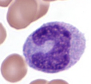
monocye, kidney shaped diff into macrophage
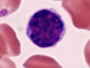
lymphocyte, only recirculating, diff into b t nk
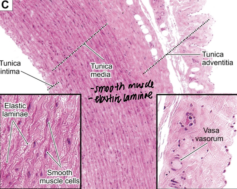
LARGE elastic arteries (aorta, brachiocephalic trunk, carotids)
tunica media has abundant elastin in fenestrated sheets to modulat BP
Cant see IEL and EEL due to elastin sheets
tunica adventitia has lots vasa vasorum (BV to supply o2)
parts of artery/vein
tunica intima: endothemium, lamina propria, IEL
tunica media: smooth muscle, elastic fibres, EEL
tunica adventitia/externa: dense irr CT, vasa casorum
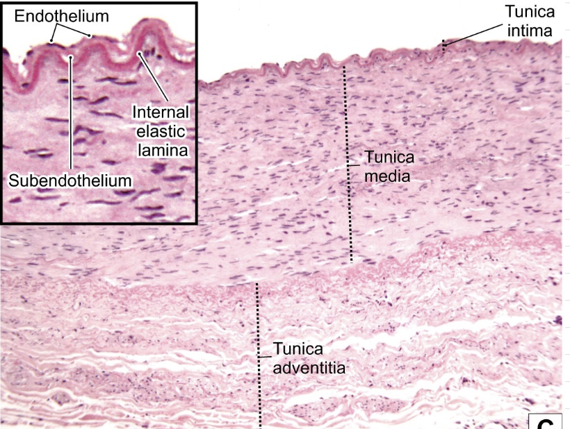
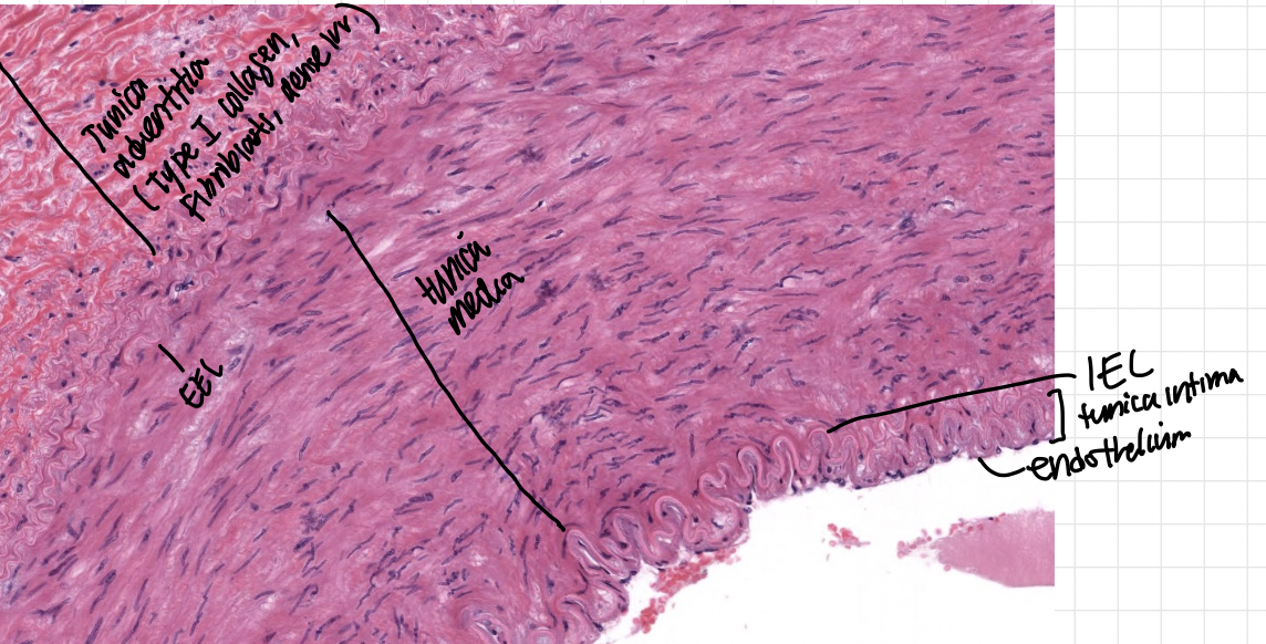
muscular artery
Tunica media has more smooth muscle, less elastic sheets
can see IEL and EEL
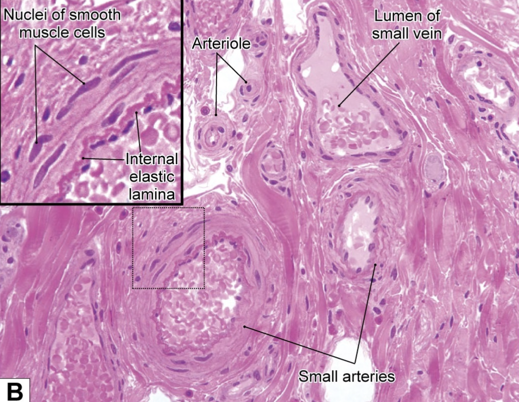
small arteries
3-6 laters smooth muscle
sometimes IEL, NO EEL
arteriole
1-2 layer smooth muscle
NO IEL EEL
thin ill defined tunica externa
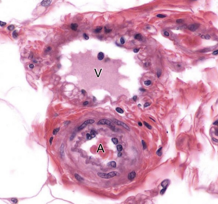
artery: thicker tunica media, smaller lumen
vein: Thicker tunica adventia, larger lumen
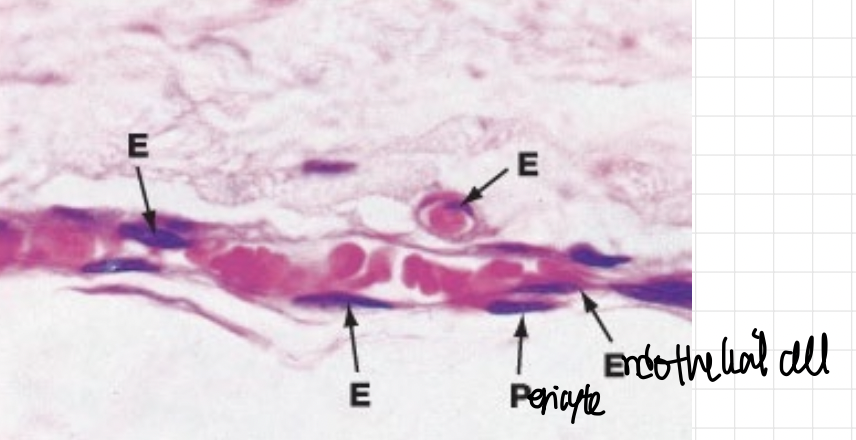
capillary has endothelial cells and pericytes mmmmmmmmmmmmmmmm
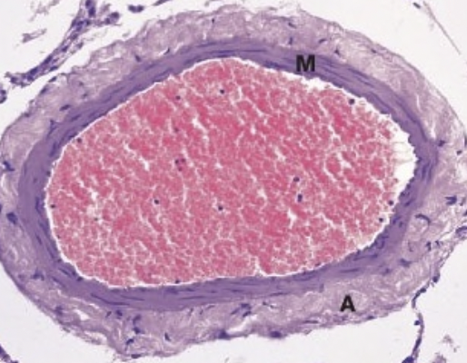
medium vein— companion to muscular artery
thinner media, thicker adventitia
veins
post capillary venule: endothelial + BM
muscular venules: thin media, thin externa
medium vein: companion to muscular artery, thinner media, thicker adventitia
larger veins: thinner media, thicker adventitia,
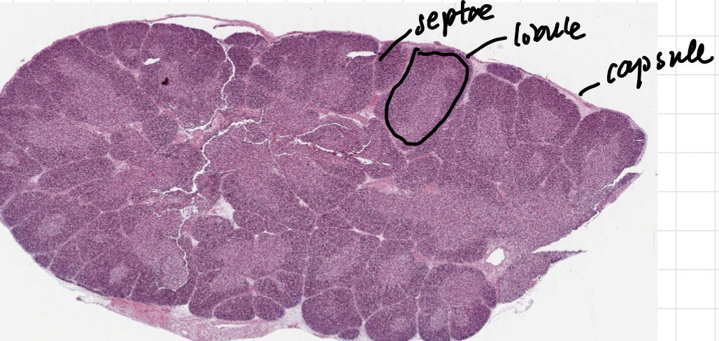
thymus
septae
lobule
capsule
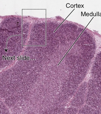
thymic cortex: darker staining with many lymphocytes (95% die in positive selection—bind MHC), epithelioreticular cells and macrophages
Medulla: lighter staining, fewer lymphocytes (5%) negative selection—bind self?, also epithelioreticular cells, T cells go thru bloodstream into blood
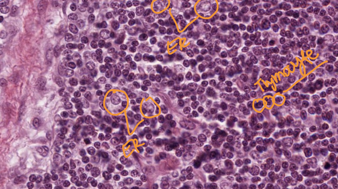
Thymic cortex
epithelioreticular cells: LARGER lighter euchromatic nucleus, joined by desmosomes to form stroma (flattened epithelioreticular cells at corticomedullary junction isolate the cortex from blood-thymus barrier
Thymocyte cells have smaller darker nucleus
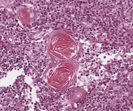
thymic medulla has hassalls corpuscles (concentric epithelioreticular cells w keratin
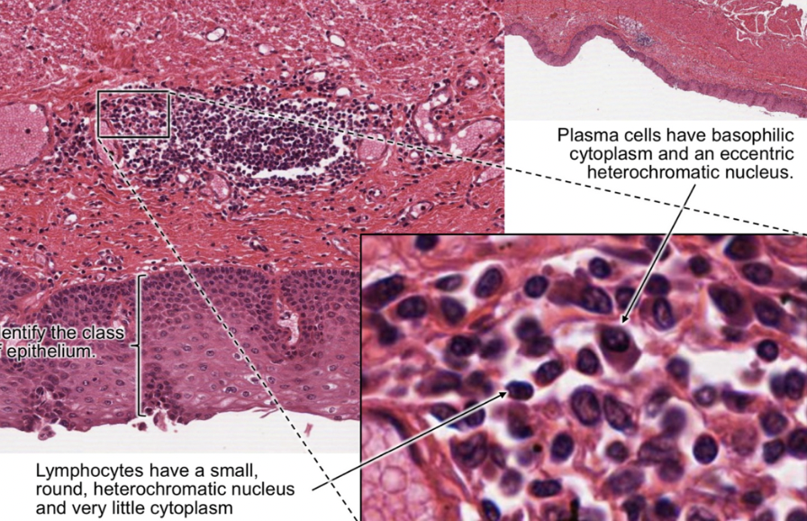
secondary lymphatic organs— MALT, lymph nodes, spleen
mucus membrane= open to exterior= epithelia+lamina propria (loose CT) + MALT
the lymphocytes have small round heterochromatic nucleus little cytoplasm
the plasma cells have eccentric nucleus and more cytoplasm
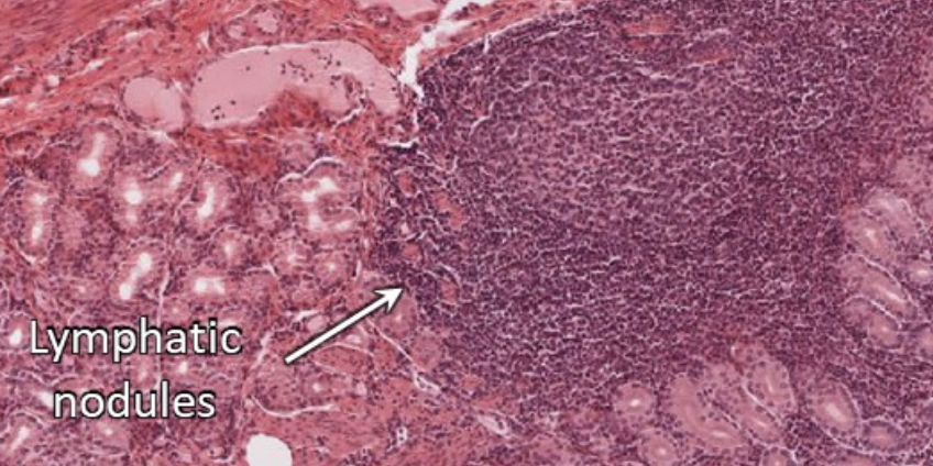
secondary lymph nodules
the mantle zone is naive lymphocytes are (DARKER)
the germinal center is where activated proliferating lymphocytes are, with more cytoplasm LIGHTER
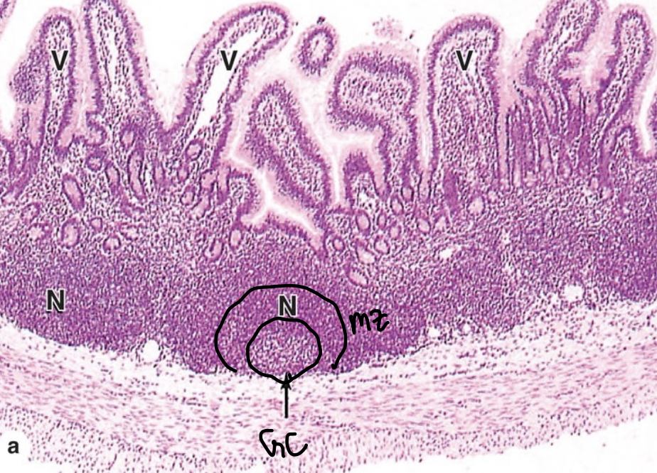
the mantle zone is darker
the germinal centre is lighter
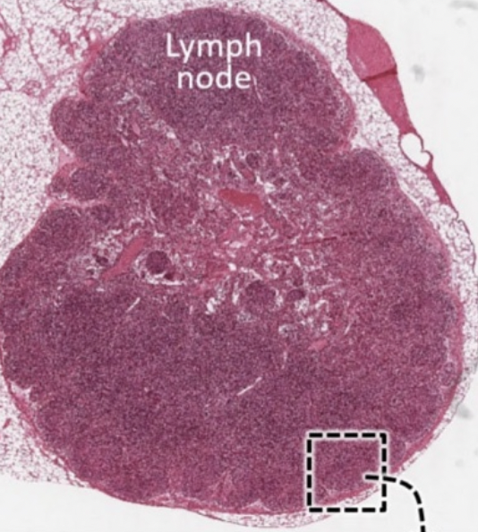
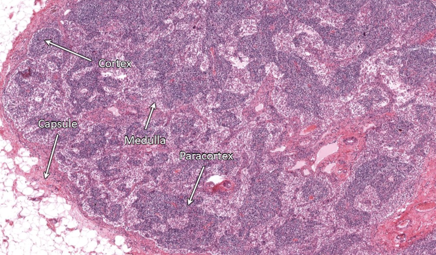
lymph node cortex
b cells form nodules— with mantle zone and germinal center(where they proliferate)
afferent lymph passes subcapsular sinus to trabecular sinus and macrophages detect and process ag to T cells in the paracortex
b cells in cortex will proliferate to plasma cells in the GC of the nodules
plasma cells migrade via the medullary cords to release Ab via efferent lymph
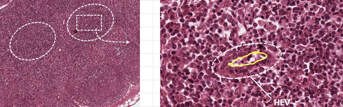
paracortex where T cells are, cuboidal endothelium
have HEV so circulating lymphocytes can bind and cross into lymph of deep cortex
activated T cells proliferate and go into the system via efferent lymph
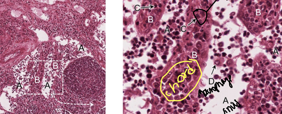
medulla
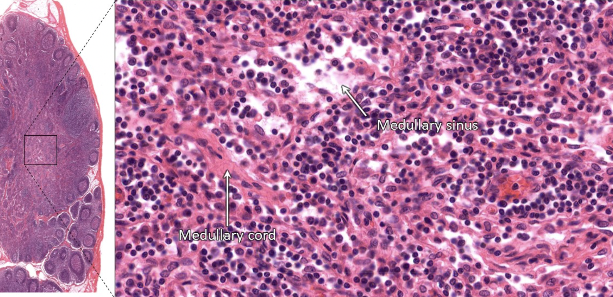
has medullary sinuses (white things) seperated by medullary chords (pink things)
medullary chords have b cells, plasma cells and macrophages
plasma cells from the germinal center, and the ab they produce leave via efferent lymph
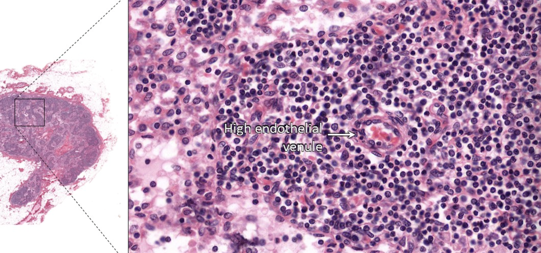
in paracortex HEV
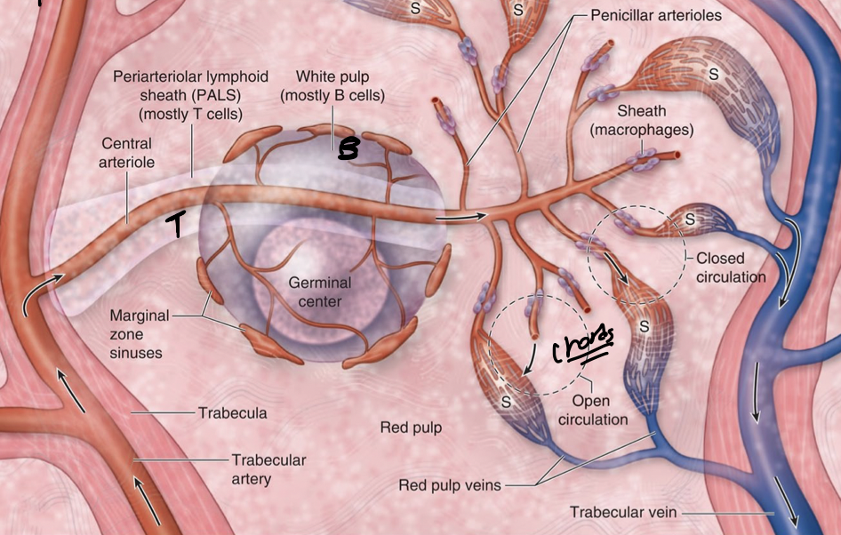
white pulp: dominated by lymphocytes, APC, immune response to blood AG, blood from splenic artery flows into central arterioles
-marginal zone sinuses b&t cells interact with ag/apc
T cells and macrophages in periarteriolar sheath (like paracortex of lymph node)
B cells in peripheral white pulp where activated follicles seen (like cortex of lymph node)
red pulp:
splenic sinusoids (blood filled space) between chords
splenic chords made of reticular fibres, lymphocytes, PC
WBC, RBS< platelet pass chords into the sinusoids
white pulp central A becomes penincellar A of red pulp, sheathed by macrophages to catch escaping Ag
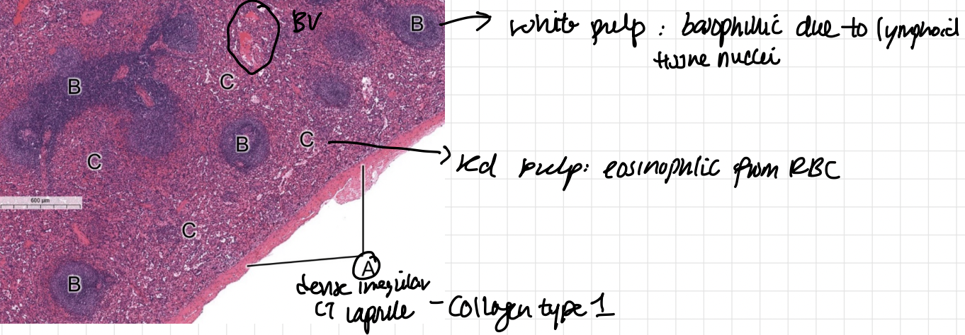
white pulp: basophilic and darker due to lymphoid tissue nuclei
red pulp: eosinophillic from RBC, lighter
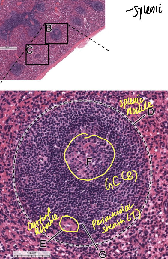
white pulp
have splenic nodules with inside GC B cells and outside periarteriolar sheath T cells (contains central arteriole)
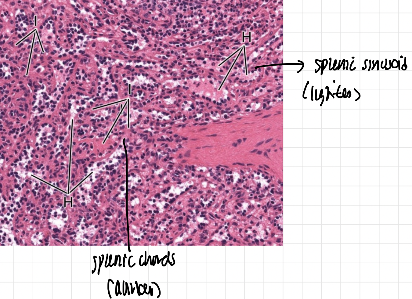
splenic sinuses are lighter (lots of open spaces and plasma)
splenic chords are darker (lots of WBC, macrophages, PC, reticular cells
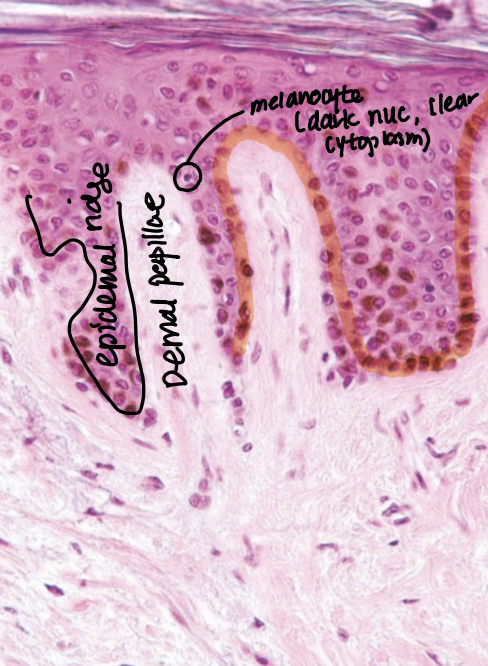
stratum basale/germinativum
have keratinocytes, merkle cells (thick), melanocytes—>spinulosum
connect via produced tonofilaments
epidermal ridge (down), dermal papilla (up) ›
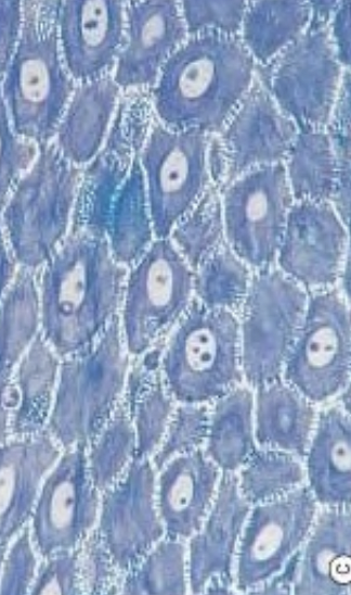
stratum spinosum— langerhan cells
connected via desmosomes to give spiny appearance
keratohyalin granules
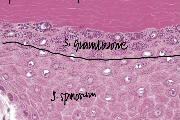
stratum granulosum
flatten granule cells with lamellar granules to make epidermal water barrier
soft keratinization turns granules into cornified cells
tonofilaments into tonofibrils
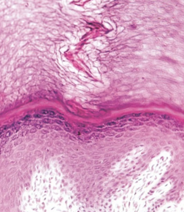
stratum lucidum— thick skin only
flatten protein packed cells
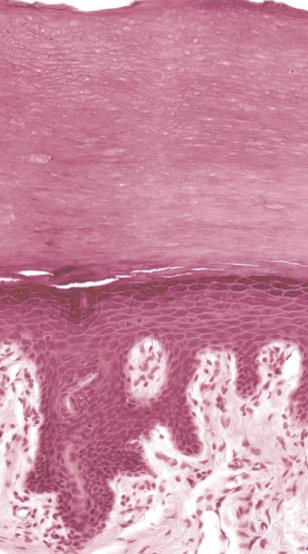
stratum corneum
soft keratin with tonofibrils
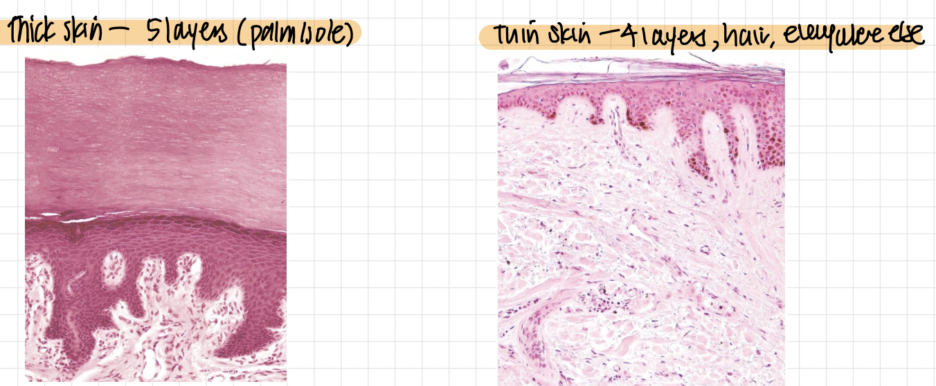
thick skin has 5 layers and merkle, hairless
thin skin has 4 layers, no merkle, hair
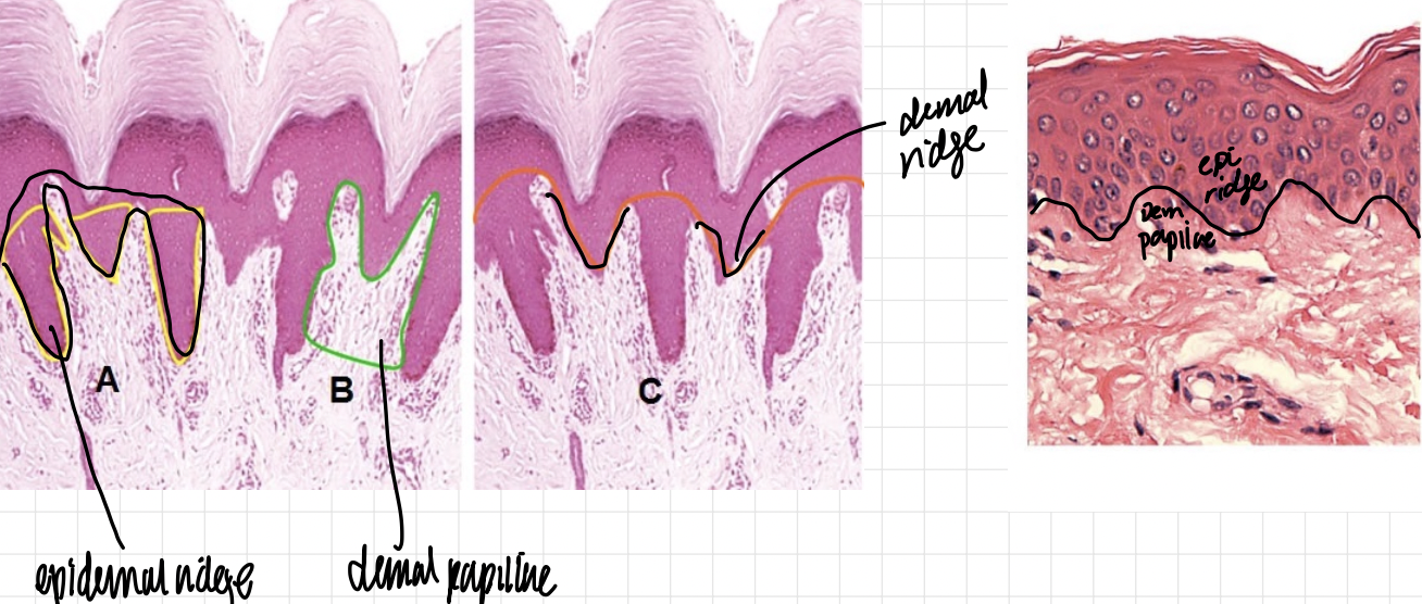
epidermal ridge—fingerprint/ dermal ridge (thick) goes down
dermal papillae go up
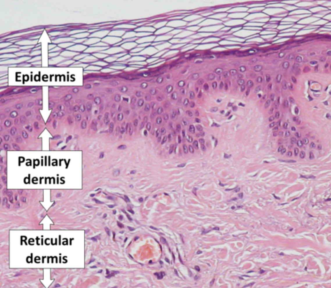
dermis:
1. papillary layer of loose CT (makes dermal papillae)
reticular layer with dense irregular CT
3. blood supply
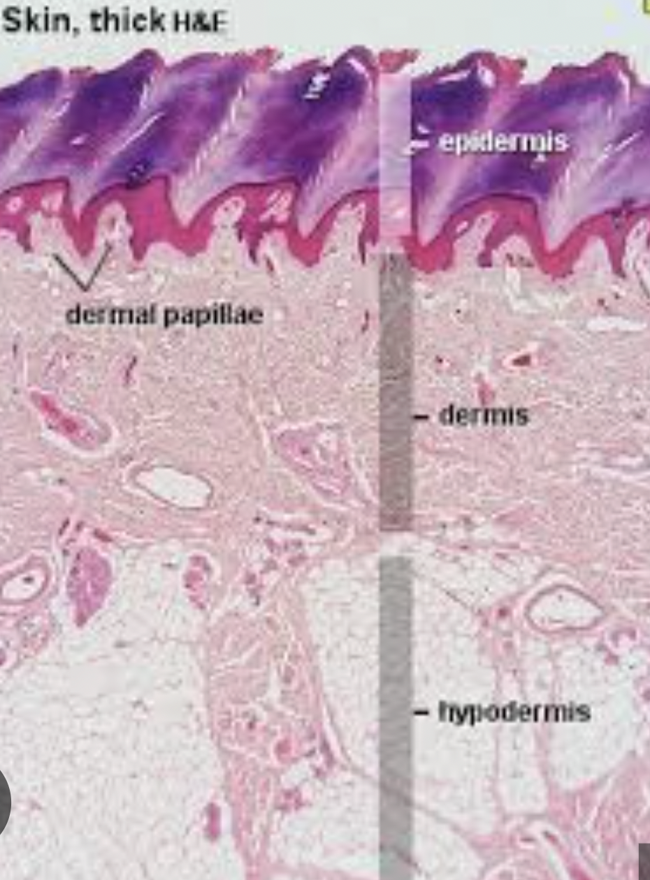
hypodermis/subcutaneous layer
mostly loose areolar to adipose tissue
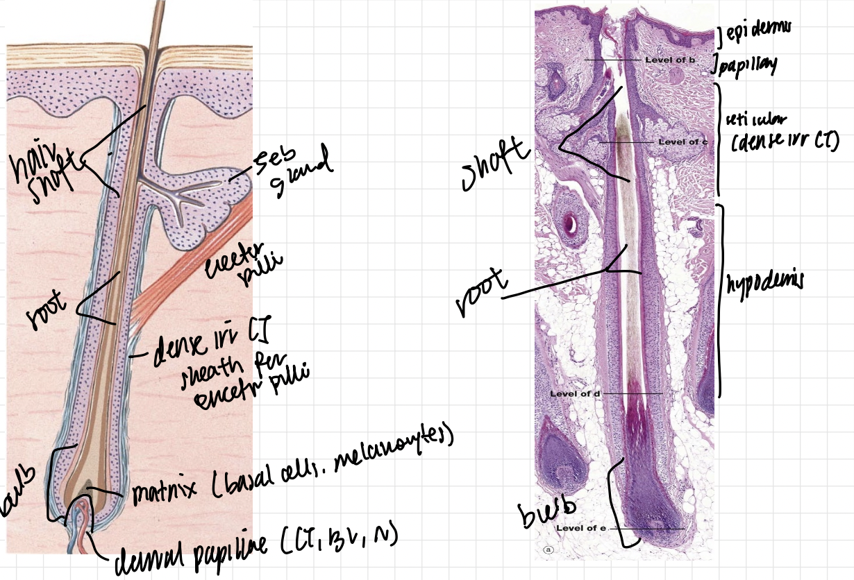
hair shaft: dermis
hair root: hypodermis
hair bulb: hypodermis
matrix—basal cells and melanocyte, dermal papillae (CT, BV, N)
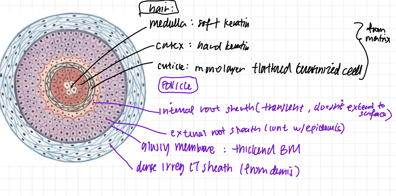
from matrix hair:
inside medulla: soft keratin
cortex: hard keratin
cuticle: monolayer flattened kertinized cell
follicle:
internal root sheath: does not extend to surface
external root sheath: continuous with epidermis
glassy membrane: thickened BM
dense irregular CT sheath from dermis
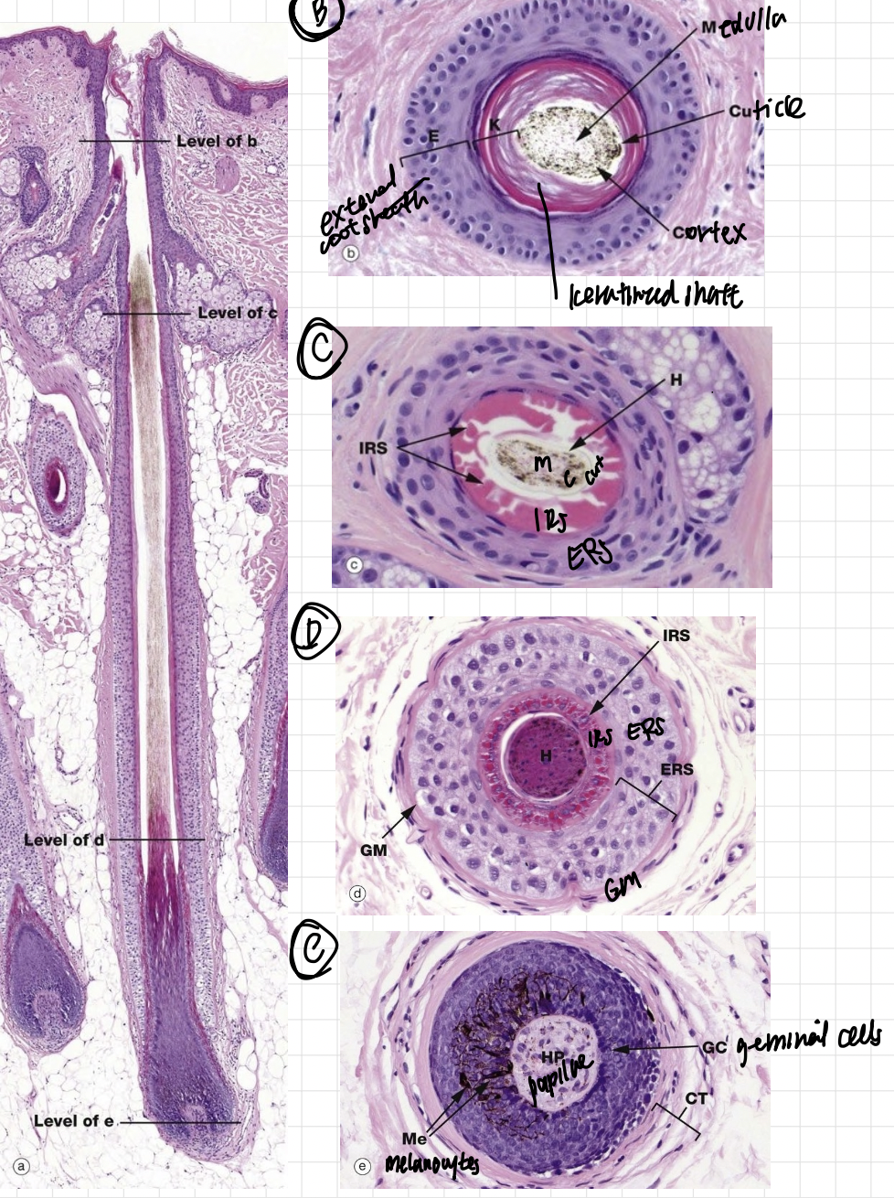
IRS: very eosiniphillic and pink, but not the liney one thats the kertinized shaft
ERS: much lighter
Glassy membrane then the dermis
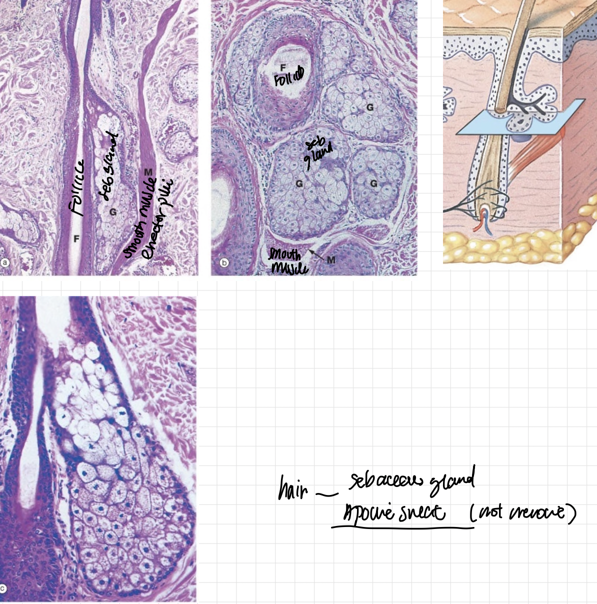
sebaceous glands are simple alveolar to simple branched alveolar glands
use holocrine whole cell secretion
erector pilli promote secretion
hair: sebacous glands and aprocrine sweat glands!!
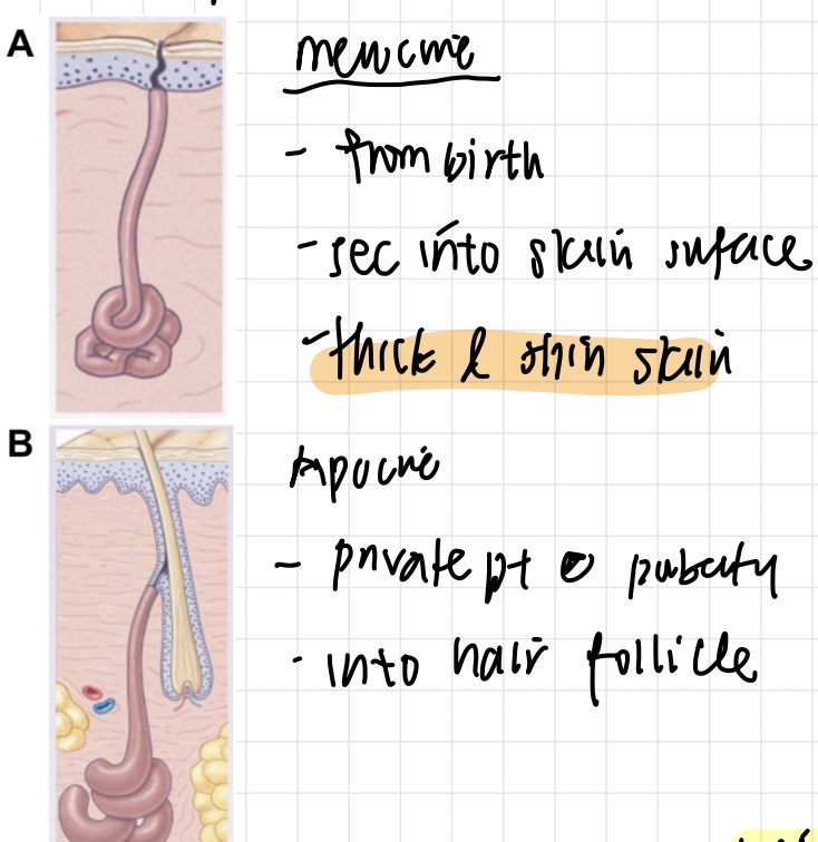
merocrine sweat gland (merocrine secretion)— watery
from birth and secrete onto surface of skin in both thick and thin skin
apocrine sweat gland (merocrine secretion)— protein rich
only in certain areas of thin skin that activate during puberty like armpits and groin and secrete INTO the hair follicle!
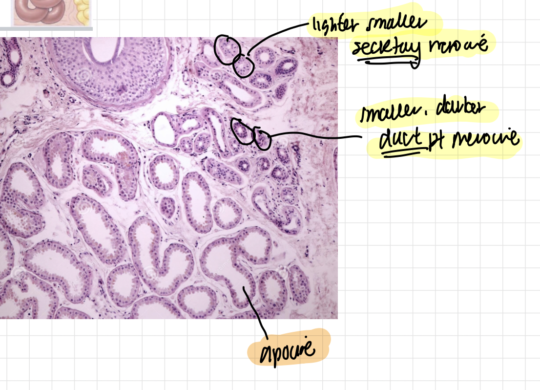
merocrine sweat glands (birth, thick, thin) are smaller!!!
the darker small ones are the duct and the lighter small ones are the secretory part
apocrine sweat glands (puberty, thin) are much larger!!!!