Abnormal findings and their indications - neurological examination
 Call Kai
 Learn
Learn Practice Test
 Spaced Repetition
 Match
Match Flashcards
 Knowt Play
1/52

Earn XP
Description and Tags
An overview of abnormal findings in the neurological examination, and whether they point to a muscle, NMJ, LMN, or UMN pathology
Name | Mastery | Learn | Test | Matching | Spaced | Call with Kai | Chat |
|---|
No analytics yet
Send a link to your students to track their progress
53 Terms
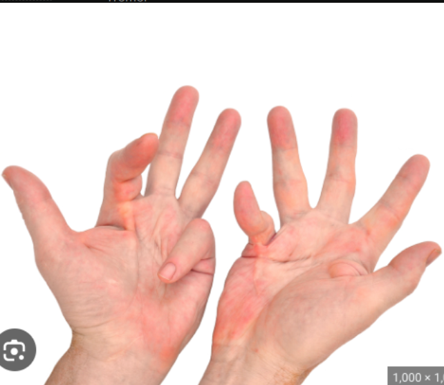
Contractures
A permanent tightening of the muscles, tendons, skin, and nearby tissues that causes the joints to shorten and become very stiff.
What scars should you be looking out for?
Spinal
Axillary
Upper limb
Lower limb
Indicates there may have been surgery / trauma there.
Involuntary movements
Pseudoathetosis - Abnormal slow writhing movements (typically affecting fingers) caused by a failure of proprioception.
Chorea - brief, semi-directed, irregular movements that are not repetitive or rhythmic but appear to flow from one muscle to the next. (Huntington’s disease)
Myoclonus - brief, involuntary, irregular twitching of a muscle or group of muscles (persistent and widespread - potentially epilepsy).
Tardive dyskinesia - Involuntary, repetitive body movements which can include protrusion of the tongue, lip-smacking, and grimacing (can develop secondary to neuroleptic medications).
Fasciculations
Involuntary rapid muscle twitches that are too weak to move a limb but are easily felt by patients and seen or palpated by clinicians. E.g eye twitch. Associated with LMN pathology. (e.g. ALS)
What units are used to describe tremors?
Hertz
Hertz
Unit of frequency - equal to 1 cycle per second
Low frequency tremors
Slow tremors (2-4 Hz), like an intention tremor
High frequency tremors
Fast tremors (14-20 Hz), like essential tremors
Different types of tremors to be aware of
Intention tremor
Essential tremor
Resting tremor
Pill rolling tremor
Intention tremors
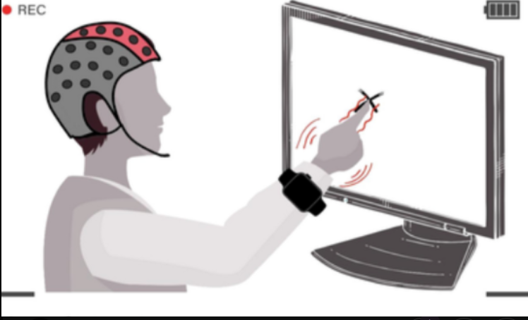
Low frequency tremor that occurs during an a direct and purposeful movement.
Essential tremor
A neurological condition that causes the hands to shake rhythmically (high frequency).

Resting tremor
Tremor that occurs when the affected body parts are at rest. It tends to be slow, and can be stopped by intentionally moving the affected part of the body.
Pill rolling tremor
A resting tremor of median amplitude (4-6 Hz) that involves rhythmical movement of the finger and thumb which looks like the patient is trying to roll a pill. It is characteristic of Parkinson’s disease.

Increased tone (hypertonia)
There is too much muscle tension meaning limb are hard to move. It can be categorised into spasticity and rigidity.
Hypotonia
Lack of muscle tone - movements of the joints are abnormally floppy and loose (more common in paediatric populations).
Clonus
Clonus is a series of involuntary rhythmic muscular contractions and relaxations that is associated with upper motor neuron lesions of the descending motor pathways (e.g. stroke, multiple sclerosis, cerebral palsy).
UMN pathology
Pathology affecting the brain or spinal cord.
LMN pathology
Pathology affecting the nerve roots. peripheral nerves, neuromuscular junction, or muscle.
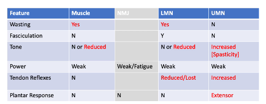
Overview of the interpretation of the motor examination in the limbs and trunk
Spasticity Vs Rigidity
Both describe increased tone (hypertonia).
Spasticity:
Pyramidal tract lesions (e.g. stroke)
Velocity dependent (faster you move the limb, the worse it is)
Worst in the first part of the movement (clasp knife spasticity)
Typically accompanied by weakness
Rigidity:
Extrapyramidal tract lesions (e.g. Parkinson’s)
Velocity independent
Cogwheel rigidity (superimposed tremor results in intermittent increases in tone) and lead pipe rigidity (uniform)
How is muscle power rated?
MRC muscle power assessment scale from 0-5

What is an abnormal response when assessing for ankle clonus?
Clonus is felt as rhythmic beats of dorsiflexion and plantarflexion. If more than 5 beats of clonus are present, this would be classed as an abnormal finding.
Babinski’s sign (plantar reflex)
Normal result: flexion of the big toe and flexion of the other toes.
Abnormal (Babinski sign): extension of the big toe and spread of the other toes (suggestive of an upper motor neuron lesion).
What clinical findings from the neurological examination indicate a patient has a muscle pathology?
Muscle atrophy
Normal / reduced tone
Weak power
Normal for everything else
What clinical findings from the neurological examination indicate a patient has a NMJ pathology?
Weak power / becomes weak with fatigue
Everything else is normal
What clinical findings from the neurological examination indicate a patient has a UMN pathology?
Increased tone (spasticity)
Weak power - pyramidal pattern
Increased tendon reflexes
Extensor plantar response
Ankle clonus
Everything else is normal (may be some disuse atrophy or contractures)
Pyramidal pattern - muscle power
Extensors are weaker than flexors in arms, and vice versa in legs.
What clinical findings from the neurological examination indicate a patient has a LMN pathology?
Muscle atrophy
Fasciculations
Normal / reduced tone
Weak power (focal patterns: proximal pattern in muscle disease, and distal in peripheral neuropathies)
Reduced / lost tendon reflexes
What can muscle atrophy indicate?
A muscle or LMN pathology (or disuse).
What can fasciculations indicate
LMN pathology
What can increased tone (spasticity) indicate?
UMN pathology
What can reduced tone indicate?
A muscle or LMN pathology.
What can reduced power indicate?
Muscle, NMJ, LMN or UMN pathology. Fatigue is quite specific to a NMJ pathology.
What does reduced / absent tendon reflexes indicate?
LMN pathology.
What do increased tendon reflexes indicate?
UMN pathology.
What does an extensor response to the plantar reflex indicate?
UMN pathology.
What nerve tracts does light touch (cotton wool) sensation involve?
The dorsal columns and spinothalamic tracts
What nerve tracts does the pin-prick (pain) sensation involve?
The spinothalamic tracts
What nerve tracts does vibration (tuning fork) sensation involve?
The dorsal columns
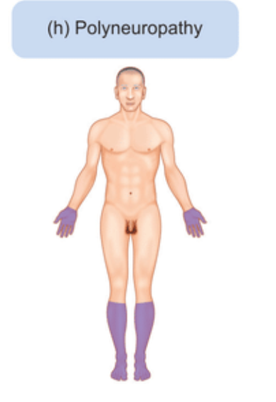
Stocking distribution of sensory loss
A pattern of sensory loss where the distal limb is more affected than the proximal limb - associated with peripheral neuropathy.
What nerve tracts does proprioception involve?
The dorsal columns.
Name the different patterns of sensory loss
Mononeuropathies
Peripheral neuropathy
Radiculopathy
Spinal cord damage
Thalamic lesions
Myopathies
Mononeuropathy (sensory loss pattern)
Localised sensory disturbance in the area supplied by the damaged nerve.
Peripheral neuropathy (sensory loss pattern)
Typically causes symmetrical sensory deficits in a ‘glove and stocking’ distribution in the peripheral limbs. The most common causes of peripheral neuropathy are diabetes mellitus and chronic alcohol excess.
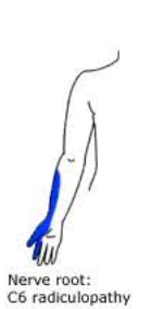
Radiculopathy (sensory loss pattern)
Occurs due to nerve root damage (e.g. compression by a herniated intervertebral disc), resulting in sensory disturbances in the associated dermatomes.
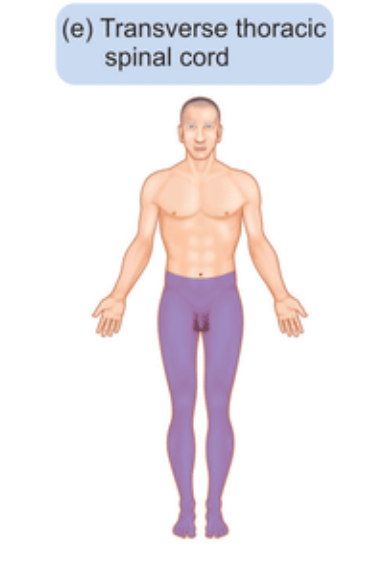
Spinal cord damage (sensory loss pattern)
This results in sensory loss both at and below the level of involvement in a dermatomal pattern due to its impact on the sensory tracts running through the cord.
Thalamic lesions (sensory loss pattern)
Result in contralateral sensory loss.

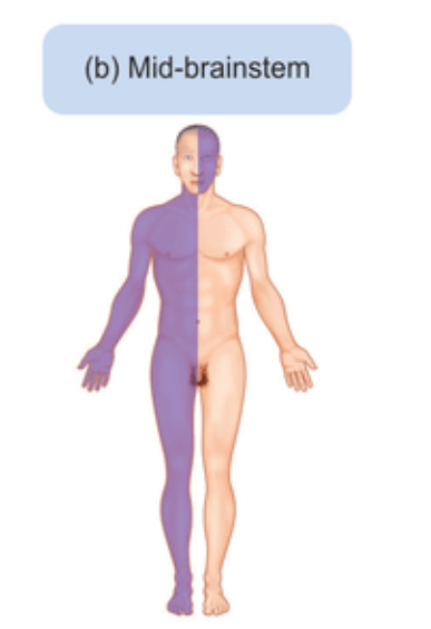
Mid-brainstem lesions (sensory loss pattern)
Results in ipsilateral sensory loss in the head, then contralateral sensory loss in the rest of the body.
Myopathies
Symmetrical proximal muscle weakness.
Interpretation of the finger-to-nose test
Dysmetria (incoordination) and an intention tremor present during this test is suggestive of ipsilateral cerebellar pathology.
Interpretation of the dysdiadochokinesia assessment
Dysdiadochokinesia is a term that describes the inability to perform rapid, alternating movements, which is a feature of ipsilateral cerebellar pathology. If the patient has this, their movements will be slow and irregular.
Interpretation of heel-to-shin test
Dysmetria - incoordination whilst attempting to perform this task is suggestive of ipsilateral cerebellar pathology. Muscle weakness can also produce apparent incoordination of this movement - so assess this before making conclusions!
What further investigations and assessments could be offered after the examination is complete?
Full neurological examination including the cranial nerves and the other limbs.
Neuroimaging - MRI spine and head