UCF PCB 3703 Exam 4
 Call Kai
 Learn
Learn Practice Test
 Spaced Repetition
 Match
Match Flashcards
 Knowt Play
1/345

There's no tags or description
Looks like no tags are added yet.
Name | Mastery | Learn | Test | Matching | Spaced | Call with Kai | Chat |
|---|
No analytics yet
Send a link to your students to track their progress
346 Terms
The fundus of the stomach has _______ cells that secrete _______ __________ and ____.
-parietal cells
-intrinsic factor (aka gastric IF)
-HCL
What does intrinsic factor of the parietal cells do?
takes B12 to the ileum for absorption
What does HCL- of the parietal cells do?
conversion of pepsinogen to pepsin
Chief cells are in the ____ of the stomach and secrete _____________.
-body
-pepsinogen
What does pepsinogen do?
interacts with HCl to convert to pepsin
Goblet cells are in the ___________ of the stomach and secrete __________.
-body
-mucus
Mucus from goblet cells ________ the stomach lining from HCl and helps form _______ by moving digested food and nutrients down the stomach.
-protect
-chyme
Chief cells are also called ____________ cells.
zymogenic
G Cells are from the ______ of the stomach and produce _____________.
-body
-gastrin
What does gastrin do?
stimulates parietal cells for HCl- secretion.
The pylorus of the stomach has _______ cells that secrete ____________ , _________ and__________________.
-paracrine
-histamine, serotonin and somatostratin
What is mucous made of?
mucins
What does histamine from paracrine cells stimulate?
stimulate parietal cells to increase HCl secretion.
What is the difference between pepsinogen and pepsin?
pepsinogen is inactive, pepsin is active.
What does somatostratin of the paracrine cells do?
- in GI tract, inhibits other hormone secretion
(is also secreted by hypothalamus which inhibits the growth hormone, and also secreted by pancreas for inhibition of glucose secretion)
What is somatostratin mainly controlled by? (not paracrine)
D-cells
An excess of histamine causes what?
gastritis
What does serotonin of the paracrine do?
similar to histamine in the way it controls parietal cells to secrete HCl-
I cells in the __________ and ____________ of the stomach secrete __________.
-duodenum and jejunum
-cholecystokinin (CCK)
What does cholecystokinin do?
-inhibits premature gastric emptying by controlling the ileocecal sphincter
-relaxes ODDI sphincter (connected to gallbladder and pancreas) to release pancreatic enzymes (like HCO3-) and bile from gallbladder
-helps contract gallbladder to increase bile secretion
S cells in the __________ and _____________ of the stomach secrete ____________.
-duodenum and jejunum
-secretin
Secretin from S cells do what?
-helps stimulate pancreatic enzyme secretion
-partial control of G-cell activity (inhibits)
-helps stimulate pancreatic secretion of bicarbonate (HCO3-)
GIP (gastric inhibitory peptide) cells from the ________ and _____________ of the stomach does what?
-duodenum and jejunum
-stimulates insulin secretion from the pancreas and decreases gastric hydrogen ion secretion.
For the GI tract to provide the body with nutrition, it requires the following activities:
-movement (of food)
-secretion (of digestive juices)
-Absorption (of water and electrolytes)
-Circulation (of blood)
-Control (of functions by CNS and hormones)
The breathing cycle before inspiration (at rest)
-alveolar pressure equals atmospheric pressure
-intrapleural pressure is negative
-lung volume is the functional residual capacity
The breathing cycle during inspiration (inhale)
-inspiratory muscles contract and cause the thorax volume to increase, causing the alveolar pressure to decrease less than the atmospheric pressure, resulting in airflow inside lungs.
-intrapleural pressure becomes more negative
-lung volume increases by one tidal volume
The breathing cycle during expiration (exhale)
-alveolar pressure becomes greater than atmospheric pressure
-intrapleural pressure returns to normal
-lung volume returns to functional residual capacity
Asthma can be caused or "triggered" by...
1) allergies
2) congenital
3) bacterial/viral (leads to inflammation of bronchi in lungs)
Main difference between asthma and bronchitis (mentioned in class)
asthma has many different causes, but bronchitis is only caused by bacterial infection
main asthma treatment
Albuterol for relaxation of the smooth muscle by beta 2 receptor.
Asthma is when the patient has difficulty during expiration because...
the internal airway pressure is too high. Asthma is an obstructive lung disease.
Bronchitis treatment
a combination of antibiotic therapy, albuterol, and cortisol
Albuterol drug
is a bronchodilator that activates the beta2-adrenergic receptors in the lungs
COPD (chronic obstructive pulmonary disease) is what?
-air that should have been exhaled does not, and air becomes trapped in chest
-a combination of chorionic bronchitis and emphysema
-decreased forced vital capacity (FVC) and decreased forced expiratory volume (FEV1)
Emphysema main cause
destruction of alveoli wall which is important for gas diffusion which acts as surface area
-obstructive
-mild hypoxemia
Pink puffers (emphysema)
-What is it? What does the patient look like? What symptoms does that patient show?
patient has mild hypoxemia and normocapnia (normal pCO2, low pO2)
-patient has dyspnea (heavy breathing), pink appearance, difficulty exhaling, thin barrel-shaped chest.
Blue bloaters (bronchitis)
-What is it? What does the patient look like? What symptoms does that patient show?
severe hypoxemia and hypercapnia (low pO2 high pCO2)
-they have right ventricular failure and systemic edema
-patient looks cyanotic (blue), has hypercapnia (increased CO2 pressure), likely has edema in systemic circulation and a muscular barrel-shaped chest.
Fibrosis
-a restrictive disease with decreased lung inspiration and damaged lung tissue
-specifically caused by overproduction of collagen and elastic fibers
Hemoglobin difference between fetal and adult
fetal hemoglobin is able to bind oxygen with greater affinity than the adult form, giving the developing fetus better access to oxygen from the mother's bloodstream.
CO2 Transport (3 ways)
1) CO2 is dissolved in plasma
2) CO2 combines with hemoglobin and forms carbaminohemoglobin
3) 90% of CO2 is converted to bicarbonate
Zone 1 of lung
-blood flow is lowest
-alveolar pressure>arterial pressure>venus pressure
-high alveolar pressure can compresses capillaries and reduces blood flow in zone 1. This occurs as result of hemorrhage
Zone 2 of lung
-blood flow is medium
-arterial pressure > alveolar pressure > venous pressure
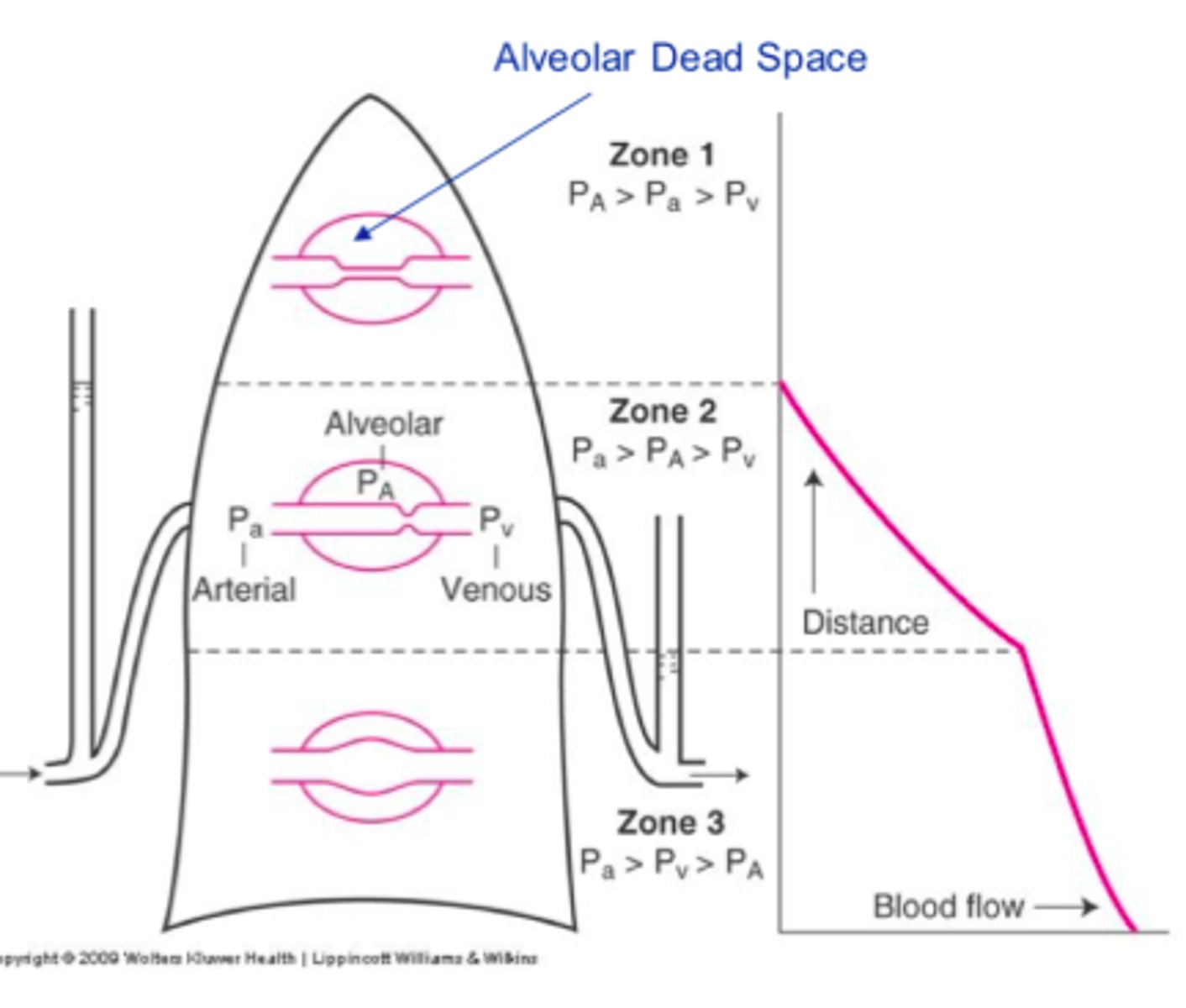
Zone 3 of lung
-blood flow is highest
-arterial pressure > venous pressure > alveolar pressure
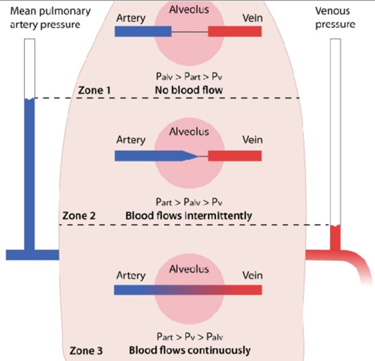
Hypoxia in lungs
the blood supply focuses on unaffected part of lungs to saturate with gas exchange, and then goes to the affected area of lungs. Vasoconstriction attempts to block the affected area
ventral respiratory group
-primarily responsible for expiration
-is not active during normal, quiet breathing, when expiration is passive
-is activated when expiration becomes active process (like exercise)
Apneustic center
-is located in the lower pons.
-stimulates inspiration, producing a deep and prolonged inspiratory gasp (apneusis).
pneumotaxtic center
-located in upper pons
-inhibits inspiration and regulates inspiratory volume and respiratory rate
alimentary tract
1) oral cavity
2) pharynx
3) esophagus
4)stomach (left side upper quadrant abdominal cavity)
5) duodenum
6) jejunum
7)ileum (connected to large intestine by ileocecal junction)
8)cecum
9)ascending colon
10)transverse colon
11) descending colon
12) sigmoid
13) rectum
14)anus
Wall of GI tract (4 layers from in to out)
1)mucus membrane
2)muscularis mucosa
3)muscle layer (circular and longitudinal muscle)
4)Serosa (adventita)
Innervation of smooth muscle
is by intrinsic nervous system meaning there are two plexus inside the wall of GI called submucosal plexus of meissner and myenteric plexus of auerbach
Submucosal plexus of Meissner and Myenteric plexus of Auerbach
-they control the relaxation and contraction of GI.
-They are controlled by the ANS
-Parasympathetic is more active, stimulation of PS causes movement of GI.
-Sympathetic decreases GI movement
Sympathetic and Parasympathetic efferent fibers
carry info from brainstem and spinal cord to GI tract
Sympathetic and Parasympathetic afferent fibers
carry sensory info (chemoreceptors, mechanoreceptors, etc.) from GI to brainstem and spinal cord
Sympathetic innervation of GI tract originates from where
spinal cord coming via abdominal splanchnic nerves (T5-L2)
Parasympathetic innervation of GI tract originates from where
Vagus nerve and pelvis splanchnic nerve (S2-C4)
Contraction of circular muscle in GI leads to
decrease in diameter of that segment of the GI tract
Contraction of longitudinal muscle of GI tract leads to
decrease in length of that segment of the GI tract
Phasic contraction is found in
esophagus, gastric antrum, and small intestine
-contracts and relaxes periodically
Tonic contraction is found in
lower esophageal sphincter, the orad stomach, and the ileocecal and internal anal sphincters
slow wave
-weak contraction of GI
-only reaches threshold line and cant past it for action potential
-weak depolarization and repolarization
Spike potentials are...
When GI tract receives stimulus a slow wave is converted to spike potential, which is a strong action potential that passes threshold
spike potentials are caused by (3 causes)
1) neurology (stimulation of CN10)
2) gastric (food was eaten)
3) hormonal (hormones were secreted)
tidal volume
is the volume inspired or expired with each normal breath
inspiratory reserve volume
-volume that can be inspired over and above tidal volume
-ex: exercise
expiratory reserve volume
volume expired after expiration of a tidal volume
residual volume
The volume of air remaining in lungs after maximum exhalation.
dead space
-anatomic: volume of the conduction airways
-physiologic: volume of the lungs that does not participate in gas exchange
What is the most important muscle for inspiration and what is it innervated by?
diaphragm
C3-C5 and the phrenic nerve
"3, 4, 5 keep the diaphragm alive"
Lung compliance in emphysema
lung compliance is increased and the tendency of the lungs to collapse is decreased
lung compliance in fibrosis
lung compliance is decreased and the tendency of the lungs to collapse is increased
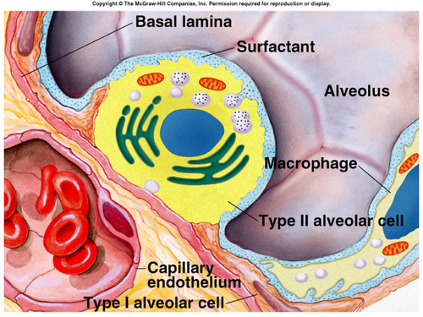
Surfactant does what?
-reduces surface tension, preventing alveoli from collapsing
-increases lung compliance
surfactant consists primarily of
dipalmitoyl phosphatidylcholine (DPPC)
surfactant is where in the lungs?
lines the alveoli
medullary respiratory center
-located in the reticular formation
1) dorsal respiratory group: inspiration
2) ventral respiratory group: expiration
An infant born prematurely in gestational week 25 has neonatal respiratory distress syndrome, what is the cause of this?
collapse of the small alveoli
In which vascular bed does hypoxia cause vasoconstriction?
pulmonary
A 12 year old boy has a severe asthmatic attach with wheezing. He experiences rapid breathing and becomes cyanotic. His arterial PO2 is 60mmHg and his PCO2 is 30mmHg.
What does this mean?
His arterial PCO2 is lower than normal because hypoxemia is causing him to hyperventilate
Normal PCO2
40 mmHg
Normal PO2 range
80-100 mmHg
A 12 year old boy has a severe asthmatic attach with wheezing. He experiences rapid breathing and becomes cyanotic. His arterial PO2 is 60mmHg and his PCO2 is 30mmHg.
Treatment should be...?
a beta 2 adrenergic agonist which is a bronchiodilator, such as albuterol
What volume remains in the lungs after a tidal volume is expired?
Functional residual capacity
When a person is standing, blood flow in the lungs is...
highest at the base (zone 3) because that is where the difference between arterial and venous pressure is greatest
What is the site of highest airway resistance?
medium sized bronchi
A 49 year old man has a pulmonary embolism that completely blocks blood flow to his left lung. As a result, what will occur reguarding his PO2 levels?
Alveolar PO2 in the left lung will be approximately equal to the PO2 in inspired air
What volume remains in the lungs after a maximal expiration?
residual volume
Compared with the systemic circulation, the pulmonary circulation has a
lower resistance
Hypoxemia produces hyperventilation by a direct effect on the
Carotid and aortic body chemoreceptors
If an area of the lung is not ventilated because of bronchial obstruction, the pulmonary capillary blood serving that area will have a PO2 that is
equal to mixed venous PO2
In the transport of CO2 from the tissues to the lungs, what occurs in venous blood?
Conversion of CO2 and H2O to H+ and HCO3- in the red blood cells
What cause of hypoxia is characterized by a decreased arterial PO2 and an increased A-a gradient?
Right-to-Left cardiac shunt
A 42 year old woman with severe pulmonary fibrosis is evaluated by her physician and has the following arterial blood gases: pH=7.48, PO2= 55mmHg, PCO2= 32mmHg. What explains the observed value of PCO2?
The decreased PO2 stimulates breathing via peripheral chemoreceptors
A 38 year old woman moves with her family from New York City to Colorado. What occurs as a result of residing at higher altitude?
hyperventilation
The pH of venous blood is only slightly more acidic than the pH of arterial blood because
the H+ generated from CO2 and H2O is buffered by deoxyhemoglobin in the venous blood
The total volume of air that can forcefully be exhaled following a maximal inhalation
Vital capacity (VC)
Surfactant
A fluid secreted by the cells of the alveoli that serves to reduce the surface tension of pulmonary fluids; it contributes to the elastic properties of pulmonary tissue, preventing the alveoli from collapsing.
Obstructive diseases
-bronchitis
-emphysema
-COPD
-asthma
caused by: pulmonary edema, infection, constriction
Restrictive diseases
-Fibrosis
caused by: tissue damage, irritation
Surfactant is secreted by pneumocyte type 2 cells which are classified as _______ type cells.
cubodial
When does the body use forced exhalation? What muscles does that include?
-exercise, coughing, sneezing
-transversus abdominus
-intercostal muscles
Frequency of slow waves
-stomach 3 per minute
-duodenum 12 per minute
-ileum 9 per minute