a + p 1 lab prac 1 - bones
1/50
There's no tags or description
Looks like no tags are added yet.
Name | Mastery | Learn | Test | Matching | Spaced | Call with Kai | Chat |
|---|
No analytics yet
Send a link to your students to track their progress
51 Terms
axial skeleton
the central core
skull, vertebral column, thoracic cage
protect vital organs + support the head, neck, and torso.
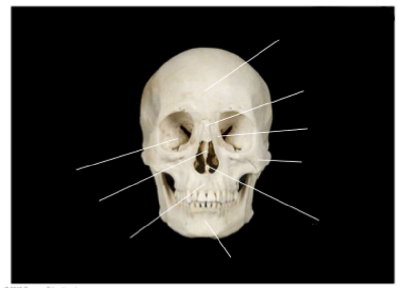
Skull, anterior
- facial and cranial bones
left to right:
sphenoid bone
ethmoid bone
maxilla
mandible
vomer bone —> voldemort bone **
zygomatic bone —> my cheekbone is what makes ppl wanna make zygotes w/me **
lacrimal bone —> lachimolala bone —> jimin bone —> eye bone
nasal bone
frontal bone
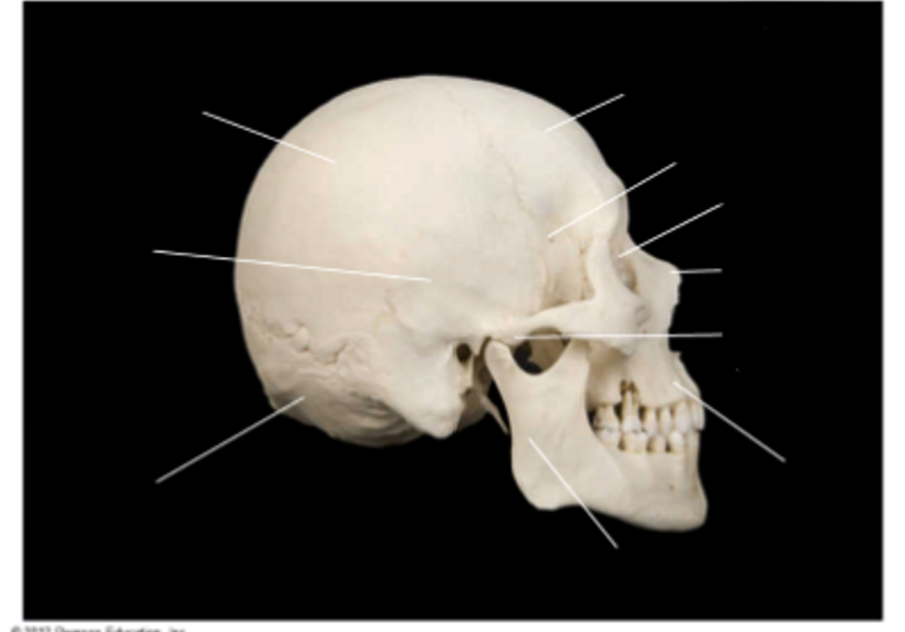
Skull, lateral view
- facial and cranial bones
left to right:
parietal bone
temporal bone
occipital bone
mandible
maxilla
zygomatic process (process = projection **)
nasal bone
lacrimal bone
sphenoid bone (side bone**)
frontal bone
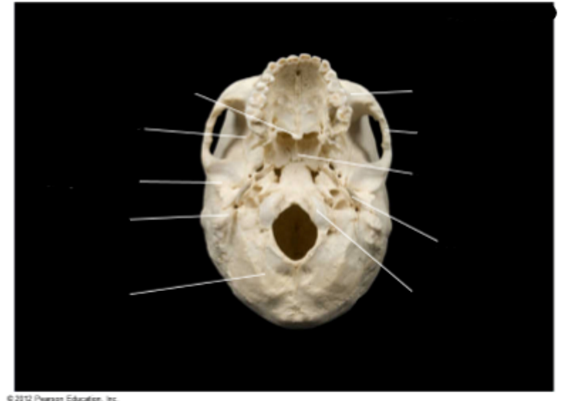
Skull, inferior view
- cranial and facial bones
palatine bone
sphenoid bone
temporal bone
mastoid process
occipital bone
occipital condyle
styloid process — style comes from within
vomer -- vomer vomit bone **
zygomatic bone
maxilla
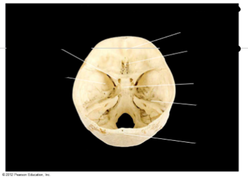
Skull, posterosuperior view of cranial cavities
- cranial cavity, cranial and facial bones
lesser wing of sphenoid
greater wing of sphenoid
occipital bone
temporal bone
sella turcica
crista galli
frontal bone
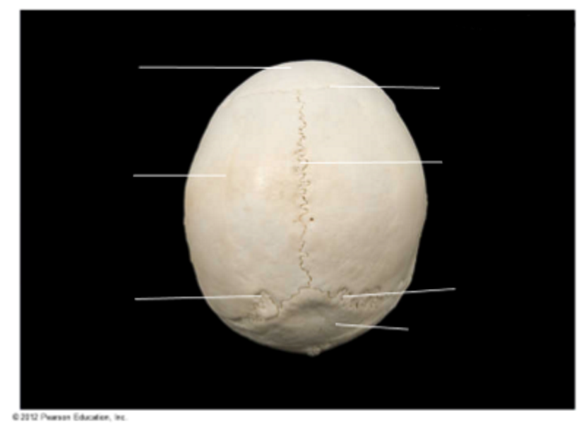
Skull, superior view
- sutures + other structures
frontal bone
parietal bone
sutural bone
occipital bone — at the back where the optic nerve goes
lambdoid suture
sagittal suture — if you have something sagging, they’d give you a big suture down the middle**
coronal suture
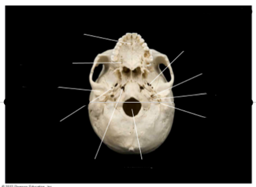
Skull, inferior view
- foramen and canals
incisive fossa
greater palatine foramen
mandibular fossa
foramen spinosum
foramen lacerum
stylomastoid foramen
jugular foramen
carotid canal
foramen ovale
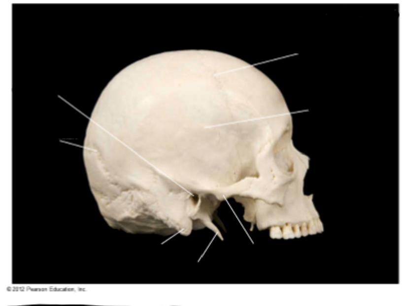
Skull, lateral view with sutures + other structures
- facial and cranial bones
external acoustic meatus
lambdoid structure
mastoid process
styloid process — pointy stylish *
zygomatic process
squamous suture — where grandma got sutures for her squamous cancer bc now she wear wig**
coronal suture
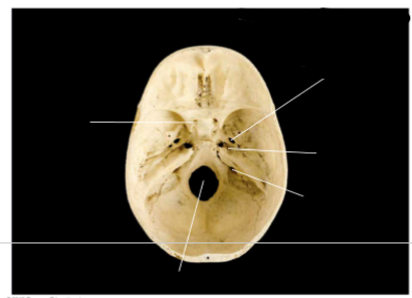
Skull, superior view of cranial cavity holes
- cranial cavity holes
optic canal
foramen magnum — need a magnum for that hole **
jugular foramen
carotid canal
foramen ovale
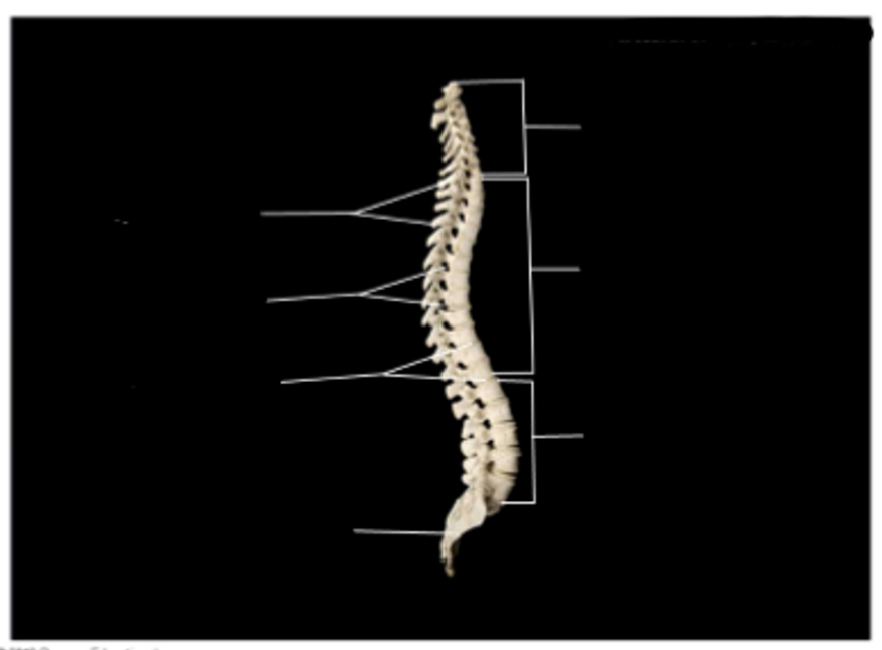
Vertebral column, articulated
spinous processes (processes PROTRUDE **)
intervertebral foramina (foramina = holes for nerves, arteries, etc. to pass through)
intervertebral discs
sacrum + sacral curvature
lumbar vertebrae + curvature
thoracic vertebrae + curvature
cervical vertebrae + curvature
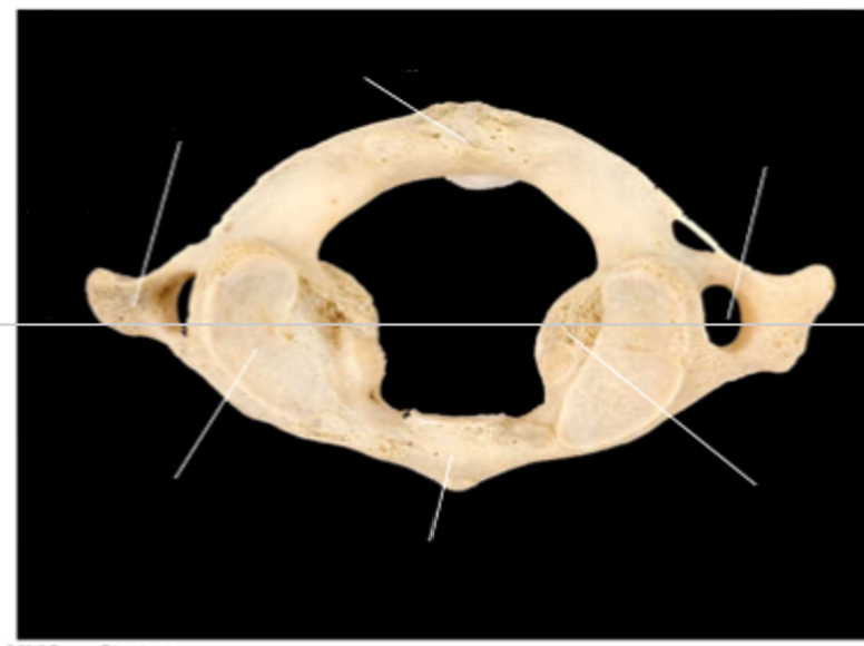
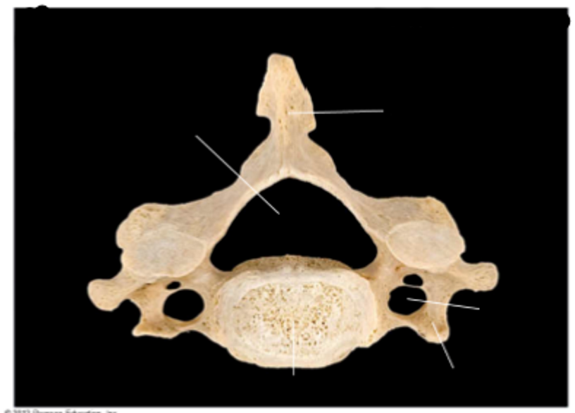
Atlas, C1, superior view
- top vertebrae of spine
transverse process (protrudes)
superior articular facet (connects vertebrae)
anterior arch
lateral mass
transverse foramen (hole)
posterior arch
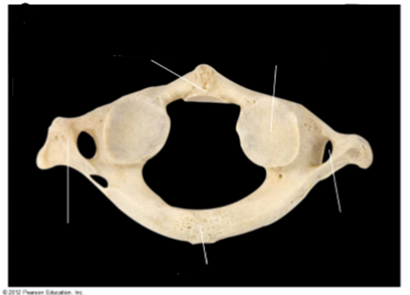
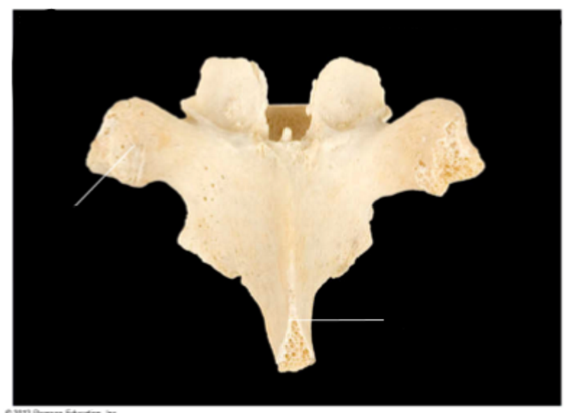
Atlas, C2, inferior view
transverse process
posterior arch
transverse foramen
inferior articular facet
anterior arch
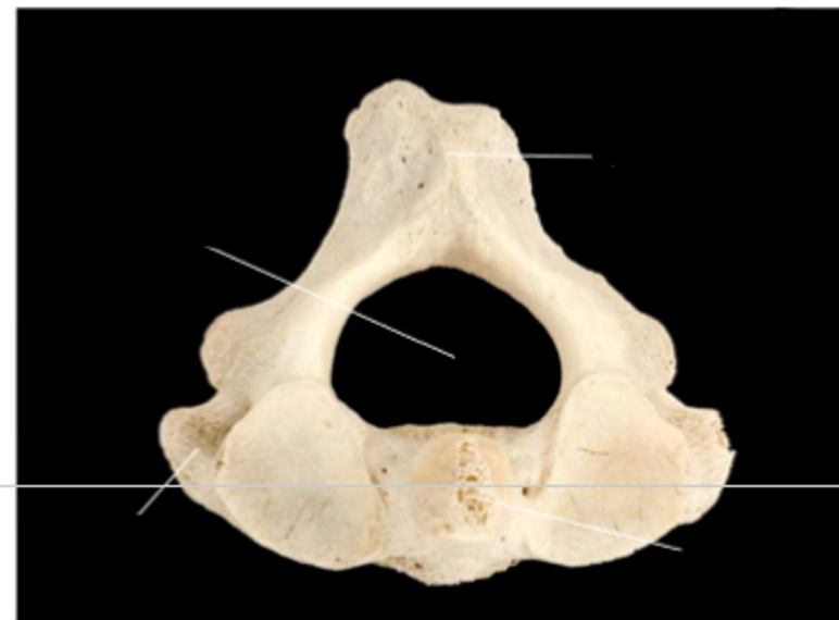
Atlas, C2, dens labeled, superior view
transverse process
dens (odontoid process)
spinous process
vertebral foramen
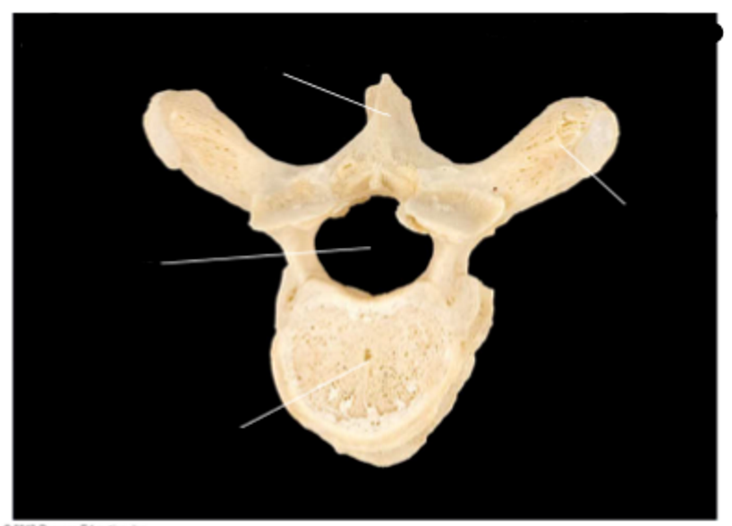
Cervical vertebrae, superior view
body
transverse process
transverse foramen
spinous process
vertebral foramen
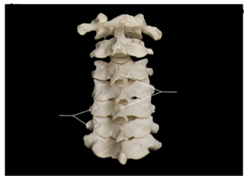
Cervical vertebrae in position
- transverse + spinous processes labeled
transverse processes
spinous processes
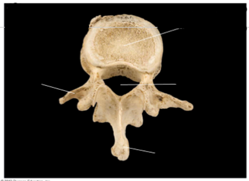
Thoracic vertebra, superior view
body
transverse process
spinous process
vertebral foramen
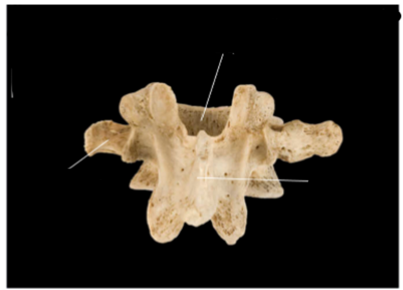
Thoracic vertebra, posterior view
transverse process
spinous process
Lumbar vertebra, superior view
transverse process
spinous process
body
vertebral foramen
Lumbar vertebra, posterior view
transverse process
body
spinous process
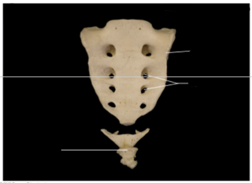
Sacrum + Coccyx
coccyx
sacral foramina
Ala — ala ocarina coccyx **
Sternum
top to bottom:
manubrium
body
xiphoid process
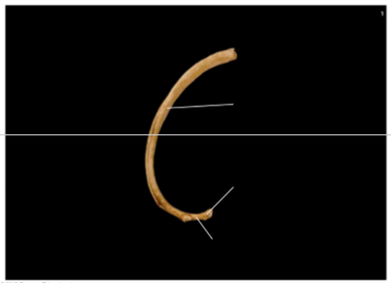
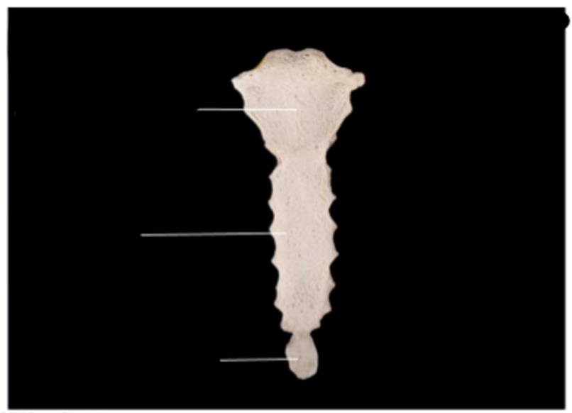
Rib
top to bottom:
body
neck
head
appendicular skeleton
the appendages
facilitate movement
upper limbs, lower limbs, girdles
girdles
The bone groups that attach the limbs to the axial skeleton
(the pectoral girdle for the shoulders and the pelvic girdle for the hips)
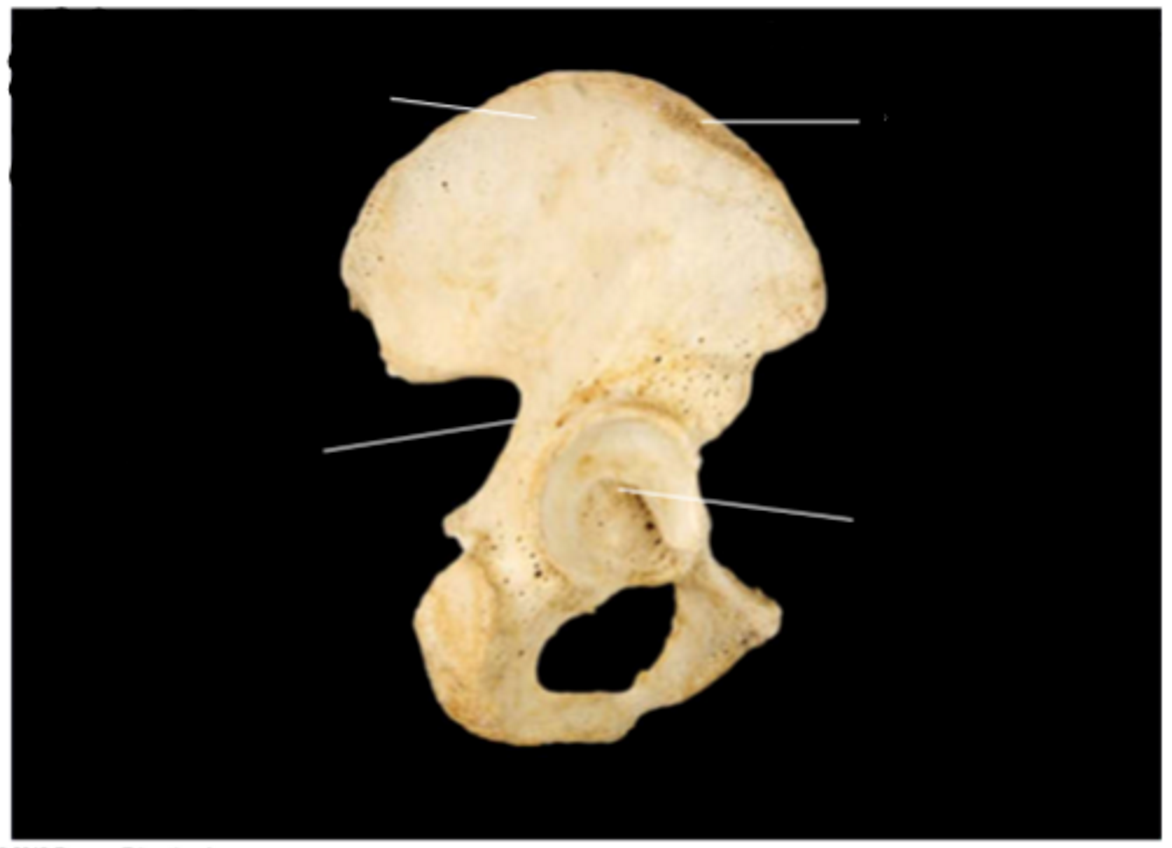
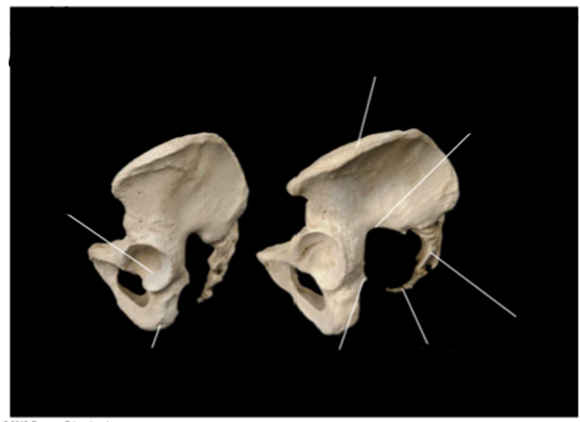
Hip bone, lateral view, right ilium
greater sciatic notch **
acetabulum — i see a your bootylum — acetabulum
iliac crest
ilium
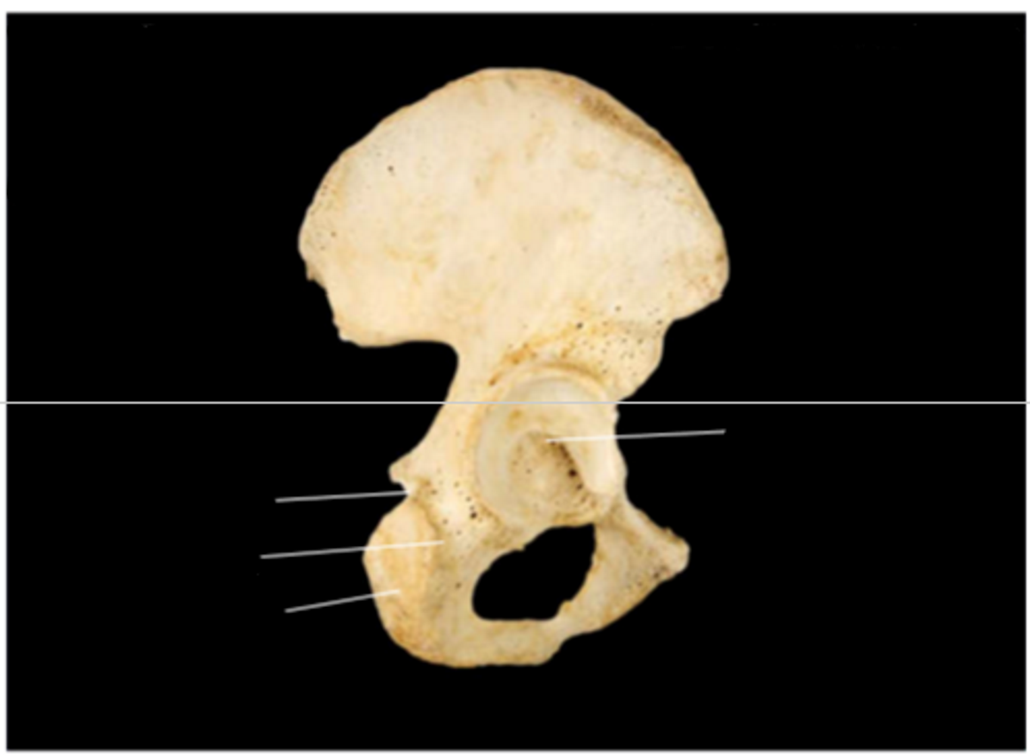
Hip bone, lateral view, right ischium
lesser sciatic notch
ischium
ischial tuberosity
acetabulum (articulates for hip joint)**
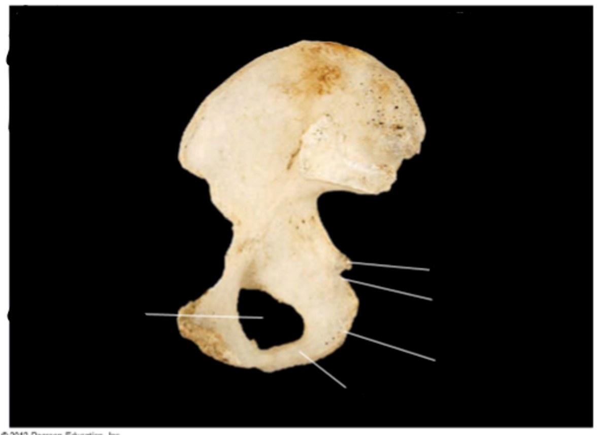
Hip bone, medial view, right ischium
obturator foramen
ischial ramus
ischial tuberosity
lesser sciatic notch
ischial spine
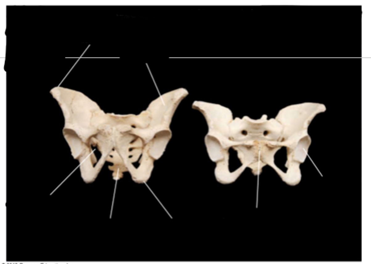
Articulated pelvis, male (left) and female (right), anterior view
obturator foramen
sacrum
ischial tuberosity
iliac fossa
iliac crest
pubic symphysis
acetabulum
male bigger, longer sacrum
female smaller, wider, shorter sacrum
Articulated pelvis, male (l), female (r), lateral view
acetabulum
ischial tuberosity
ischial spine
coccyx
sacrum
greater sciatic notch
iliac crest
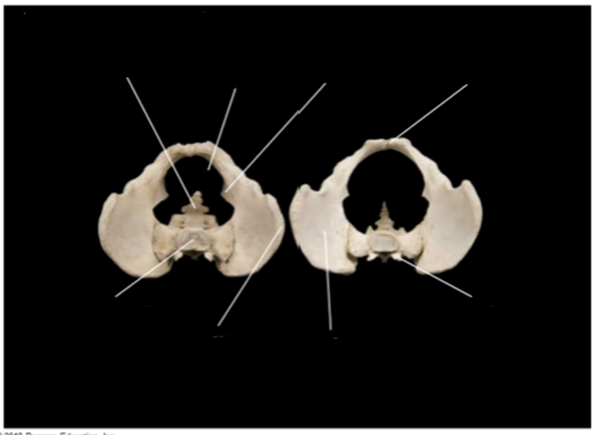
Articulated pelvis, male (l), female (r), superior view
body of sacrum
iliac crest
ischial spine
pelvic brim
coccyx
iliac fossa
superior articular process
pubic crest
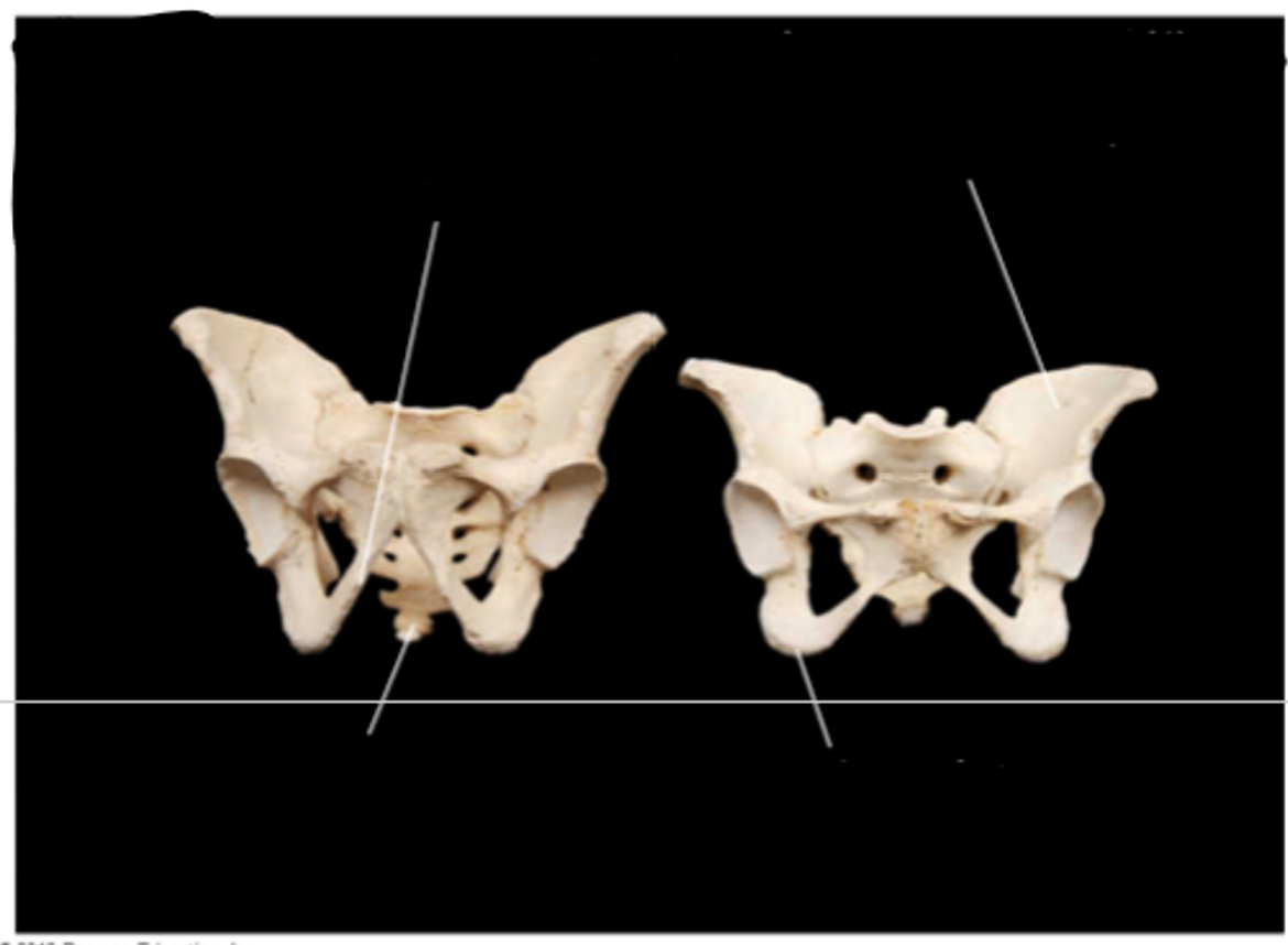
Articulated pelvis, male (l), female (r), demonstrating angles
coccyx in male = more ventral
pubic angle in male = more acute
ilium of female = flared
ischial tuberosities in female = farther apart
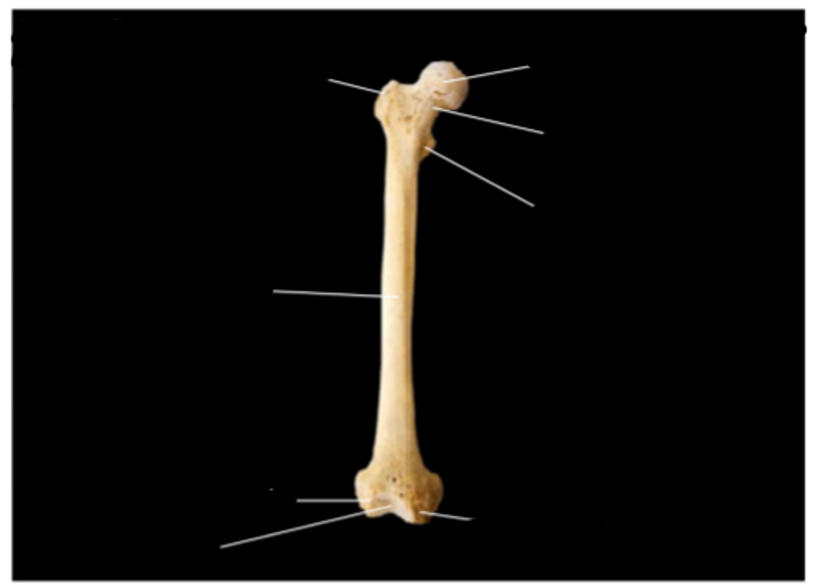
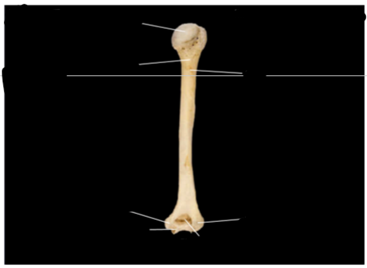
Femur, anterior view, right side
shaft
lateral condyle (the rounded prominence at the end of a bone that helps form a joint. Covered in smooth cartilage, it allows bones to articulate (connect and move) smoothly with one another.)
patellar surface
medial condyle
lesser trochanter (inner) (a large, bony prominence on the upper part of the thigh bone (femur) where major muscles attach)
neck
head — looks like the head of a penis **
greater trochanter (outer)
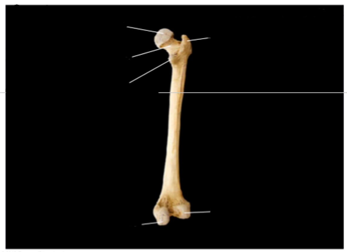
Femur, posterior view, right side
medial condyle
lateral condyle
greater trochanter
head
neck
lesser trochanter
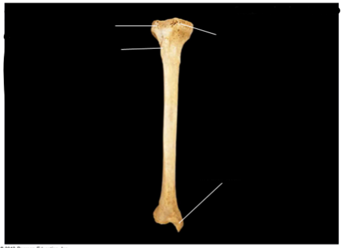
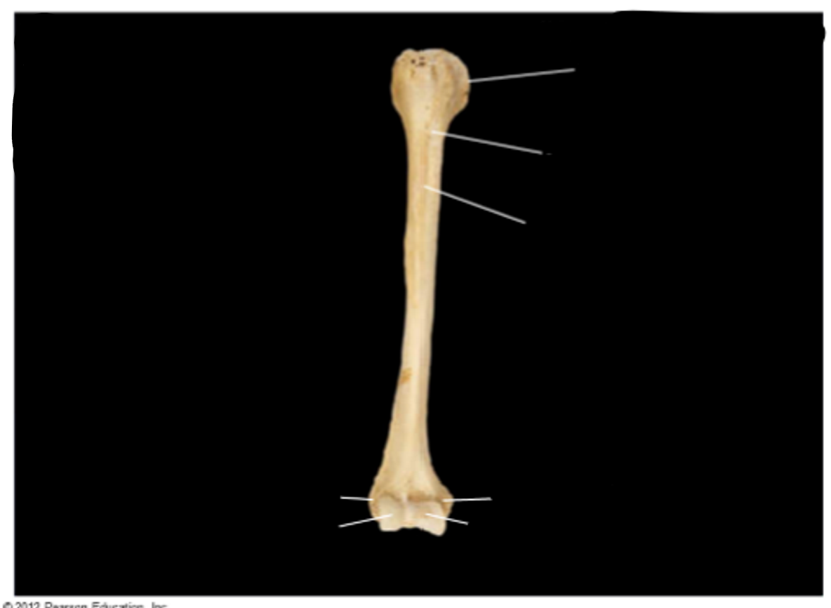
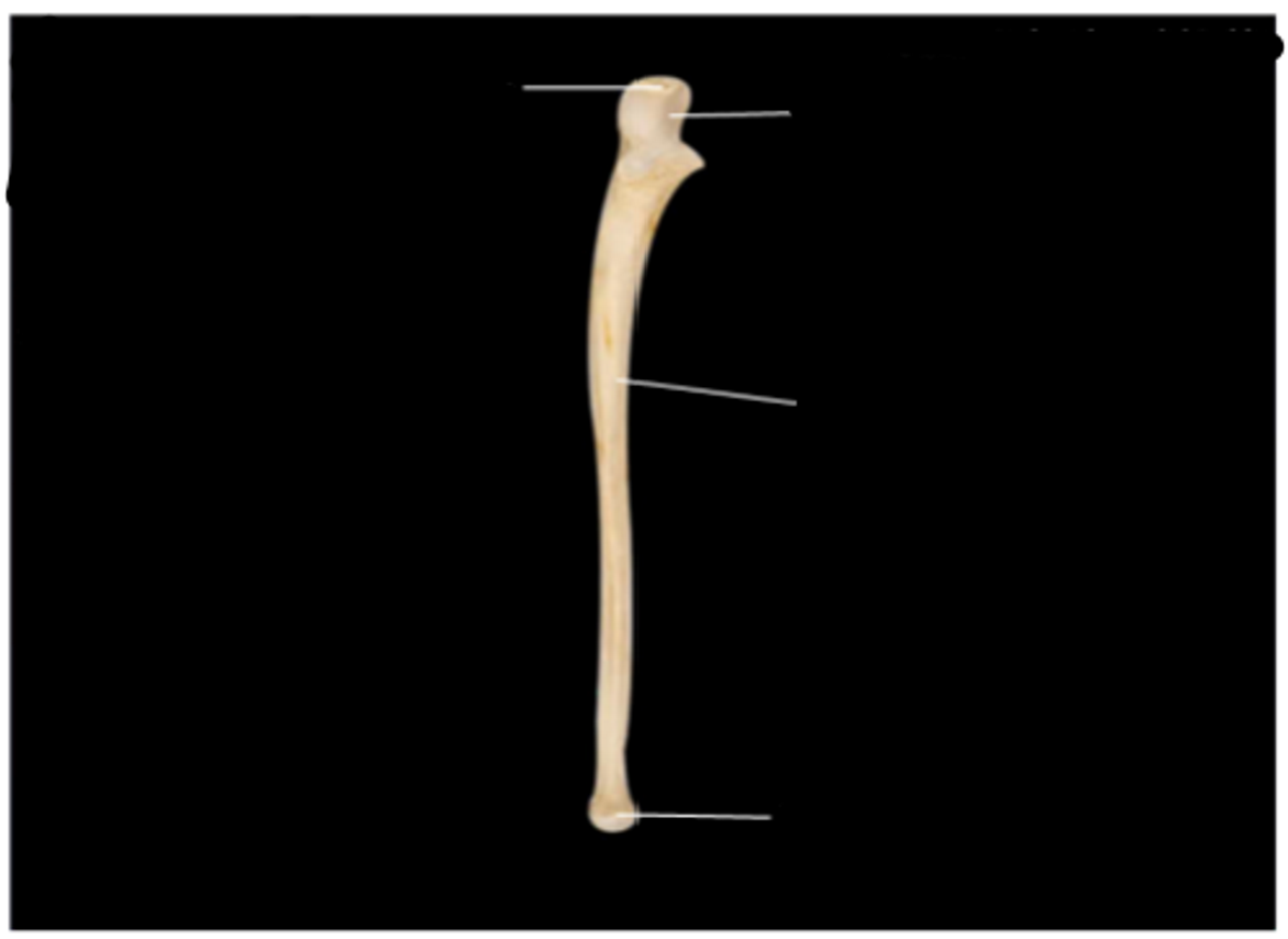
Tibia, anterior view, right side
medial malleolus (prominent, bony bumps on either side of the ankle)
medial condyle
lateral condyle
tibial tuberosity (a large, rounded, and typically roughened prominence on a bone. Its primary function is to serve as a strong, secure attachment point for muscles, tendons, and ligaments)
Tibia, lateral view, right side
medial malleolus
tibial tuberosity
lateral condyle
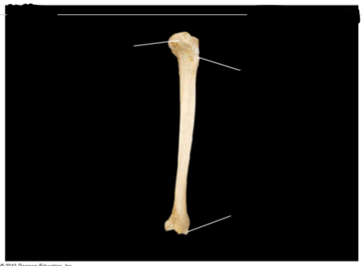
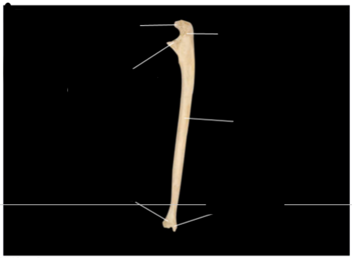
Fibula, anterior view, right side
lateral malleolus
head
fibula is on outside of leg and thin
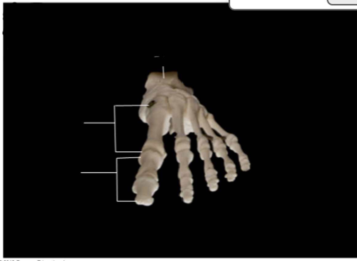
Foot, anterior view
talus
metatarsals (bones before toe tips)
phalanges
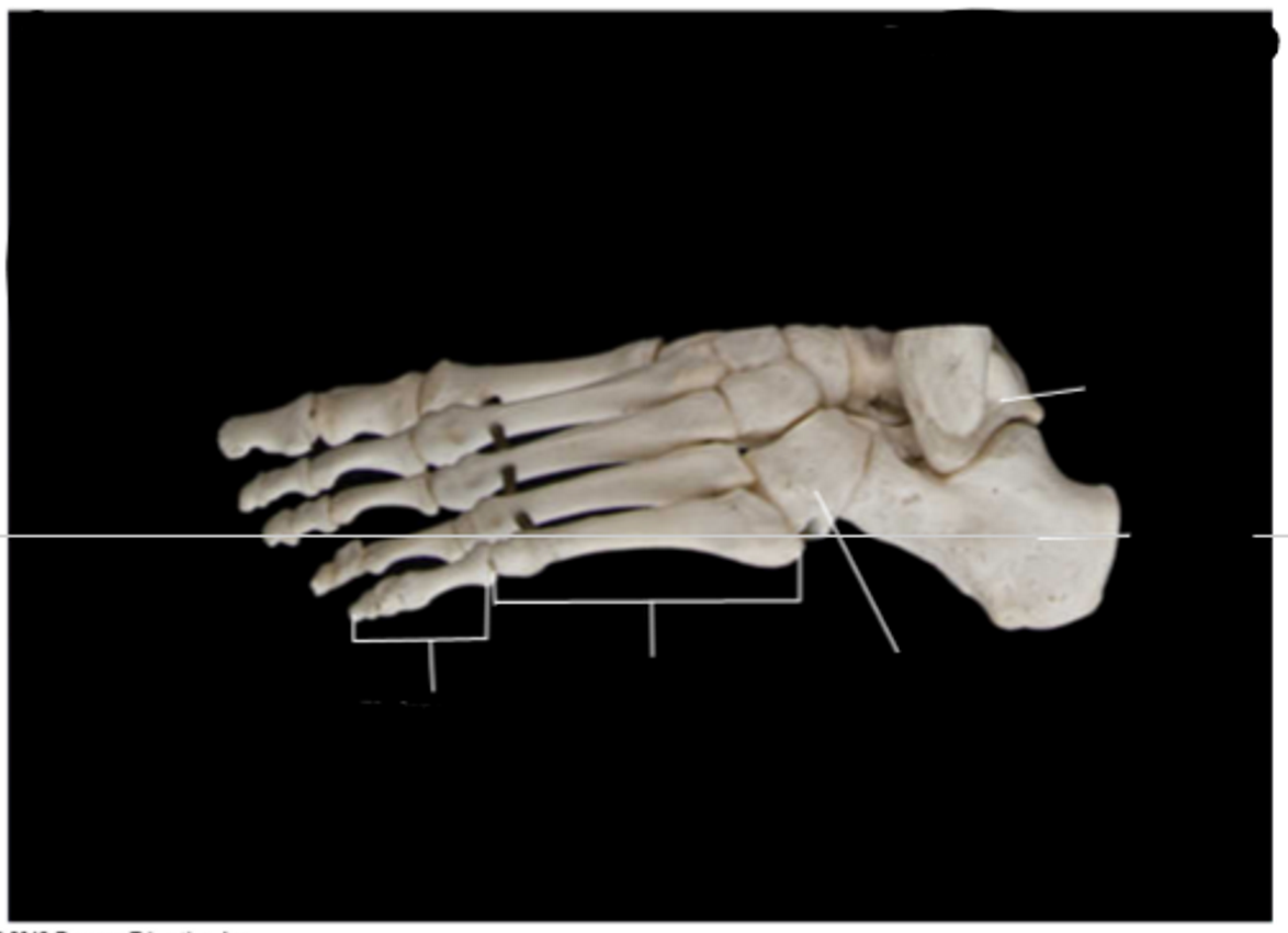
Foot, lateral view
phalanges
metatarsals
cuboid
calcaneus
talus
Humerus, anterior view, right side
lateral epicondyle (a raised, rounded bony prominence located on or above a condyle)
capitulum
trochlea
medial epicondyle
shaft
surgical neck
head
Humerus = upper arm bone
Humerus, posterior view, right side
medial epicondyle
trochlea
olecranon fossa (a broad, shallow depression or hollow space, typically found on the surface of a bone. Derived from the Latin word meaning "ditch" or "trench", fossae serve crucial roles as attachment points for muscles, vessels, or nerves, and to cradle other organs) — crucial for elbow joint
lateral epicondyle
shaft
surgical neck
head
Humerus = upper arm bone
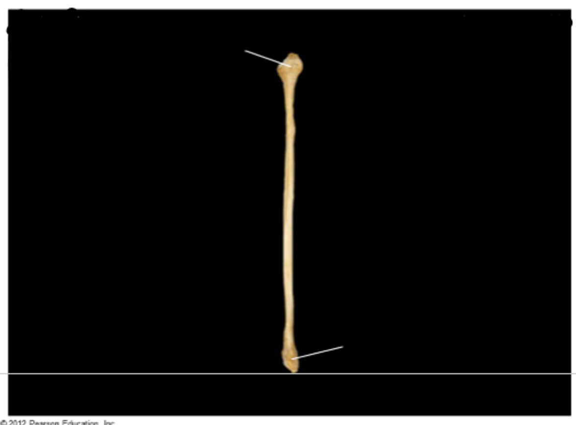
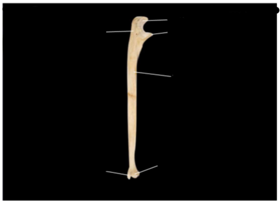
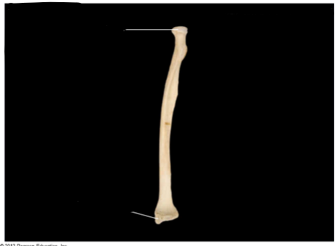
Ulna, anterior view, right side
head AT BOTTOM***
shaft
trochlear notch
olecranon process
Ulna = little inner forearm bone
Ulna, lateral view, right side
styloid process
head
shaft
coronoid process
trochlear notch
olecranon process
Ulna, medial view, right side
head
styloid process
shaft
olecranon process
trochlear notch
coronoid process
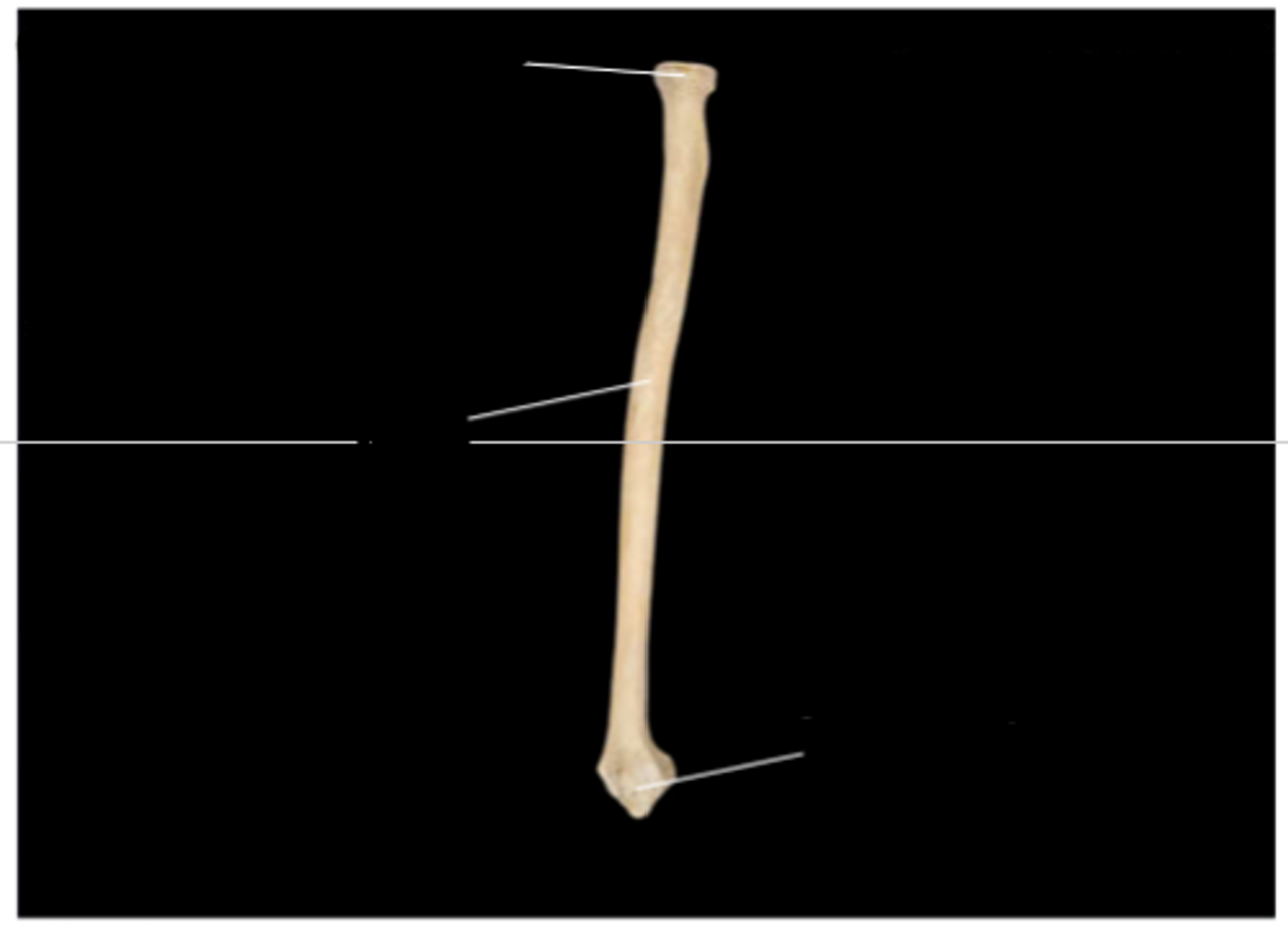
Radius, anterior view, right side
styloid process
head AT TOP**
Radius, lateral view, right side
styloid process
shaft
head
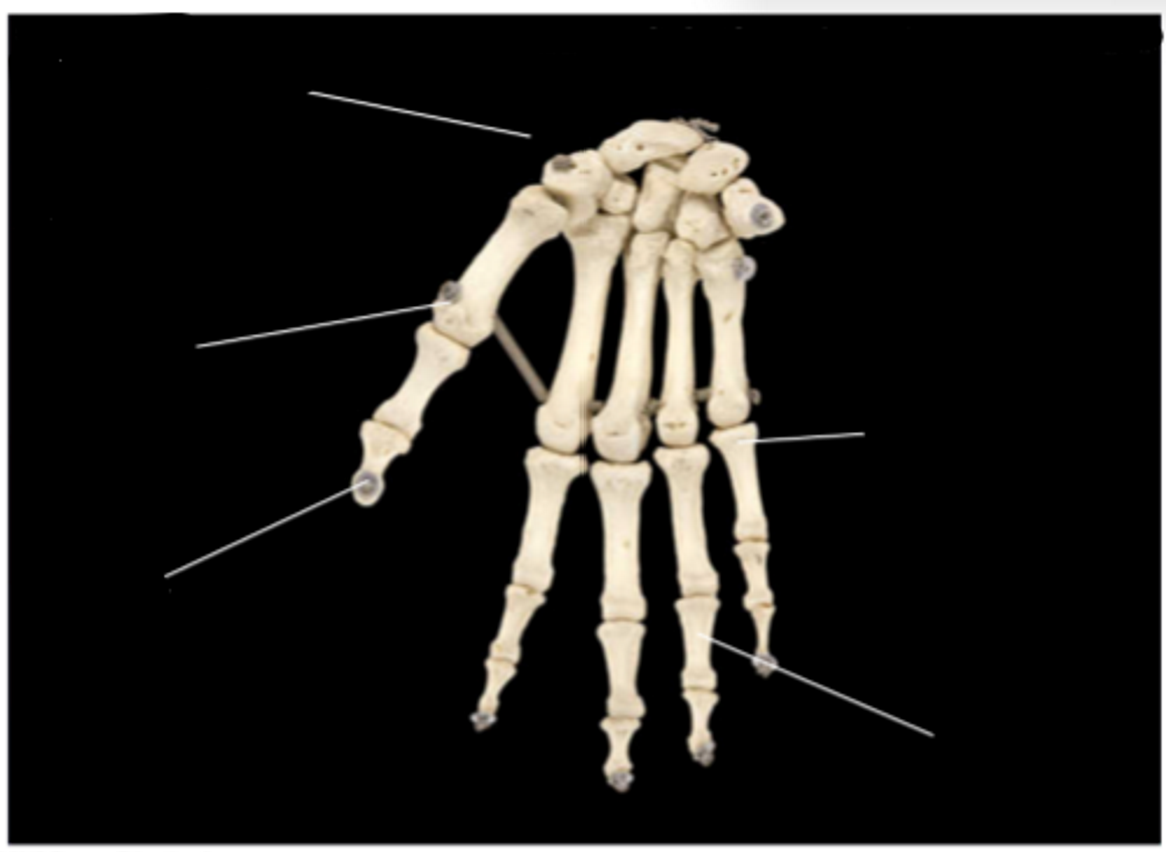
Hand, overview, anterior view, right side
carpals
metacarpals
distal phalanges (far from trunk)
middle phalanges
proximal phalanges (close to trunk)
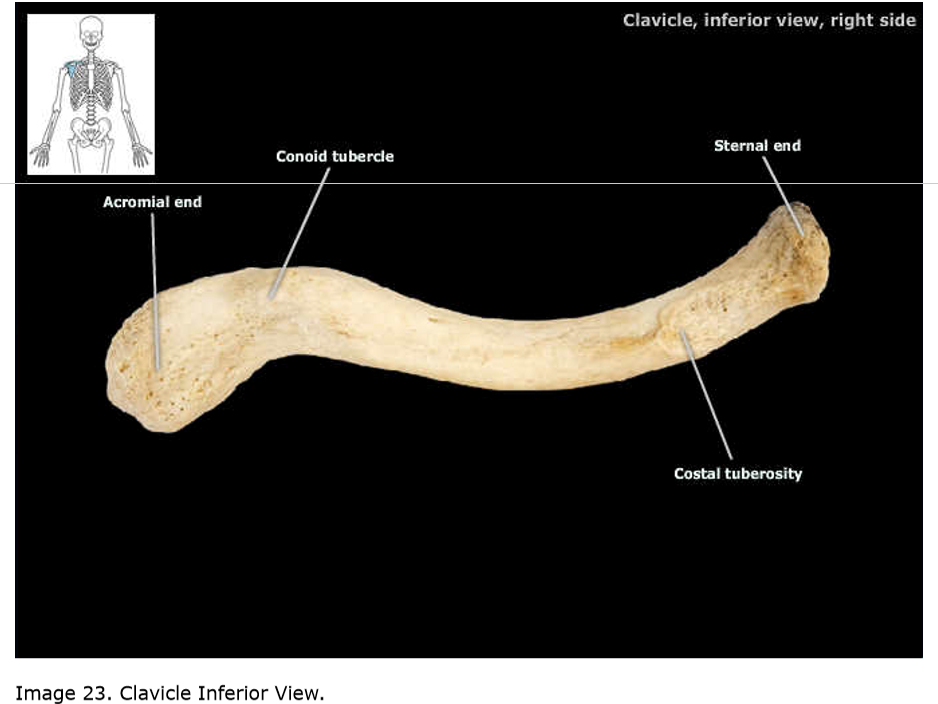
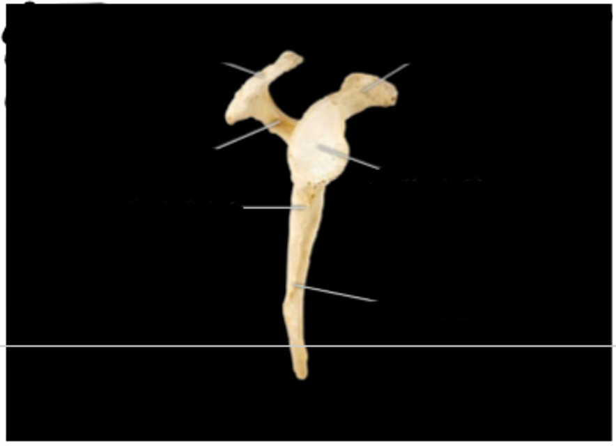
Clavicle, inferior view, right side
acromial end (outer end)
conoid tubercle
costal tuberosity
sternal end (medial end)
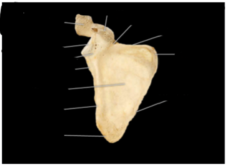
Scapula, anterior view, right side
acromion
glenoid fossa (cavity)
lateral angle and neck
infraglenoid tubercle
lateral (axillary) border
inferior angle
medial (vertebral border)
superior angle
superior border
suprascapular notch
coracoid process
subscapular fossa
Scapula, lateral view, right side
acromion (projection from collarbone)
spine
infraglenoid tubercle
lateral (axillary) border
glenoid fossa (cavity)
coracoid process
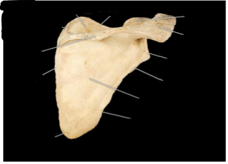
Scapula (shoulder), posterior view, right side
medial (vertebral) border
inferior angle
lateral (axillary border)
infraspinous fossa
infraglenoid tubercle
glenoid fossa (cavity)
acromion
coracoid process
superior border
supraspinous fossa
superior angle
new terms
condyle — the rounded prominence at the end of a bone that helps form a joint. Covered in smooth cartilage, it allows bones to articulate (connect and move) smoothly with one another — on ankles and knee — joint articulation
epicondyle — epicondyle is a bony prominence or rounded eminence located just above or upon a condyle (the smooth, articular surface of a joint). Its primary function in the skeletal system is to serve as a vital anchoring site for muscles, tendons, and ligaments. — elbow
trochanter — a large, bony prominence on the upper part of the thigh bone (femur) where major muscles attach — greater and lesser — on femur
tuberosities — a large, rounded, and typically roughened prominence on a bone. Its primary function is to serve as a strong, secure attachment point for muscles, tendons, and ligaments. — attach muscles and tendons — on legs
fossa — a broad, shallow depression or hollow space, typically found on the surface of a bone. Derived from the Latin word meaning "ditch" or "trench", fossae serve crucial roles as attachment points for muscles, vessels, or nerves, and to cradle other organs (like the brain)
transverse — a horizontal cross-section that divides the body or organs into upper (superior) and lower (inferior) portions — There are two on each vertebra (one on the left, one on the right), extending laterally (outward) from the sides of the bone — attach back muscles and ligaments and ribs to them
spinous — Projects directly backward from the center of the vertebra — lever for muscles + rotate the spine
vertebral — related to the vertebrae — the individual interlocking bones that stack together to form the human spine
process — any prominent projection or outgrowth of tissue from a larger body structure
foramen — any natural opening, perforation, or hole in a bone or tissue. Its primary function is to act as a passageway, allowing nerves, blood vessels (arteries and veins), and other soft tissues to safely travel from one part of the body to another.