rt second week
 Call Kai
 Learn
Learn Practice Test
 Spaced Repetition
 Match
Match Flashcards
 Knowt Play
1/96

There's no tags or description
Looks like no tags are added yet.
Name | Mastery | Learn | Test | Matching | Spaced | Call with Kai | Chat |
|---|
No analytics yet
Send a link to your students to track their progress
97 Terms
Low Flow Device - Nasal Cannula
1-6 liters per minute (lpm)
22-44% FiO2
1 lpm = 24%
2 lpm = 28%
3 lpm = 32%
4 lpm = 36%
5 lpm = 40%
6 lpm = 44%
Low Flow Device - Simple Mask
5-10 lpm
35-60% FiO2
No Reservoir Bag
Low Flow Device - Non-rebreather Mask
10-15 lpm
60-90% FiO2
Has (3) valves - 1 reservoir bag, 2 exhalation valves
Valves keep room air from coming in
Do NOT let bag collapse
Low Flow Device - Partial Pressure Rebreather Mask
6-15 lpm
40-70% FiO2
Bag should NEVER completely collapse
Does NOT have exhalation (2) valves on the sides of masks
Has reservoir bag
High Flow O2 Device/Precise FiO2 - Venturi Mask
24%
28%
31%
35%
40%
50%
Chain of what device to start with
Nasal Cannula
Simple Mask
Partial Rebreather
Non-rebreather
OxyMask (has many holes in it)
1-15 lpm
24-90% FiO2
Over 4L for nasal cannula
use humidifier
nasal cannula lpm
1-6
nasal cannula FiO2
22-44%
nasal cannula 1 lpm %
24%
nasal cannula 2 lpm %
28%
nasal cannula 3 lpm %
32%
nasal cannula 4 lpm % fio2
36%
nasal cannula 5 lpm %
40%
nasal cannula 6 lpm %
44%
simple mask lpm
5-10
simple mask FiO2
35-60%
what does a simple mask lack
no reservoir bag
non-rebreather mask lpm
10-15
non-rebreather mask FiO2
60-90%
what does a non-rebreather mask have?
3 valves - 1 reservoir bag, 2 exhalation valves. valves keep room air from coming in.
bag: do not let bag collapse
partial pressure rebreather mask lpm
6-15
partial pressure rebreather mask FiO2
40-70%
what does a partial pressure rebreather mask have and not have?
has a reservoir bag (should never collapse).
does not have exhalation (2) valves on the sides of masks
Inspection: What You See
•Observe respiratory rate
•Work of breathing
•Chest shape
•Accessory muscle use
•Skin color and patient position
Types of chest abnormalities
barrel chest
kyphosis, scoliosis, kyphoscoliosis
lordosis, pectus excavatum, pectus carinatum
barrel chest
rounded, bulging chest shape that makes the rib cage look like it is permanently stuck in a deep breath. Common in people with COPD.
Increased anteroposterior (AP) diameter.
Kyphosis
Abnormal curvature of the upper spine, the anterior- to- posterior curvature gives the patient the (Humpback) shape. People with chronic (lasts a long time) lung disease develop this.

Scoliosis
Lateral curvature of the spine that causes the vertebrae in the affected area to rotate flattening the rib cage anteriorly. Again, this curvature interferes with the patient's ventilation because they are not able to move air in and out of the lungs.
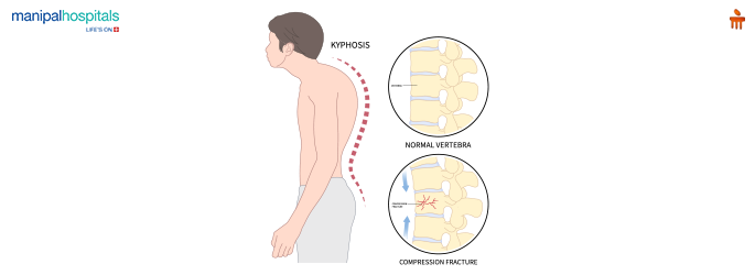
Kyphoscoliosis
Combination of kyphosis and scoliosis. This extremely affects the patient's ability to inhale volume greatly affecting the patients respiratory effort. Because the heart is not able to pump effectively and the lungs are not able to help in breathing, fluid backs up into the lungs and cause congestive heart failure. So essentially it will feel as if the patient is drowning. Many patients with this pass early in life from respiratory infections.

Lordosis
Inward curvature of the lumbar spine, results in a sway back appearance
Pectus Excavatum
This is a congenital (born with) deformity
Funnel chest (the breastbone sinks inward into the chest at the sternum and xyphoid process, creating a caved or sunken look)
If this is severe enough, it may result in decreased lung volumes because the patient can not expand their lungs like a person who does not have this condition.
Pectus Carinatum
(congenital deformity) breastbone and push outward, giving the chest a bird-like or pointed look.(pigeon chest) Even though it looks like it hurts, the patient does not suffer from any respiratory complications
Digital Clubbing
The angle between the nailbed and finger become increased giving the fingers and toes a concave look. Usually the result or hypoxemia (not enough oxygen in the blood) or circulation issues. You will find it difficult at times to place your pulse oximeter on the patients fingers due to the shape, when that happens you may also grab your SpO2 measurement from the patient's ear, forehead, or toe.
Bony landmarks on the Anterior chest
•Clavicle (collar bones)
•Suprasternal notch
•Sternal angle
•4th rib
Bony structures on the Posterior chest
•Clavicle
•C7 vertebra prominens
•T1
•Scapula (Shoulder blades)

Vertical Divisional Lines
•Anterior Chest- midsternal line
•Midclavicular line
•Anterior midaxillary line

Lateral chest
•Anterior axillary line
•Midaxillary line
•Posterior axillary line
Posterior chest
•Right and left midscapular lines
•Vertebral line
Palpation: What You Feel
•Assess chest expansion
•Evaluate symmetry
•Check tactile fremitus
•Relate findings to disease processes
What is Palpation?
Physical assessment of the chest by the sense of touch
Symmetry
Place your hands on both sides of the chest (right and left) you are observing the motion of the patients chest wall. As the pt. inhales deeply, our hands move apart equally in theory. If not and one hand moves more than the other, this could be a clue to consolidation (airless, fluid-filled uncollapsed lung tissue), pleural effusion (fluid in the pleural space), atelectasis (loss of air in the lung tissue), or a pneumothorax (air in the pleural space).
Tactile fremitus
Say “99” while the RT palpates the patients chest. This is the vibration felt as the pt is speaking.
•Increased: Pneumonia, Consolidation
•Decreased: Pleural Effusion, Pneumothorax
Subcutaneous Emphysema
Presence of air beneath the skin in the subcutaneous tissues. Usually a complication of tracheostomy placement, pneumothorax, or from the ventilator.
Percussion: What You Hear (RHDF)
•Resonant = Normal
•Hyperresonant = Too much air
•Dull = Tissue or fluid
•Flat = No air
2 methods to percuss the chest, direct and indirect
Direct is when you tap on the chest with a finger using a sharp, short, stroke. Indirect is placing a finger firmly against the chest and tapping on that finger.
You are listening for different sounds. Hyperresonance, resonance, dullness, and flatnesss.
Hyperresonance
Air trapping, air in an enclosed cavity (pneumothorax)- loud, low-pitched sound
Resonance
Heard when you percuss normal lung tissue. Low pitched sound with a long duration. Equal parts of air and tissue.
Dullness
Medium intensity and pitch with a short duration. More tissue or fluid than air. Consolidation, atelectasis, fluid in the pleural space
Flatness
Low amplitude and pitch. More tissue than air. Pleural effusion
Disease Connections (EPPP)
•Emphysema → Hyperresonant
•Pneumothorax → Hyperresonant + Decreased Fremitus
•Pneumonia → Dull + Increased Fremitus
•Pleural Effusion → Dull + Decreased Fremitus
Classifications of medical gases
•Medical gases are classified as laboratory, therapeutic, or anesthetic gases
•Medical compressed gases are classified as either: Nonflammable (do not burn), Nonflammable but supportive of combustion (oxidizing), Flammable (burns readily, potentially explosive)
Laboratory gases
used for equipment calibration and diagnostic testing.
Therapeutic gases
used to relieve symptoms and improve the oxygenation of patients with hypoxemia
Anesthetic gases
combined with oxygen (O2) to provide anesthesia
medical gas safety systems
Oxygen Delivery Systems
Vacuum Systems
Medical Air Systems
Oxygen Delivery Systems
These systems provide a controlled supply of oxygen to patients, essential for respiratory support. They are designed to deliver oxygen at various flow rates, accommodating different clinical needs, such as low-flow systems for stable patients and high-flow systems for those requiring intensive care.
Vacuum Systems
used for airway suctioning, which is vital in emergency situations and for patients with respiratory issues. They help clear secretions from the airway, ensuring unobstructed breathing and preventing complications.
Medical Air Systems
These systems supply a mixture of gases used to power pneumatic equipment and support various medical procedures. They are integral in anesthesia delivery and other applications where a clean, dry air source is necessary.
Medical gas alarm panel
Medical gas alarm panels are essential safety devices in healthcare settings. They monitor the status of medical gas and vacuum systems, ensuring that gases are available and within safe operational limits.
The normal ranges are usually listed on the bottom of the panel with the designated normal values of the pressures it is measuring
Medical gas alarm panel Key Functions:
Monitoring
Alerting
Location
These panels are crucial for maintaining patient safety and ensuring that medical staff can respond quickly to any gas supply issues.
Medical gas alarm panel - monitoring
Continuously checks pipeline pressures and operational status of medical gases.
Medical gas alarm panel - alerting
Generates alarms when parameters exceed set thresholds or when faults are detected, alerting healthcare staff to potential issues.
Medical gas alarm panel - location
Typically installed near points of use, such as patient rooms or operating rooms, or in central locations like nurses' stations.
Oxygen (O2): what you need to know
Colorless, odorless, transparent, tasteless
Heavier than air
Nonflammable BUT ACCELERATES COMBUSTION
Used to treat low oxygen levels in the blood (hypoxemia), acute conditions like pneumonia, COPD, and with traumas or surgeries before, during, and after operations
Oxygen Advantages
Quickly restores normal oxygen levels in the organs like the brain and heart
Improves breathing because it reduces the patient's shortness of breath reducing the workload placed on the heart and lungs
Enhances recovery by helping patients heal faster, and increases mobility while using portable O2 tanks.
Oxygen Limitations
Does not treat the cause, it fixes the hypoxemia but does not fix the underlying disease, some can not afford the portable tanks and need to stay home near their oxygen concentrators
Dryness and irritation to the nose, sinuses, and airways
Patients run a risk of toxicity (too much Oxygen) over a long period of time, this can damage the lung tissue especially in patients who have chronic disease.
Oxygen Safety Issues
Fire hazards, never smoke or use open flames near o2 equipment.
Pressure dangers are also on the list of limitations because compressed gas cylinders are under high pressure. They can explode or become dangerous projectiles if damaged or dropped,
Medical Air: what you need to know
Colorless, odorless, dry, purified gas
21% oxygen and 78% nitrogen (manufactured drug)
Processed through OIL-FREE compressor systems (to eliminate moisture, oils, and toxic particulates before being delivered to the patients.)
Prescribed for human respiration and respiratory equipment calibration
Used as a base gas to dilute pure oxygen to give an exact Fraction of Inspired Oxygen(FiO2) tailored to a patients oxygenation needs (OR).
Used in neonatal and infant care because they are highly vulnerable to hyperoxia (state of being exposed to high levels of O2)
Used in aerosol and nebulizer therapy to convert liquid medications into an inhaled fine mist,
Used as a carrier gas to transport anesthetic agents
medical air advantages
Mitigates oxygen toxicity (meaning it reduces the harmful effects O2 can have
Low fire risk (nonflammable) safer to use near sources of ignition
Medical Air limitations
Inadequate for acute hypoxemia because it only contains 21% oxygen
Medical Air safety issues
system contamination (contaminates and moisture can enter the respiratory pipeline fostering microbial or bacterial growth in the hospital pipes)
toxic gas exposure can be a problem
pressure fluctuations can disrupt the performance of critical mechanical ventilators,
gas misconnections can also occur, and the result can be catastrophic.
Carbon Dioxide (CO2) what you need to know
non-flammable
highly soluble in blood
rapidly cleared by the lungs
used as insufflation gas to inflate and stabilize the abdominal or body cavities
radiographic contrast agent serving as an alternate for patients who are allergic to iodine contrast
used as a respiratory stimulant in patients with respiratory depression because it activates the central and chemoreceptors in the brainstem that drive the urge to breathe (not used as often due to already dangerously high levels the patient could have.)
carbon dioxide advantages
Low risk of allergic reactions, it can enhance blood flow and improve oxygen delivery in tissues, less expensive than other contrast agents
carbon dioxide limitations
Potential for hypercapnia (excess CO2 in the blood), limited use in certain conditions mentioned above
carbon dioxide safety issues
Monitoring required complications can occur if CO2 is being used for insufflation or as a contrast, RISK for embolism if CO2 enters the blood stream
Heliox (Helium) what you need to know
Odorless, tasteless, nonflammable, nonreactive with other elements, resists physical stress
Produced by liquification
Must be mixed with at least 20% O2 to sustain life for therapeutic purposes
Can manage severe airway obstruction/asthma attacks/stridor
Monitor patient for adequate oxygenation, it does not provide oxygen itself
Only works when helium concentration is high (80:20) or (70:30) mixtures
Limited benefits when FiO2 requirements increase
It helps to manage the airway because it is less dense than air or oxygen allowing gas to flow smoothly through tight or blocked airways (laminar flow)
Nitric Oxide (NO) what you need to know
Vasodilatory properties
Colorless, nonflammable, toxic gas that SUPPORTS combustion
Forms brown fumes
Strong respiratory irritant (chemical pneumonitis)
Can cause a fatal form of pulmonary edema
Methemoglobinemia
methemoglobinemia
blood disorder where an abnormal amount of methemoglobin builds up in the blood. This prevents red blood cells from effectively releasing oxygen to body tissues. Blood looks like chocolate. (treatment is methylene blue) IV quickly converts iron back to its normal state.
Nitrous Oxide (N2O) what you need to know
Colorless gas
Sweet odor and taste
Anesthetic agent (depresses the central nervous system) conscious sedation
Can support combustion, but can’t support life
Must ALWAYS be mixed with 20% O2
Known as Laughing gas
Conscious sedation is when you are awake and still able to talk
Oxygen tank Sizes
H cylinder
E cylinder
E-AA are considered small and used for patient transport
G and H are for stationary patients
Cylinder Safety Factor (E)
E cylinder 0.28 L/psi
Formula for calculating how long an “E” cylinder will last
Duration(minutes)= PSIG off tank x 0.28 (tank factor)/ Flow L/M
For every psi of pressure in the cylinder, there are about 0.28 liters of oxygen available. Crucial for calculating how long the oxygen will last based on the cylinders pressure and the flow rate being used.
Cylinder Safety Factor (H)
H cylinder 3.14 L/psi
Formula for calculating how long an “H” cylinder will last
Duration(minutes)= PSIG off tank x 3.14 (tank factor)/ Flow L/M
storage precautions of tanks/cylinders
Ventilation: Ensure storage areas are well-ventilated to prevent the accumulation of gases.
Temperature Control: Store cylinders in a cool, dry place away from heat sources and direct sunlight.
Secure Storage: Cylinders should be secured upright to prevent tipping and should be protected from physical damage.
transportation precautions of tanks/cylinders
Proper Securing: Cylinders must be secured in an upright position during transport to prevent movement and potential damage.
Leak Checks: Regularly inspect cylinders for leaks before and during transportation.
Avoiding Heat Sources: Keep cylinders away from flames, heat, and oil during transport.
Compliance with Regulations: Follow Department of Transportation (DOT) regulations for transporting hazardous materials.
general safety practices of tanks/cylinders
Personal Protective Equipment (PPE): Use appropriate PPE, such as gloves and goggles, when handling cylinders.
Training: Ensure personnel are trained in oxygen safety protocols.
Pressure Regulators
E cylinders have 2 prongs that line up for the tank and an “O” ring. Use pin-index regulators. Support lower flow rates up to 15 LPM
H tanks have use large-bore threaded regulators. Can handle higher flow rates, up to 25 LMP or more
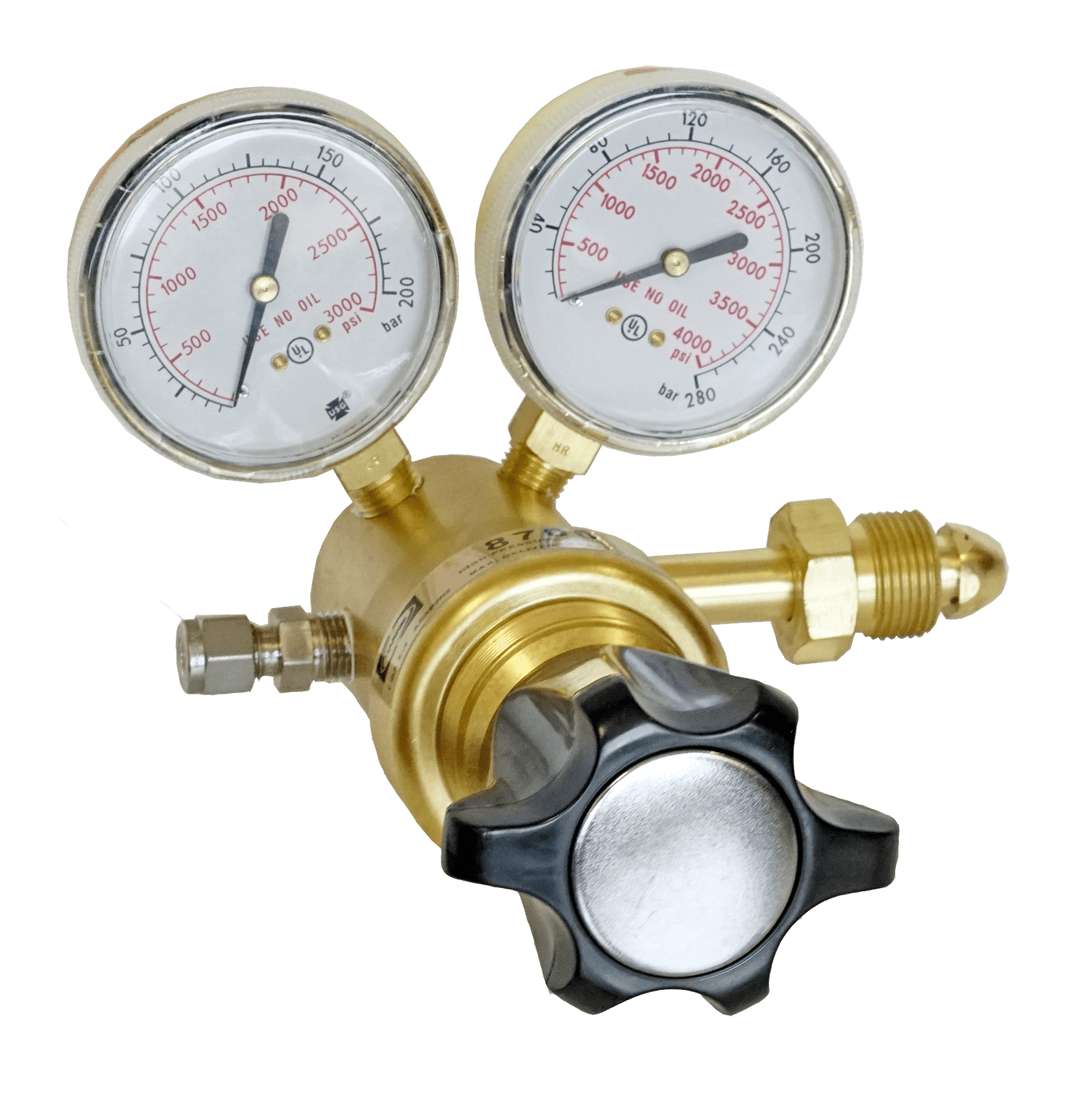
pressure regulators a.k.a reducing valves
reduce cylinder pressure to a safe usable working pressure. The unit of measure is psi (pounds per square inch)
Manage force/pressure

flowmeters
Measure or regulate the exact volume or speed of fluid/gas passing through a system per unit of time. The unit of measure is lpm (Liters per minute)
Manage volume/speed

Thorpe Tube flowmeter
Flow meter device attached to a 50 psig gas source
Facilitates accurate flow adjustments/compensated
Calibrated in liters per minute
Not ideal to transport patients
Gravity Dependent- The tube must remain completely vertical to give an accurate reading.
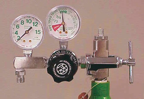
Bourdon Gauge flowmeter
Uses a fixed orifice/operates under variable pressures that can be adjusted with a pressure reducing valve
When delivering a medical gas, use this whenever a flowmeter cannot be maintained in an upright position
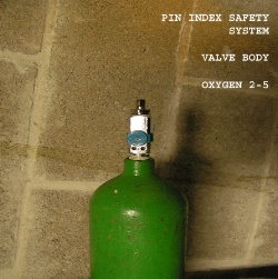
ASSS American Standard Safety System
used with “H” cylinders- threaded high pressure connections

PISS Pin Index Safety System
used with “E” cylinders. Made with pins to align with valve outlet

DISS Diameter Index Safety System
Used with low pressure medical gas connections

Oxygen Analyzer
Device that measures the concentration of oxygen in a gas mixture
Displayed as a percentage
What is the atmosphere made up of
Nitrogen (N2)- 78%
Oxygen (O2)- 21%
Argon (Ar)- 1%
N2- 760 x .78=592.8
O2- 760 x .21= 159.6
Ar- 760 x .01= 7.6
Add all these up and it equals 760mmHg at sea level