RCM VARIATIONS / SUBTYPES
1/63
There's no tags or description
Looks like no tags are added yet.
Name | Mastery | Learn | Test | Matching | Spaced | Call with Kai |
|---|
No analytics yet
Send a link to your students to track their progress
64 Terms
How is restrictive cardiomyopathy classified under AHA cardiomyopathies?
What are the 3 possible general causes of RCM?
RCM is classified as a mixed primary cardiomyopathy and is typically non-hypertrophied and non-dilated.
What are the 3 possible general causes of RCM?
Secondary
Idiopathic
Genetic
Secondary cardiomyopaties (systemic causes)
Infiltrative name 4
storage name 3
Endomyocardial name 2
inflammatory (granslomatues) name 1
Autoimmune / collagen name 1
Infiltrative: Amyloidosis (primary, familial, autosomal dominant), Gaucher disease, Hurlers disease, hunters disease
Storage: Hemochromatosis, Fabrys disease, Glycogen strorage disease
Endomyocardial: Endomyocardial Fibrosis, Hypereosinophilic syndrome (Loeffler’s endocardities)
inflammatory (granslomatues) Sarcoidosis
Autoimmune / collagen, Scleroderma
Explain how infiltrative diseases cause restrictive cardiomyopathy.
Abnormal substances are deposited _________within the what These deposits cause the ventricular ____to become progressively what list two?
Abnormal substances are deposited extracellularly within the myocardial tissue. These deposits cause the ventricular walls to become progressively rigid and impair ventricular filling.
List the 5 infiltrative diseases or disease groups discussed.
Amyloidosis
Hurler syndrome, Hunter syndrome, and Gaucher disease
Chagas disease
(Other diseases that deposit abnormal material in the myocardium)
What type of material is deposited in amyloidosis?
Protein deposits.
What type of deposits are associated with Hurler syndrome, Hunter syndrome, and Gaucher disease? List 2
Genetic disease or Sugar fatty deposits
(Sugar or fatty deposits related to genetic disease.)
Hurler syndrome, Hunter syndrome, and Gaucher disease are genetic storage diseases. A person is born with a problem in an enzyme that normally breaks down certain sugars or fats inside cells.
Because the enzyme does not work correctly:
Sugars or fats build up inside the cells → tissues become damaged, scarred, and stiff
What causes myocardial involvement in Chagas disease?
Parasitic invasion.
Which infiltrative disease Most common cause of RCM in the US?
AMYLOIDOSIS
List the 3 major facts about amyloidosis.
Is Amylodiosis Primary or secondary CM?
How can somone get Amylodiosis list 2 ?
what deposits does amyloidosis have and where is it enerting ?
It is a secondary cardiomyopathy
It may be idiopathic or familial
Protein is deposited extracellularly (entering in the myocardium)
In amyloidosis, abnormal proteins collect outside the heart muscle cells, in the spaces between them.
The heart may look thick on echo, but it is not true muscle growth. It looks thick because protein is packed into the heart walls. The squeezing function may remain normal early on, but filling becomes difficult.
List the 10 echo findings associated with cardiac amyloidosis.
Gradual deterioration of systolic function
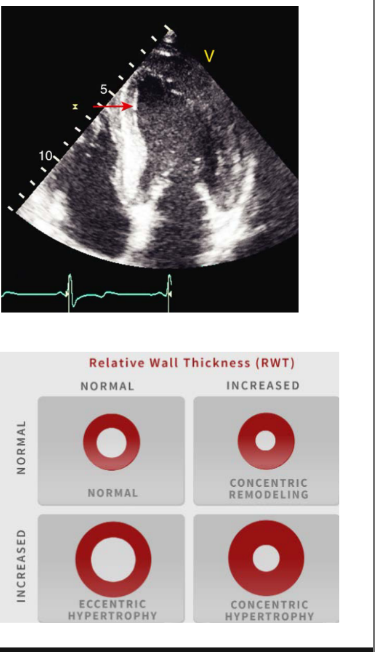
Increased LV wall thickness
Normal to small LV cavity size
Ground-glass appearance of the myocardium*****
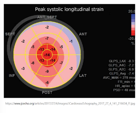
Apical sparing with a “cherry-on-top” strain pattern******
RV hypertrophy
Pulmonary hypertension
Valvular thickening and regurgitation
Progressive diastolic dysfunction and possible pericardial effusion
What is the ground-glass appearance in amyloidosis?
The myocardium has a bright, speckled appearance caused by protein deposits reflecting ultrasound waves differently.

What is apical sparing in cardiac amyloidosis?
The basal and midventricular segments have reduced strain, while the apex remains relatively preserved. - APEX IS AFFECTED LAST
( THATS WHY WE GET A CHERRY ON TOP) (mechanism is idopathtic)
(differental from HCM blue berry)
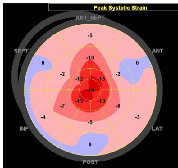
What strain appearance is produced by apical sparing?
A “cherry-on-top” pattern.
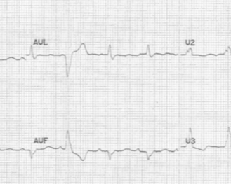
How does the EKG in amyloidosis differ from the EKG in HCM?
Amyloidosis usually has a low-voltage QRS, while HCM usually has a high-voltage QRS.
Simple reminder:
Amyloidosis may look thick on echo but have low electrical voltage on EKG.
List the 4 treatment approaches for amyloidosis.
Supportive medical measures (treating their symptoms)
Treat the underlying disease process
Chemotherapy
Heart transplant (Later stage of amyloidosis)
what causes Gaucher disease?
Rare,
inherited condition that leads to the Fatty substance in the tissue / **myocardium, the buildup causes overall damage different tissues and organs as well not just the heart, spleen, liver, and bones are the most affected, but can affect the myocardium as well, these lipids deposite can acculmate in the pericardium or pericardum space so thats why there is pericardial changes can see thickening, effusion, clafication of the pericardial
What causes Chagas disease, and where is it primarily found? - Where in the world?
Chagas disease is caused by invasion of a parasite into the myocardium and is found primarily in Latin America.
Explain how storage diseases cause restrictive cardiomyopathy.
Deposits occur within the where? causing what list 2?
Deposits occur within the cell membrane causing fibrosis and rigidity
Storage diseases cause abnormal substances, such as fat, iron, or sugar, to build up inside the heart’s cells.
This buildup causes:
Fibrosis: scar tissue forms
Rigidity: the heart muscle becomes stiff
So, simply:
Material builds up inside heart cells → causes scarring and stiffness → the heart cannot relax and fill properly
List the 3 storage diseases associated with RCM.
Hemochromatosis
Fabry disease
Glycogen-storage disease, including Pompe disease
Explain the cause of hemochromatosis.
Hemochromatosis may be an what condition or a what disease - think classifcation.
caused by what? list 3
Hemochromatosis may be an autosomal-recessive condition or a secondary disease caused by excessive dietary iron, blood transfusions, or liver disease.
Hemochromatosis means the body stores too much iron.
Autosomal-recessive: The person inherits an abnormal gene from both parents, causing the body to absorb too much iron.
Secondary: Another problem causes iron buildup, such as too much iron in the diet, many blood transfusions, or liver disease.
hemochromatosis:
What happens when Excessive iron is absorbed by the ______ causes increased what within the _____; including the ____
Excessive iron absorption by the intestines causes increased iron deposits within the organs, including the heart
List the 4 cardiac findings associated with hemochromatosis.
what type of phenotype with what EF?
how is the diastolic function?
list 1 rt heart side affect
how does the late stage appear as?****
Restrictive phenotype with preserved EF
Severe diastolic dysfunction
Pulmonary hypertension
Late stage appears as dilated cardiomyopathy ****
The heart muscle is stiff and cannot relax or fill properly, but it can still squeeze out a normal percentage of blood.
Restrictive phenotype: stiff ventricles with poor filling during diastole.
Preserved EF: the ejection fraction is still normal or near normal.
In the late stage, long-term iron buildup damages and weakens the heart muscle. The ventricle then starts to:
stretch and enlarge
lose its squeezing strength
pump less blood forward
So it can change from a stiff, restrictive heart into a dilated, weak heart, which looks like dilated cardiomyopathy with a reduced EF.
List the 2 treatment options for hemochromatosis.
Phlebotomy therapy
Heart transplant in severe cases
Phlebotomy therapy = Remove blood → remove excess iron → prevent more damage to the heart, liver, and other organs.
what catogory does Fabry disease fit into from like how people get the condtion?
Caused by accumulation of _______in ______-, causing what?
Fabry disease is associated with what disease?
Fabry disease is an X-linked recessive autosomal disease caused by accumulation of sphingolipids in lysosomes, causing fibrosis of the tissue
Fabry disease is associated with Kidney disease.
(lipid and fatty deposits)
Too much fat collects inside the cells, which damages them and makes the tissue scarred and stiff.
Fatty material builds up in cells → cells get damaged → tissue becomes scarred and stiff
Their cells are missing a “clean-up tool” called an enzyme. Because of that, a fatty substance called sphingolipids builds up inside the cell’s recycling centers, called lysosomes.
List the 6 echo findings associated with Fabry disease.
Concentric LV hypertrophy (increased wall thickness, increased mass)
Thickened papillary muscles
Initial preserved LV systolic function
Grade II diastolic dysfunction
“Bilayered” appearance of the endocardium
Inferolateral strain impairment with severe LA strain impairment
SKIP
What type of disease is sarcoidosis? (explain what it is)
Sarcoidosis is a granulomatous (Cluster of immune cells) inflammatory disease.
How does sarcoidosis affect the myocardium? List 2
Cardiac granulomas produce what^
Possibly due to abnormal _____ response?
Cardiac granulomas produce myocardial fibrosis and regional wall-motion abnormalities.
Possibly due to abnormal immune response.
CHAT: Small clumps of inflamed cells form in the heart muscle. These clumps damage the muscle and cause scar tissue. Because some areas become scarred, parts of the heart wall may not squeeze normally.
Granulomas: small clumps of inflammatory cells
Myocardial fibrosis: scarring of the heart muscle
Regional wall-motion abnormalities: certain sections of the heart wall move or squeeze abnormally
What is the typical demographic pattern of sarcoidosis? which age is this most common in and occurs how much in which gender more?
It is most common in young patients around 10–40 years old and occurs approximately twice as often in females.
Which organs are commonly involved in systemic sarcoidosis? list 3
What other test can confrim sarcoidosis?
typically involves lung, skin, eyes
Biopsy can confrim sarcoidosis
List the 6 cardiac echo findings associated with sarcoidosis.
Increased wall thickness
Regional wall-motion abnormalities (NON Coronary issue, the coronary arteries are not blocked due to disposits)
Valvular regurgitation
LV aneurysms in late disease with eventual wall thinning
Pulmonary hypertension***
RV dysfunction from granulomatous involvement of the pulmonary arteries**
(*****sarcoidosis has RT heart, involment PHTN, thickening forms in PA/PV, RV enlatgement, trduce function of RV, PA enlargement, septal flattening can be seen in systole and diastole volume and pressure overload)
An LV aneurysm is a weakened area of the left ventricle that bulges outward instead of squeezing normally.
So, “LV aneurysms in late disease with eventual wall thinning” means that in advanced disease, damaged areas of the left ventricle may become thin, weak, and bulge outward.
List the 2 endomyocardial diseases (disease within the myocardium) associated with RCM.
Hypereosinophilic syndrome (Loeffler’s endocarditis)
Endomyocardial fibrosis
What diagnostic blood finding is associated with hypereosinophilic syndrome? and how many months…
Persistent eosinophilia greater than 1,500 cells/mL for more than 6 months.
(cell issue)
It means the person has had too many eosinophils in their blood for a long time.
Eosinophils = a type of white blood cell
Greater than 1,500 cells/mL = the eosinophil count is abnormally high
For more than 6 months = it is ongoing, not just temporary
In simple terms:
The blood has had too many allergy- and parasite-fighting cells for at least six months. Over time, these extra cells can release chemicals that damage organs, including the heart.
How does hypereosinophilic syndrome cause restrictive cardiomyopathy?
Accumulation of ______within the_____tissue causes ______
Accumulation of Eosinophils within the endomyocardial tissue cause fibrosis.
(scarring of the myocardium)
Because eosinophils release strong chemicals and enzymes to fight parasites and control inflammation.
When there are too many, those chemicals can also attack normal tissue by mistake. In the heart, this can cause:
inflammation → heart muscle damage → blood clots → scar tissue → a stiff heart
So the eosinophils are trying to protect the body, but in large numbers they can end up damaging the heart.
List the 5 echo findings or complications of hypereosinophilic syndrome.
what is happening with the wall and especially where, what is associated findings with this?
Eosinophiles accumulate on where list 3 causing what?
list 3 echo findings
Endocardial wall thickening, especially at the apex
Mural thrombus
Eosinophil accumulation on the papillary muscles, posterior mitral leaflet, and anterior tricuspid leaflet causing regurgitation
Biatrial enlargement, diastolic dysfunction, and pericardial effusion
What major complication can occur from the mural thrombus?
Thromboembolic events.
Where is endomyocardial fibrosis the most common cause of RCM? - in the world
It is the most common cause of RCM in tropical regions, especially Africa.
(remember fibrosis means thicken and scared)
The tropics are the warm areas of the world near the equator.
Explain how endomyocardial fibrosis causes restrictive physiology.
Fibrotic obliteration of LV and RV apices from organized thrombus causing restrictive physiology
A blood clot forms at the pointed ends of the left and right ventricles. Over time, the clot becomes hardened and turns into scar tissue. This scar tissue fills in or blocks the tips of the ventricles, making the heart stiff and unable to fill normally.
Fibrotic obliteration means scar tissue builds up and fills in or closes off a space.
Fibrotic = made of scar tissue
Obliteration = the space is filled in or blocked
In this case, scar tissue fills in the tips of the ventricles, making the chambers smaller and stiffer.
How are the AV valves affected by endomyocardial fibrosis?
The AV valves become tethered, causing regurgitation
List the 6 additional findings associated with endomyocardial fibrosis.
Biatrial enlargement
Diastolic dysfunction
Atrial fibrillation
Pericardial effusion
Pleural effusion
AV valve regurgitation / tethering
What is the prognosis of endomyocardial fibrosis?
The prognosis is poor, with approximately 30–50% mortality within 2 years.
Autoimmune:
What is another name for scleroderma, and what causes its cardiac effects?
Scleroderma is also called systemic sclerosis.
It causes excessive accumulation of connective tissue. (affects the bones, tissue, muscle and tendons)
List the 6 echo findings associated with scleroderma.
Regional wall-motion abnormalities
LV hypertrophy
Valvular sclerosis
Pulmonary hypertension or pulmonary arterial hypertension
Pericardial effusion or pericarditis
Diastolic dysfunction
Valvular sclerosis means a heart valve becomes thick, stiff, and scarred
Valve tissue hardens → valve movement becomes limited.
List the 4 miscellaneous causes of restrictive cardiomyopathy.
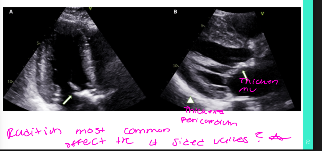
Post-radiation changes
Carcinoid disease
Chemotherapeutic agents
Malignancy
List the 4 early or structural effects of post radiation on the heart. (name one acute and one chronic)
Interstitial myocardial fibrosis
Pericardial fibrosis
Acute pericarditis
Chronic cardiac disease that may appear years or decades after treatment
Interstitial myocardial fibrosis: Scar tissue forms between the heart muscle cells, making the heart stiff.
Pericardial fibrosis: Scar tissue forms in the sac surrounding the heart, which can restrict the heart’s movement.
Acute pericarditis: A sudden inflammation of the sac around the heart, often causing sharp chest pain.
List the 5 chronic cardiac complications of radiation therapy.
Coronary artery disease
Pericardial disease
Cardiomyopathy
Valvular disease
Conduction abnormalities
List the 7 echo findings associated with post-radiation disease.
Reduced cardiac mass
Decreased LV dimensions
Reduced wall thickness
Decreased LV function
Diastolic dysfunction
Myocardial interstitial fibrosis, especially in the RV (echognic)
Valvular fibrosis - radiation most commonly affects the Left sided valves
Myocardial interstitial fibrosis means scar tissue forms between the heart muscle cells.This scarring makes the right ventricle stiff, so it cannot relax and fill with blood normally. Myocardial interstitial fibrosis means scar tissue forms in the spaces between the heart muscle cells.
How does carcinoid disease damage the heart?
Neuroendocrine tumors release tryptophan and serotonin, creating a toxic effect on the endothelial lining.
Why does carcinoid disease mainly affect the right heart?
The chemicals are usually deactivated in the lungs before reaching the left heart.
When can carcinoid disease affect the left heart?
When there is a right-to-left shunt or a lung mass.
List the 4 main valve effects of carcinoid disease.
Fibrotic thickening of the tricuspid valve
Immobility of the tricuspid valve
Possible pulmonary valve involvement
Tricuspid malcoaptation and fixed causing tricuspid stenosis and severe tricuspid regurgitation
Simple reminder:
Carcinoid causes fixed TV leafleat, not coapting b/c they are fibrotic, gap, decreased function of the RA & RV, terriontial TR, thickened right-sided valves, especially the tricuspid valve.
How can chemotherapeutic agents damage the heart? and causes what list 2
They can damage blood vessels and produce extensive myocardial and interstitial fibrosis.
Chemotherapeutic agents are medicines used to treat cancer.
Some of these medicines can injure the heart’s blood vessels and heart muscle. The damaged areas may heal by forming scar tissue, called fibrosis.
Extensive fibrosis: a large amount of scarring
Interstitial fibrosis: scarring between the heart muscle cells
List the 4 consequences of interstitial fibrosis caused by chemotherapy.
Myocyte cell death
Latent ischemia
Thrombosis
Endothelial damage
Latent ischemia means there is reduced blood flow and oxygen to part of the heart, but it may not cause obvious symptoms yet.
Latent = hidden or not noticeable
Ischemia = not enough blood and oxygen reaching tissue
What symptoms or events may result from chemotherapy-related interstitial fibrosis? list 2
Chest pain or myocardial infarction.
What happens when pericardial fat is replaced by what? List 3
The patient may develop myocarditis, pericarditis, or pericardial effusion.
When the fat around the heart is replaced by thick scar-like connective tissue, the tissue around the heart becomes stiff and irritated.
This can lead to:
Myocarditis: inflammation of the heart muscle
Pericarditis: inflammation of the sac around the heart
Pericardial effusion: fluid collecting around the heart
In simple terms: the normal soft fat is replaced by hard scar tissue, which can inflame the heart or cause fluid to build up around it.
List the 3 additional cardiovascular effects of chemotherapeutic agents.
Hypertension or hypotension
Conduction abnormalities
When can chemotherapy-related cardiotoxicity occur? & what is the most important thing to check on echo
It may occur at the start of therapy or several weeks after treatment.
important LV function and strain ***
List the 3 findings that may occur several weeks after chemotherapy.
Heart failure
Decline in myocardial function
Decreased LVEF and longitudinal strain
What monitoring is required when a chemotherapy drug is known to be cardiotoxic? list 3
Serial cardiac monitoring of LV systolic function, including LVEF and longitudinal strain
List the 2 cardiac tumors included under malignancy.
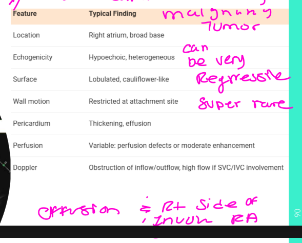
Angiosarcoma
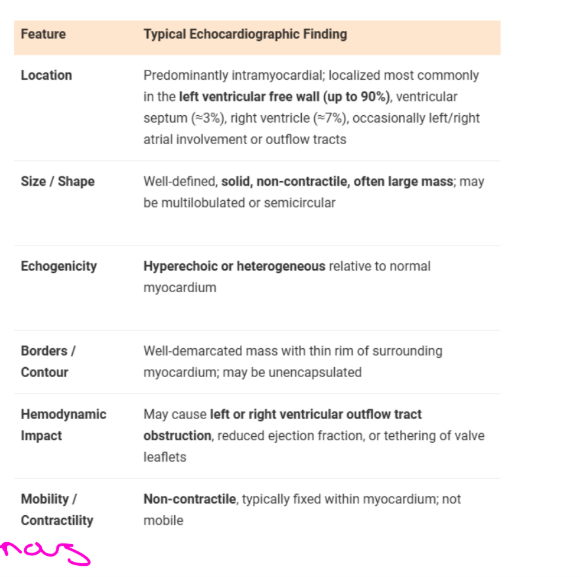
Fibroma
How can malignancy produce restrictive physiology?
Tumor infiltration between myocardial cells can interfere with normal myocardial relaxation and filling.
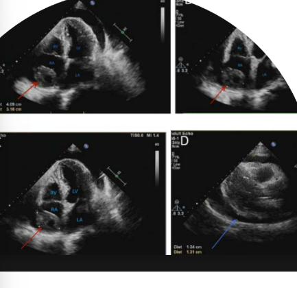
Which type is the most common maligancy?
Angiosarcome is the most common primary maligant tumor (can be very regressive and super rare) (mostly in the RA located) rt side involment with RA, effusion
where can you find the Fibroma ?
In the LV free wall 90% of tumor are located here
Match the following 6 classic findings with the correct disease.
Ground-glass myocardium and apical sparing —
Iron deposition —
Concentric LVH with inferolateral strain impairment —
Granulomas and regional wall-motion abnormalities —
Apical fibrosis and mural thrombus —
Fixed right-sided valves with severe TR —
Ground-glass myocardium and apical sparing — Amyloidosis
Iron deposition — Hemochromatosis
Concentric LVH with inferolateral strain impairment — Fabry disease
Granulomas and regional wall-motion abnormalities — Sarcoidosis
Apical fibrosis and mural thrombus — Hypereosinophilic syndrome/endomyocardial fibrosis
Fixed right-sided valves with severe TR — Carcinoid disease