TEAS 7 Cardiovascular system
 Call Kai
 Learn
Learn Practice Test
 Spaced Repetition
 Match
Match Flashcards
 Knowt Play
1/37

Earn XP
Description and Tags
Anatomy & physiiology
Name | Mastery | Learn | Test | Matching | Spaced | Call with Kai | Chat |
|---|
No analytics yet
Send a link to your students to track their progress
38 Terms
Function of system
Gas exchange, delivers nutrients, delivers hormones, & waste removal
Blood
Red liquid that circulates in the arteries & veins of humans & vertebrate animals
Transportation
Carries oxygen & carbon dioxide to and from the tissues of the body
Characteristics
Always red in color
Brighter red = more oxygen
Function
Maintains Homeostasis
PH, Temperature, Osmostic pressue
Transports
Hormones
nutrients
Gases
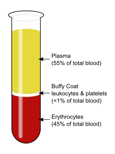
Blood composition
TOP: Plasma 55%
Liquid: Lipids, salt, proteins, water
MID: Luekocytes + platelets 1%
WBC- fight infection
Platelets- Blood clotting (think scraping knee)
BOT: Erythrocytes 44%
RBC- transport gases
Hemoglobin - Iron rich protein in RBC (makes blood red)
Heart
Pump that pushes blood through the arteries
Arteries
Blood vessels that carry blood away from the heart
Veins
Blood vessels that carry blood to the heart
Capillaries
Blood vessels that exchange material between blood & cells
Site of gas exchange
Smallest blood vessels
Nemonic memorization for blood flow through the heart
A= AWAY
Arteries
Carry blood away from the heart
Oxygen rich
V= VERB= Veins efficiently return blood
Veins
Carry blood away from the heart
Oxygen poor (except in pulmonary circulation)
Capilaries
SIte
The heart: 3 tissue layers
Epicardium(outer layer): Protects the heart & secretes lubricating serous fluid
Myocardium (middle layer): Contracts to pump blood
Endocardium (inner layer): Lines the chambers & valves
The heart: 4 chambers
Right atrium: Recieves blood form vena cava
Left atrium: Recieves blood from pulmonary veins
Right ventricle: Pumps blood into the pulmonary trunk
Left ventricle: Pumps blood into the aorta
Atria- thin walls
Ventricle- thick walls
Memory trick: A comes before V = atria on top, ventricles on bottom
Heart valves
Heart valves prevent backflow of blood
-Tricupsid valve (AV): Prevents backflow into the atrium when the ventricle contracts
-Pulmonary semilunar valve: Prevents return of blood into the right ventricle
-Bicupsid valve (Left AV): Prevents blood from entering the left atrium when the ventricle contracts
Coronary Blood flow
Coronary Arteries:
Originate from the aorta and delivers nutrients & oxygen to the heart
Coronary Veins:
Deoxygenated blood returns to the right atrium from the coronary sinus
Cellular respiration
Glucose is burned or combined with oxygen
The oxygen allows cells to transfer energy in glucose to ATP
O2, CO2, waste, & nutrients
Oxygen: Moves down partial pressure gradient from air to blood of alvelor capillaries
O2 binds to hemoglobin in RBC or diffueses in RBC
Nutrients (glucose, fats, amino acids): Circulate & are absorbed (target cells)
Hormones: released by endocrine glands in the same way ^^
Lipid soluble molecules: Require use of carrier protein
WASTE
Carbon dioxide: Transported away from tissues & diffuses out of alvelor capillaries
CO2 CAN bind to hemoglobin or diffuse in blood; usually travels in the form of bicarbonate ions (similar to oxygen)
Other waste (like urea) are brought to the kidneys and filtered
Kidneys also regulate fluids & ions in blood
Thermoregulation
Human body: 98.6 degree F
Sensory neurons (thermoreceptors) detect temp changes »
Send impulses to hypothalamus »
to effectors to»
smooth muscles so they relax with heat
Endothelial cells
Thin inner lining of blood vessels, inside of heart and entire circulatory system
Squamous endothelial cells connected by tight junctions & adherens junctions
Selectively permeable barrier between the blood & surrounding tissue
Secrete chemicals which inhibit coagulation of blood
Systolic & diastolic pressure
Blood pressure: Force per unit area exerted by blood on vessel walls
Systolic pressure: Maximum pressure exerted in arteries
Contraction of the heart
LUB sound
Under 120
Diastole: Lowest pressure in arteries
Relazation of the heart
DUB sound
Under 80
Ventricles relax & blood pressure is at lowest point
Ideal Blood Pressure = 120 over 80
Systemic circuit
Delivers oxygen rich blood to body, oxygen poor blood to the heart
Longest body circuit
Higher blood pressure than pulmonary circulation
Path: Left ventricle » aorta » arteries » arterioles » capillaries » venules » veins » superior & inferior vena cava » right atrium
What happens?: Oxygen & nutrients leave the blood & enter body cells, carbon dioxide & wastes enter blood
Blood returning to the heart is deoxygenated (low O2)
Arteries oxygen blood content
Oxygen- rich blood
Veins oxygen blood content
Oxygen- poor blood
Pulmonary circuit
Get rid of carbon dioxide & pick up oxygen carries blood from the heart to lungs back to the heart
Path: Right ventricle » lungs » left atrium
Pulmonary arteries: Carry deoxygenated blood
Pulmonary veins: Carry oxygenated blood
(Opposite of systemic)
Electrical conduction system
The hearts natural pacemaker
SA node (sinoatrial): Starts the electrical signal, atria contracts » P wave on ECG (60 to 100 BPM)
Bachman bundle:
signal from SA node to left atrium
Internodal pathways
Anterior, middle, posterior
Signal from SA node to AV node
Secondary pacemaker
AV node (Atrioventricular): Shows signal briefly, delays signal from SA node, contracts atria, gives ventricle time to fill with blood » PR on ECG (40 to 60 BPM)
Bundle of his
ONLY route between atria & ventricles
Right bundle branch: Signal to right ventricle
Left bundle branch: Signal to left ventricle
Purkinje fibers- Last ditch pacemaker if SA & AV node fail
Connect with myocytes
Initialize depolarization » contraction (20 to 40 BPM)
Path: Bundle of his » bundle branches » purkinje fibers (carries electrical impulse to ventricles that depolarize & contract (QRS complex))
Electrical conduction system mnemonics
Strong: SA node (sinoatrial) 60-100 BPM
Arteries: AV node (atrioventricular) 40-60 BPM
Benefit: Bundle of his
Bodys: Bundle branches
Performance: Purkinje fibers 20-40 BPM
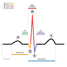
The cardiac cycle (ECG waves simplified)
QRS complex: Ventricles squeeze, Ventricle depolarization (electrical activation)
Inverted V on chart (memory trick V = ventricles)
Conceals atria repolarization
P wave: Atria squeeze, Atria depolarization (electrical activation)
T wave: Ventricles relax, ventricle repolarization
Depolarization = Contract
Repolarization = Relax
Aorta blood pressure
Blood pressure is highest in the aorta
Walls of blood vessels (EXCEPT capillaries) 3 layers
Tunica intima (innermost)
Tunica media (middle)- smooth muscle cells & elestci fibers
Tunica adventitia (outer)
Arterioles (Atrial systems)
Structure
Tiny vessels lead to capillary beds
Tunica media is thin but mostly smooth muscle cells
Fucntion:
Primary vessels involved in vasoconstriction/ vasodilation. Control blood flow to capillaries
Elastic arteries (Atrial systems)
Structure:
Aorta + major branches
Tunica media has most elastin
Largest vessel in artial system
Function:
Stretch when blood is forced out of heart, recoils under low pressure
Muscular arteries (Atrial systems)
Structure
Arteries that branch off of elastic arteries
Tunica media has more smooth muscle cells
Function
Regulate blood flow by vasoconstriction/vasodilation
Venules (Venous systems)
Structure:
Tiny vessels exit capillary beds
Thin pourus walls
Few muscle cells & elastic fibers
Function
Empty blood into larger veins
Veins (Venous systems)
Structure:
Thin tunica media & intima
Wide lumen
Valves prevent backflow of blood
Function:
Carry blood back to the heart
Capillary bed
Interconnected capillaries that facillitate gas exchange & solutes between blood and interstital
Continuous capillaries
Fenstrated capillaries
Sinusoidal capilaries
Blood flow through the heart
Deoxygenated blood
Superior vena cava (upper half of body blood ) /inferiior vena cava (lower healf of body blood)
Right atrium
Tricupsid valve
Right ventricle
Pulmonic valve
Pulmonary artery
Lungs
^ Oxygenated after this and pumped to heart
Oxygenated blood
Lungs
Pulmonary vein
Left atrium
Bicupsid/mitral valve
Left ventricle
Aorta
Septal defects
Opening in the septum (wall) that separates the hearts upper and lower chambers.
Septum defects are often congential (from birth)
Septum function in heart: Separtes oxygen rich blood from oxygen poor blood between the chambers
Interatrial septum: Thin, muscular structure that consists of 2 parts- fossa ovalis & limbus of fossa ovalis. (Separate right & left atria)
An opening here would cause- Atrial septal defect (ASD)
-Congenital heart defect where there’s an abnormal opening, allowing blood to flow betweem the 2 atria
Interventricular septum: Thick, muscular wall that consists of 2 parts- a membranous & muscular portion (Separate the right & left ventricle)
An opening here would cause- Ventricular septic defect (VSD)
-Congenital heart defect charactered by 1 or more holes in the interventricular septum, allowing blood to mix between the ventricles
WBC leukocyte
granulocytes
monocyte
lymphocyte
Blood types
blood composiiton
erythrocute production destruction function