NPTE : TMJ + Spine (joints , actions , ranges , etc)
1/71
There's no tags or description
Looks like no tags are added yet.
Name | Mastery | Learn | Test | Matching | Spaced |
|---|
No study sessions yet.
72 Terms
Lower TMJ joint
type of joint
articulation between
Hinge Joint
- mandibular condyle
(covered in fibrocartilage + dense fibrous CT)
+
- inferior disc surface.

Upper TMJ Joint
type of joint
articulation between
Gliding Joint
- superior disc surface
+
- articular eminence
What is unique about the two joints of the TMJ compared to other synovial joints?
Articular Surfaces are covered in fibrocartilage
rather than hyaline cartilage
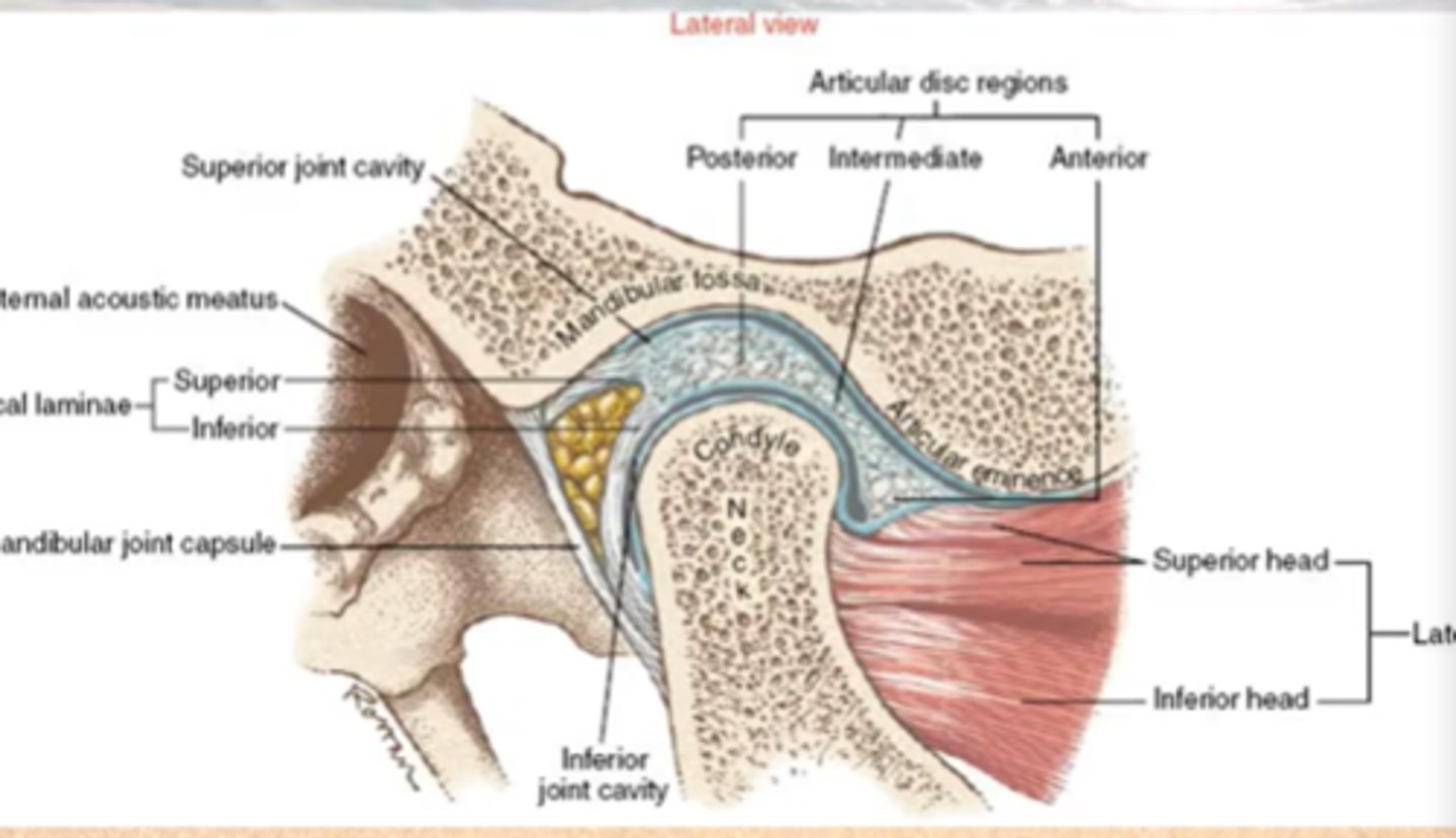
Weighbearing surface of the TMJ Intraarticular Disc
compare this potion of the disc to the rest of the disc
Intermediate : between condyle and temporal bone
- thinnest portion
- NOT innervated

How does the intra-articular disk of the TMJ move
moves WITH the condyle most of the time

intra-articular disc of the TMJ : attachments
Anterior
- bone
- joint capsule
- tendon of lateral pterygoid muscle (superior head)
Posterior = bilaminar retrodiscal tissue

Anterior Attachment of Disc of TMJ
tendon of lateral pterygoid muscle (superior head)
Posterior Attachments of Disc of TMJ
Posterior = bilaminar retrodiscal tissue

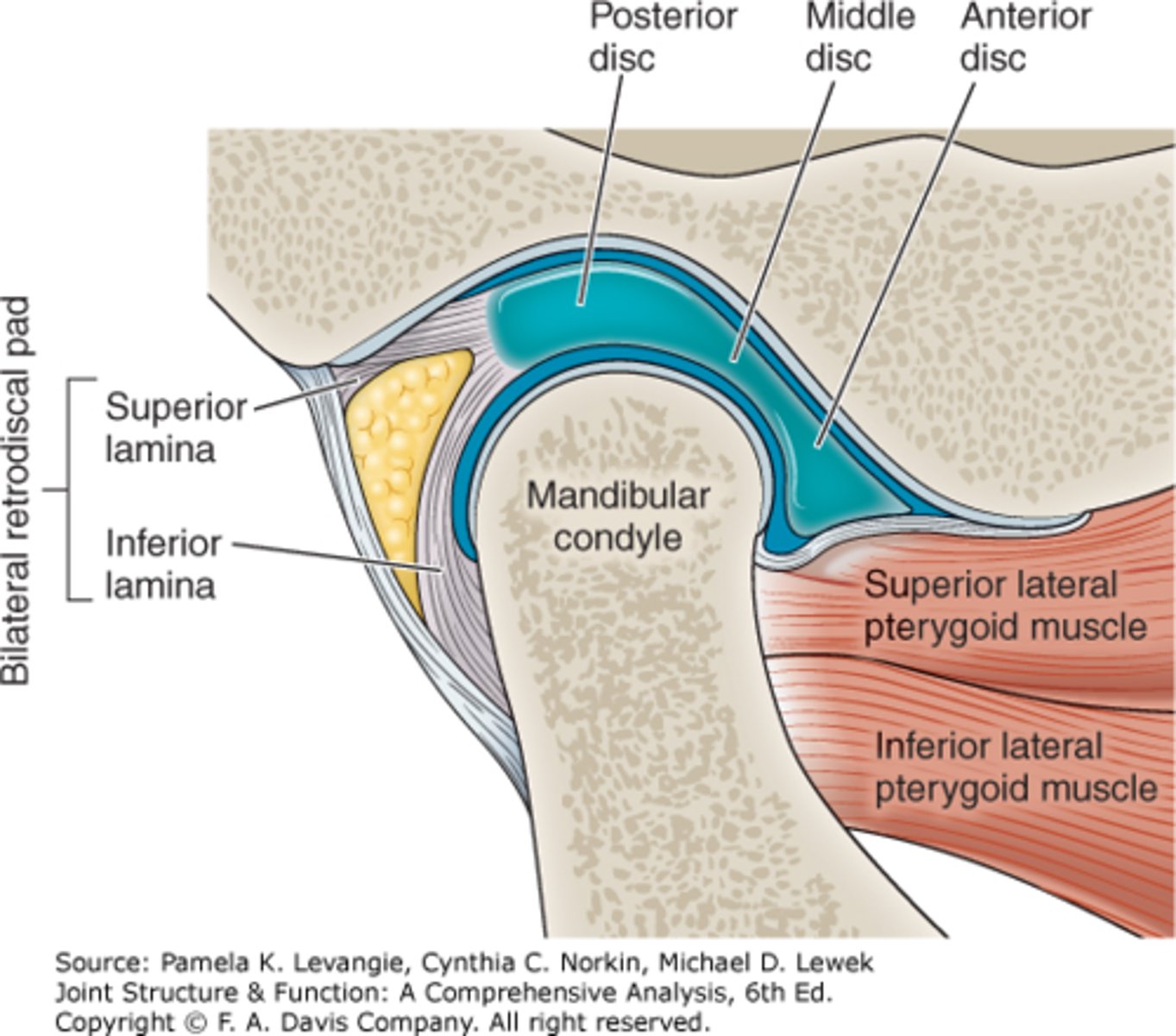
Bilaminar Retrodiscal Tissues of TMJ
location + function
superior vs. inferior
- attachments
- composition
posterior anchor to limit anterior translation of the disc
Superior Lamina = elastin rich
- attaches to the temporal bone
Inferior Lamina = collagen rich
- attaches to the neck of the mandibular condyle

Limits Anterior Translation of the Intraarticular disc of the TMJ
parts?
Bilaminar Retrodiscal Tissues
Superior Lamina = elastin rich
- attaches to the temporal bone
Inferior Lamina = collagen rich
- attaches to the neck of the mandibular condyle

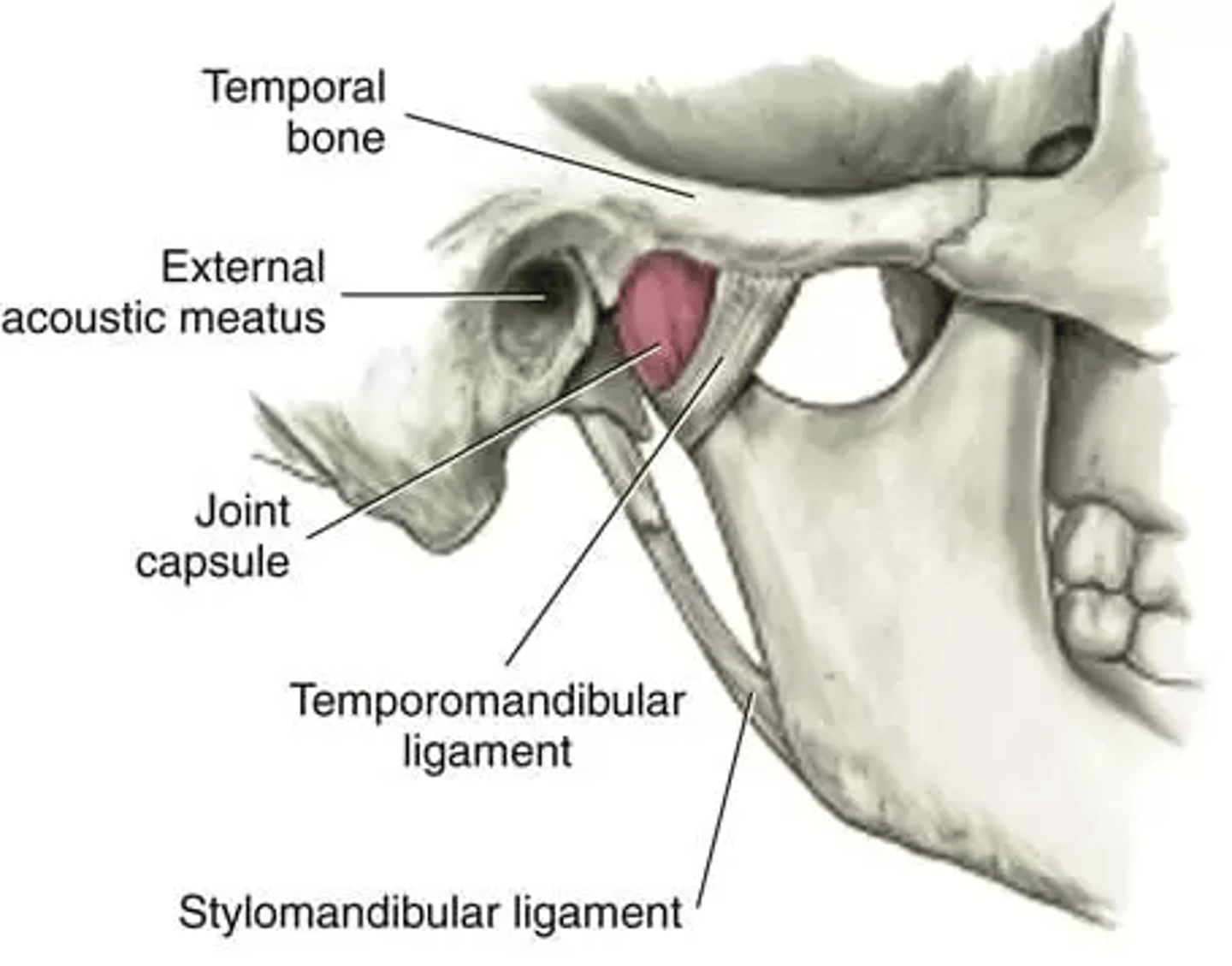
TMJ Joint Capsule
attachments
is it vascularized / innervated?
compare its tightness A-P vs. M-L
- why is this important
superiorly
= temporal bone
inferiorly
= neck of mandibular condyle.
Highly vascularized + innervated
Loose (lax) A-P
- easier opening + closing
Tighter Med-Lat
= for lateral stability with chewing
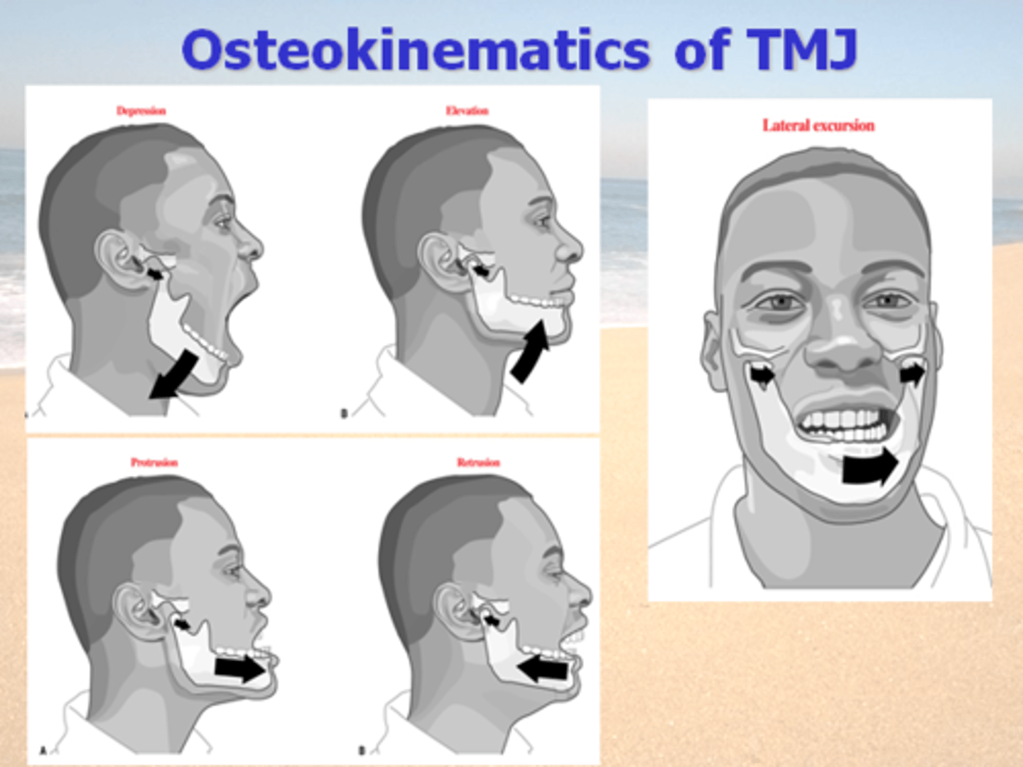
OSTEOKINEMATICS: TMJ
movements available (5)
- distances of each in mm
mandibular movements
Depression (opening): 35-50 mm
-requires protrusion
Elevation (closing)
- requires retrusion
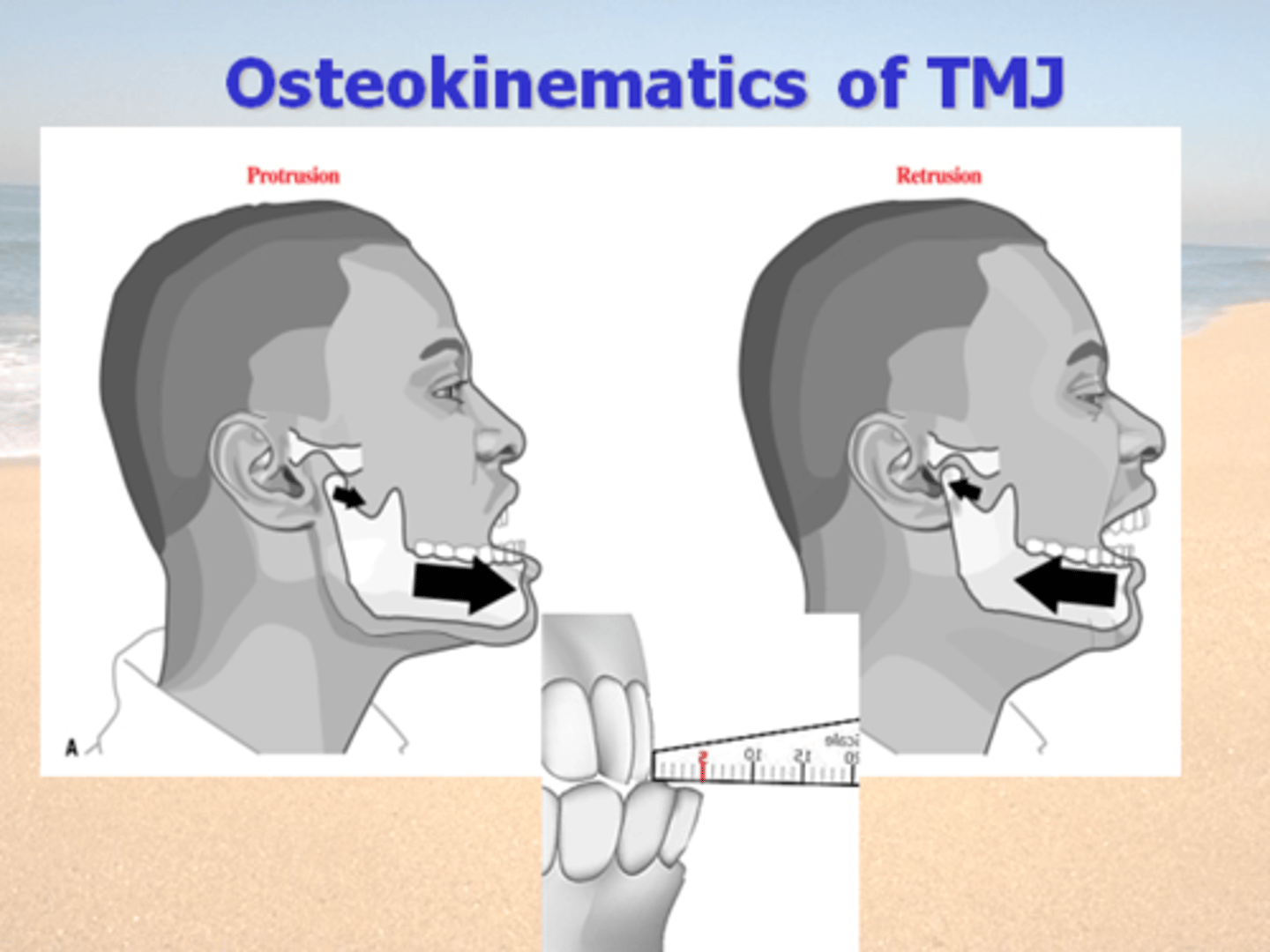
Protrusion (chin forward): <8 mm
Retrusion
(chin backward; aka: Retraction): <5 mm
Lateral Deviation
(lateral jaw excursion) 10-15mm
Protrusion vs. Retrusion
average motions
Paired with what other motions
Protrustion = <8 mm
= depression
Retrusion = <5 mm
= elevation
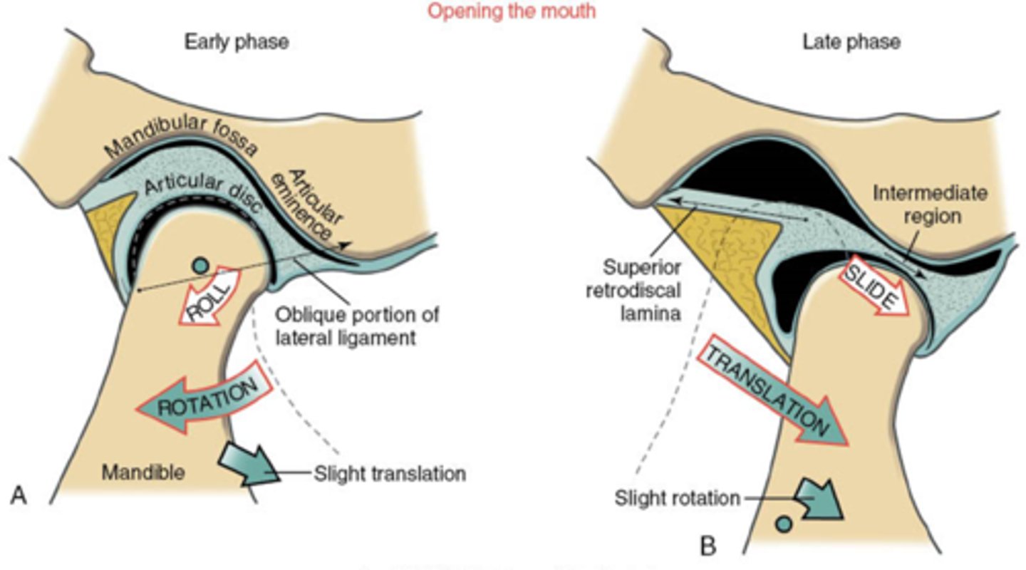
ARTHROKINEMATICs of TMJ : Depression vs. Elevation
2 main principals
where does each occur
when does each occur
describe each TMJ motion
1) Rotation = lower joint space
mandibular condyles rolls on inferior surface of disc
= first < 50% of opening
2) Translation = upper joint space
condyle AND disc slide together
= added to rotation during last 50+% for full opening.
TMJ elevation (closing) = reverse order.
TMJ protrusion and retrusion
= all translation of condyle-disc complex.
TMJ lateral deviation
= mostly translation of condyle-disc complex
+ some multiplane rotation (mostly horizontal plane)
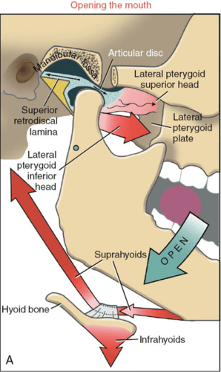
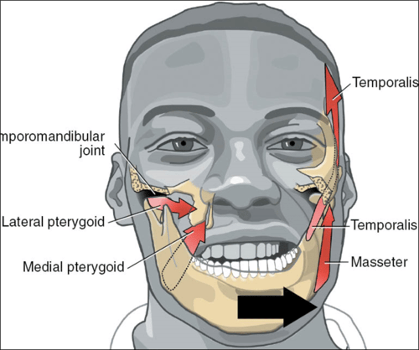
TMJ Musculature : Depression
(opening)
bilatateral
lateral pterygoids (inferior head)
suprahyoids
(submandib ms)
TMJ Musculature : Protrusion
paired with what other motion
Paired with Depression
bilateral
primarily lateral pterygoids
+
- masseters
- medial pterygoids

TMJ Musculature : Retrusion
paired with what other motion
Paired with Elevation
bilateral
temporalis
+
suprahyoids

TMJ Musculature : Lateral Deviation
Contralateral Pterygoids
+
Ipsilateral
- temporalis
- masseter
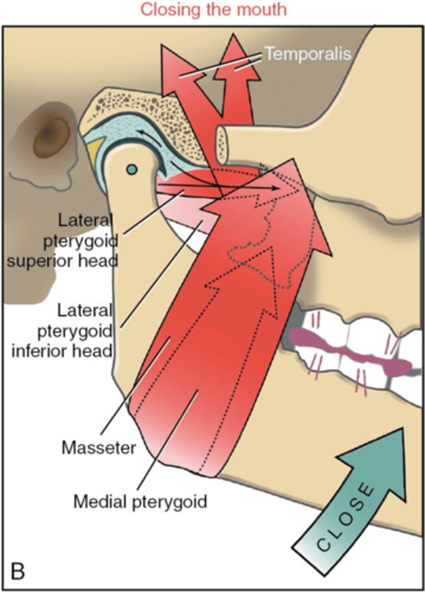
Muscles attached to TMJ Disc
Superior head of lateral pterygoid
note : eccentric action during closing of mouth to manage disc position
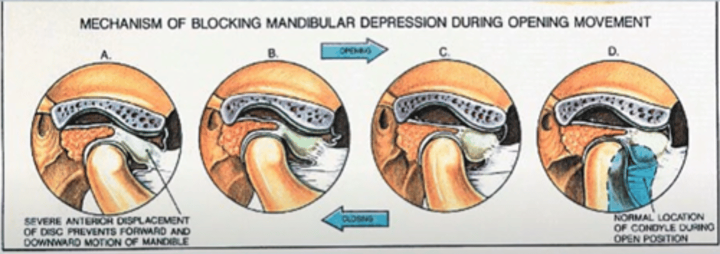
TMJ Hypomobility (+ misalignment)
- what is hypomobile?
- what does it result in?
= anteriorly displaced disk (= stuck)
- limited ROM (blocked by the disk)
+
- pain (loading on improper area of the disk)
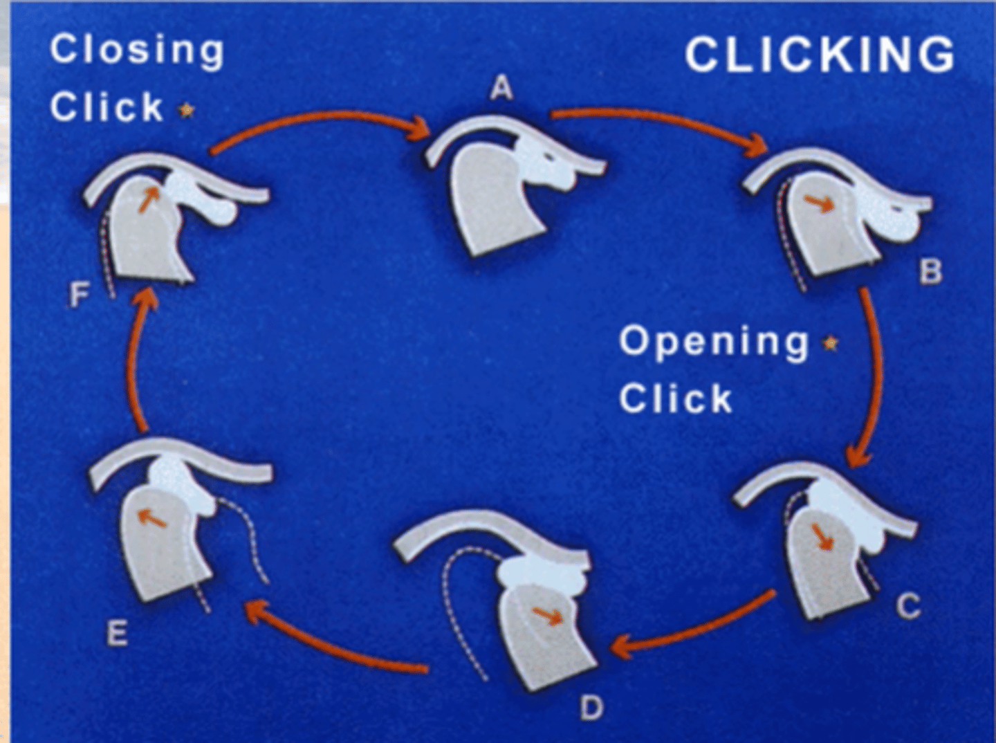
TMJ Hypermobility
(popping) disc
(typically anteriorly displaced)
NOT STUCK = moves too much
= pain and degeneration of disk.
Classic scenario:
- opening pop of the disk
and
- closing pop of the disk
as it snaps in and out of place on the condyle.
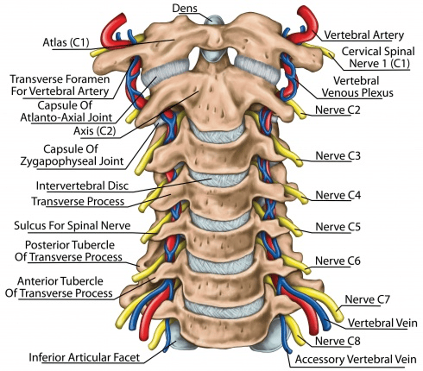
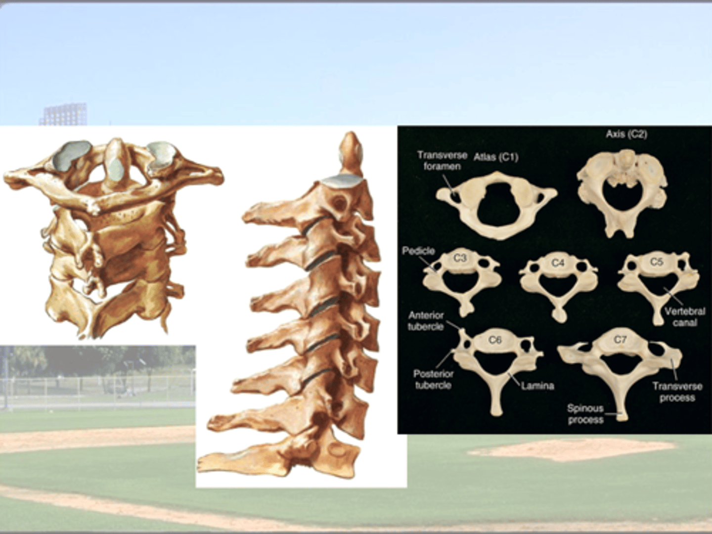
Anatomy of the Spine
how many vertebrae (including fused)
what are the regions (+ how many vertebrae in each)
how many spinal nerves
33 vertebrae (9 fused)
Cervical = 7
Thoracic = 12
Lumbar = 5
Sacral = 5 (fused)
Coccyx = 4 (fused)
How many IVDs
23
none between:
9 fused vertebrae
+
Occipital - C1
+
C1 - C2
how many spinal nerve
how are they named
31 pairs
C1-C7 = comes out above vertebrae
C8
T1-S5 = comes out below vertebrae
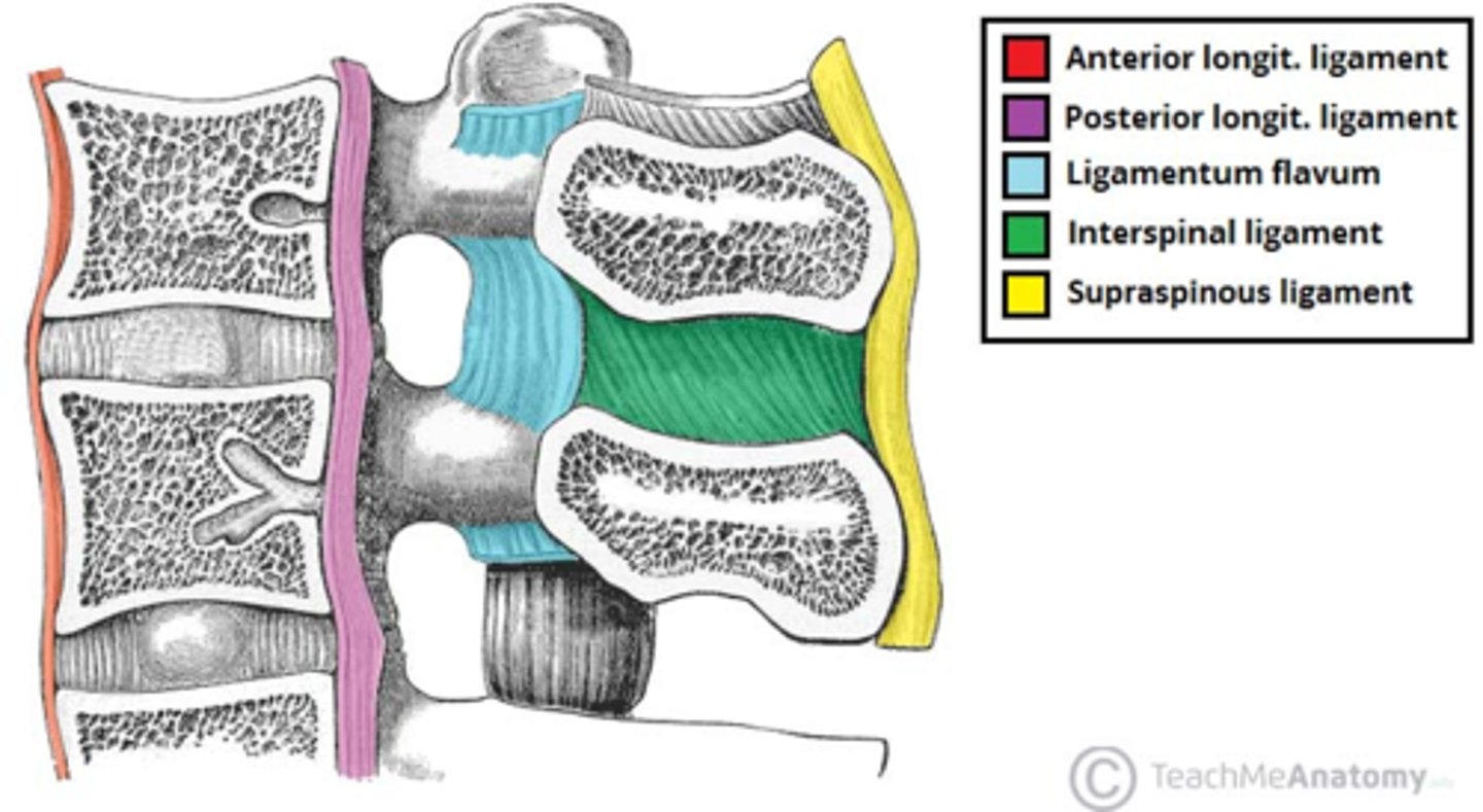
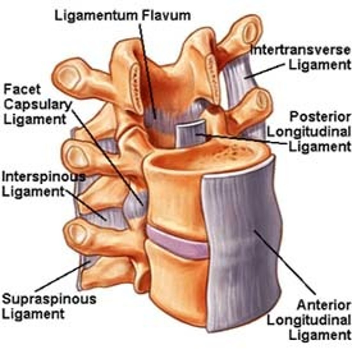
Major Ligaments of the Spine
what do they prevent
NARROW vs. BROAD
Anterior Longitudinal Ligament
C1 - sacrum;
= tightens w/ extension
= narrow (c-spine) -> Wide (L-spine)
Posterior Longitudinal Ligament
C2 - sacrum
= tightens w/ flexion
= Wide (c-spine) - Narrow (L-spine)
Ligamentum Flavum
C2 - sacrum = lamina to lamina
= tightens w/ flexion
Interspinous Ligament
– tightens w/ flexion; weaker
•Supraspinous Ligament
C7 - sacrum
(= Ligamentum Nuchae in C-spine)
– tightens w/ flexion; weaker
Only spinal ligament to prevent EXTENSION
where does it run from/to
describe its shape from c-spine --> L-spine
Anterior Longitudinal Ligament
C1 - sacrum
narrow (c-spine) --> wide (L-spine)
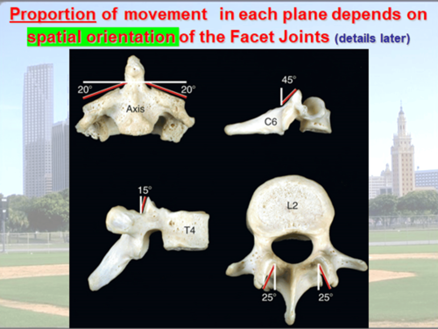
What determines the AMOUNT of movement between two vertebrae
vs.
The Direction of Movement (ie in what plane)
Amount of movement = Intervertebral Jt.
vs.
Direction of movement in each plane = Facet Jts
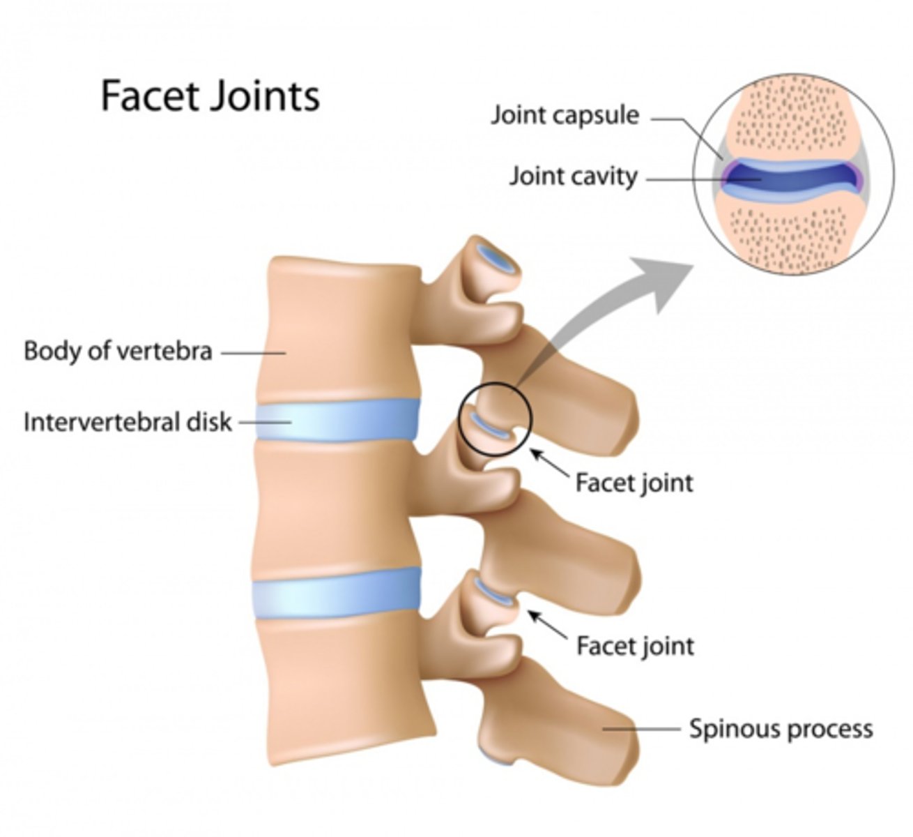
Typical Spinal Segment :
- how many joints
- name the joints + what type are they
2 vertebrae
2 facet joints (synovial)
1 intervertebral (cartilaginous)
3 total
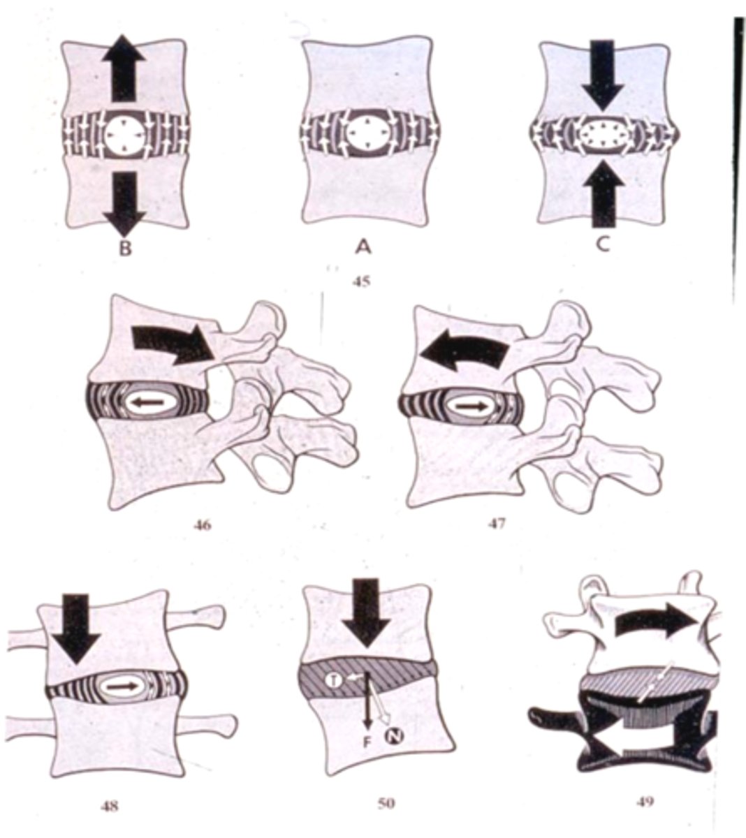
When a spinal segment is in extension
- where is the tension (stretch) in the anulous fibrosis (AF) and which direction is the nucleus pulposous (NP) pushed (due to compression)
What about when a segment is in Flexion?
AF = tension is Anterior
NP = pushed Anterior
flexion = opposite
What determines the AMOUNT of movement between two vertebrae
Intervertebral Jt.
The PROPORTION of movement in a given plane
Facet Jts
Spinal Segment
= made of up _________
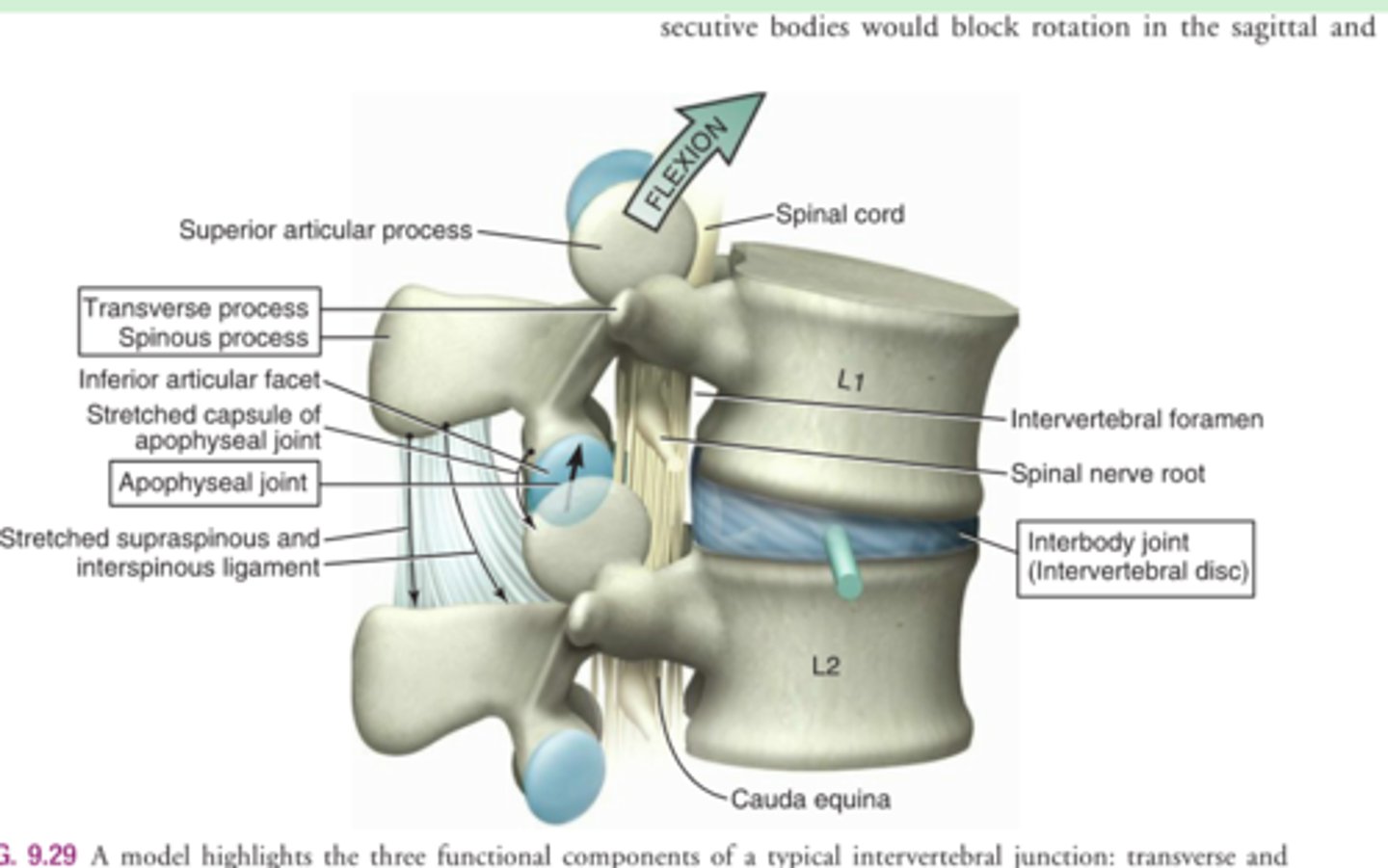
Arthrokinematics of Spinal Segment Motion
= 2 vertebrae, disk, & facet joints
“Top moving on Bottom” motion
Facets glide/slide
(arthrokinematics)
Flexion
anterior-superior slide (top moving)
= open pack position.
Extension
posterior-inferior slide (top moving)
= closed pack position
Rotation & Sidebending
= also top-on-bottom
Coupling of Motion
Which motions are coupled together
Type I vs. Type II
Rotation and Lateral Flexion (Sidebending)
at most but not all regions of the spine.
All assume NEUTRAL starting position
Type I Motion Coupling Pattern:
- Rotation + sidebending
occur together
BUT
opposite directions
Where do this occur
- O/A
(not A/A - does rotation only)
- mid & lower T-spine
- all of L spine.
Type II Motion Coupling Pattern: Rotation and sidebending
together + in same direction
Where does this occur
- lower C-spine
- upper T-spine

Type I Motion Coupling
what motions
same or opposite directions
where does it occur
Rotation + sidebending
occur together
BUT
opposite directions
Where :
- O/A
(not A/A - does rotation only)
- mid & lower T-spine
- all of L spine.

Type II Motion Coupling
what is it
where does it occur
Type II Motion Coupling Pattern: Rotation and sidebending
together + in same direction
Where does this occur
- lower C-spine
- upper T-spine
Sub regions of the Cervical Spine (2)
define (inc how many segments in each)
Suboccipital (upper C-spine)
= occipital bone, C1, & C2
(2 spinal segments)
Lower Cervical Spine
bottom of C2 - Top of T1
(6 spinal segments)
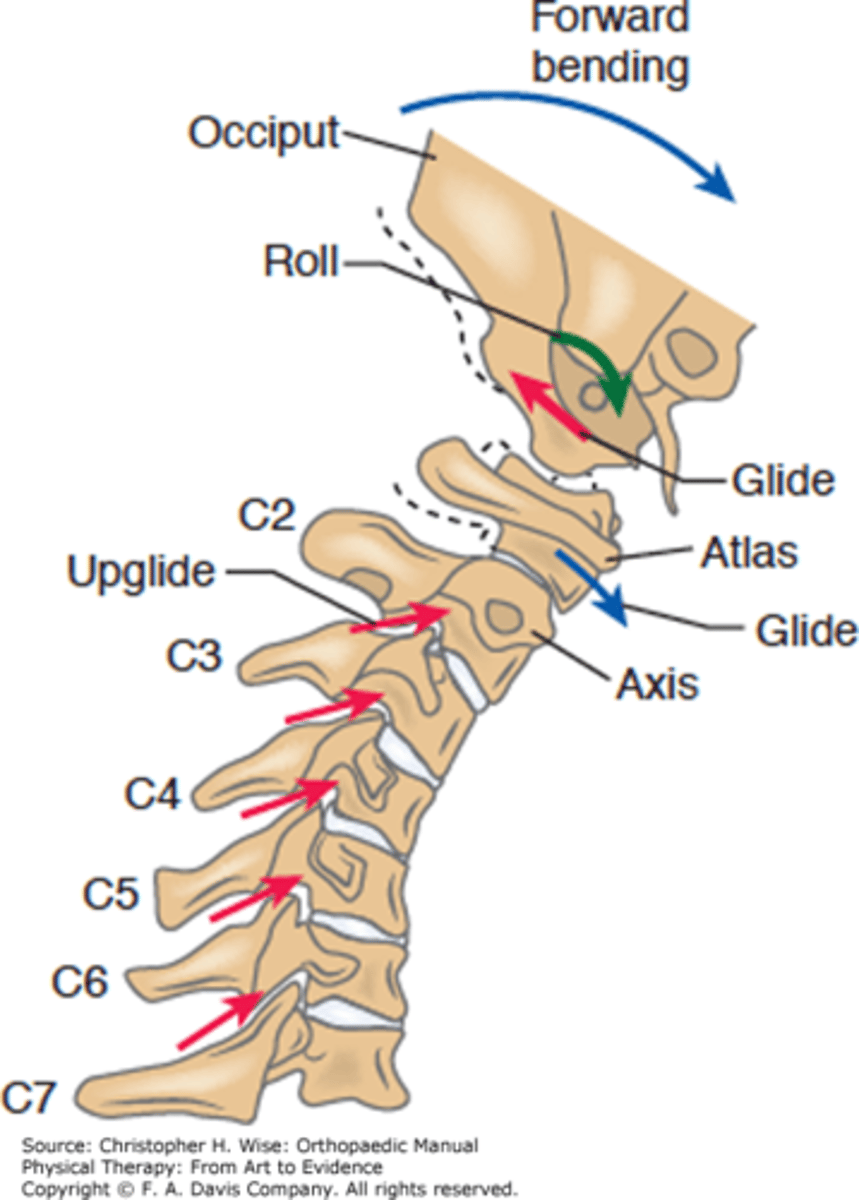
Suboccipital Joint Motions : upper joint
name the joint
articulating surfaces
arthrokinematics
motions (inc ranges)
why is it unique
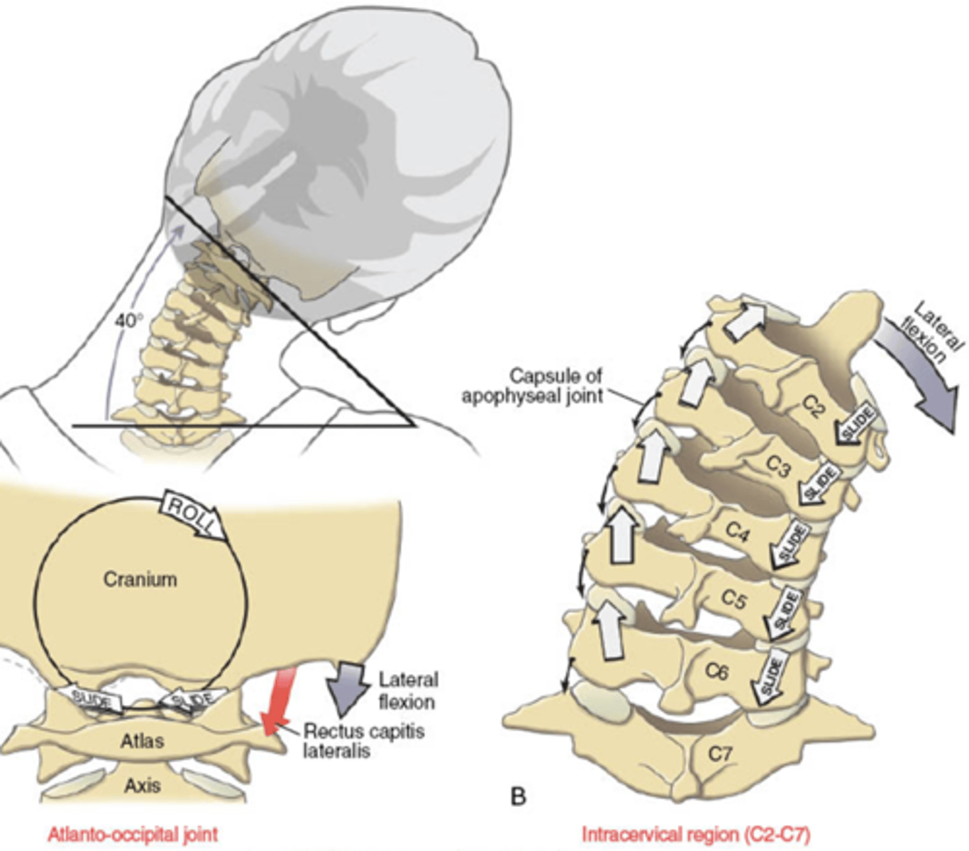
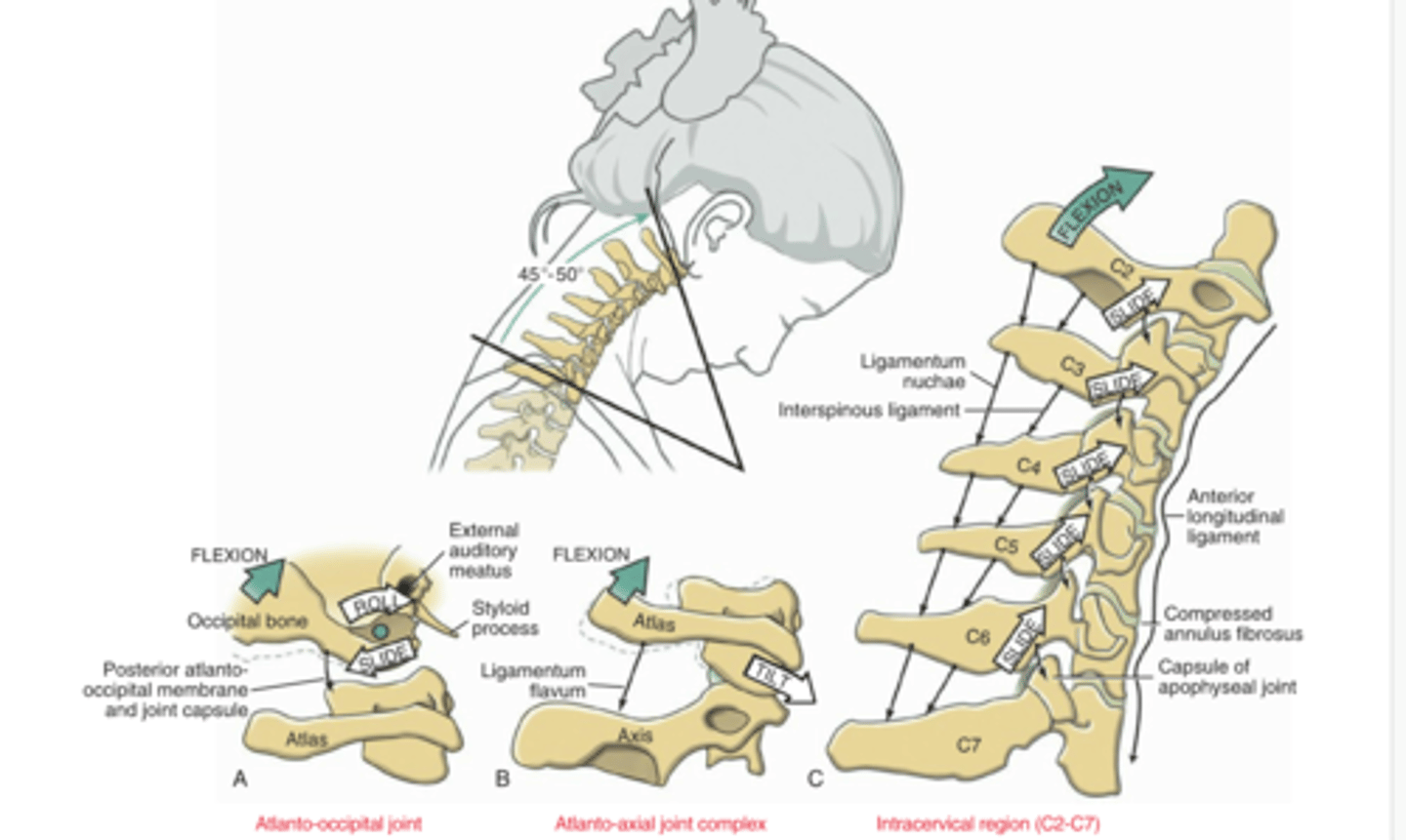
Occipito-Atlantal Joint (OA):
2 x occipital condyles (convex)
+
2 x articular surfaces of atlas (CC)
NO DISK
Motion
- Flexion/Extension ~15°of each (nodding of head)
+ small amounts of
- Lateral Flexion & Rotation
(Type I motion)
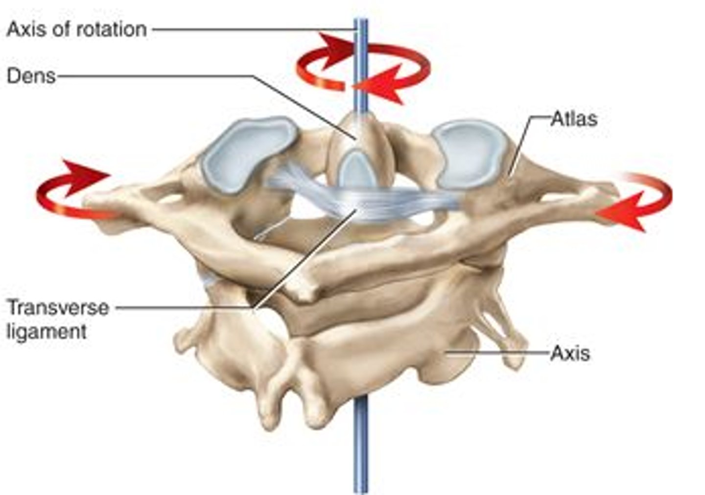
Suboccipital Region : lower joint
name the joint(s)
articulating surfaces (3)
arthrokinematics
motions (inc ranges)
why is it unique
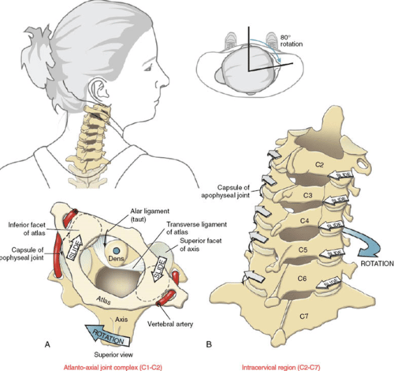
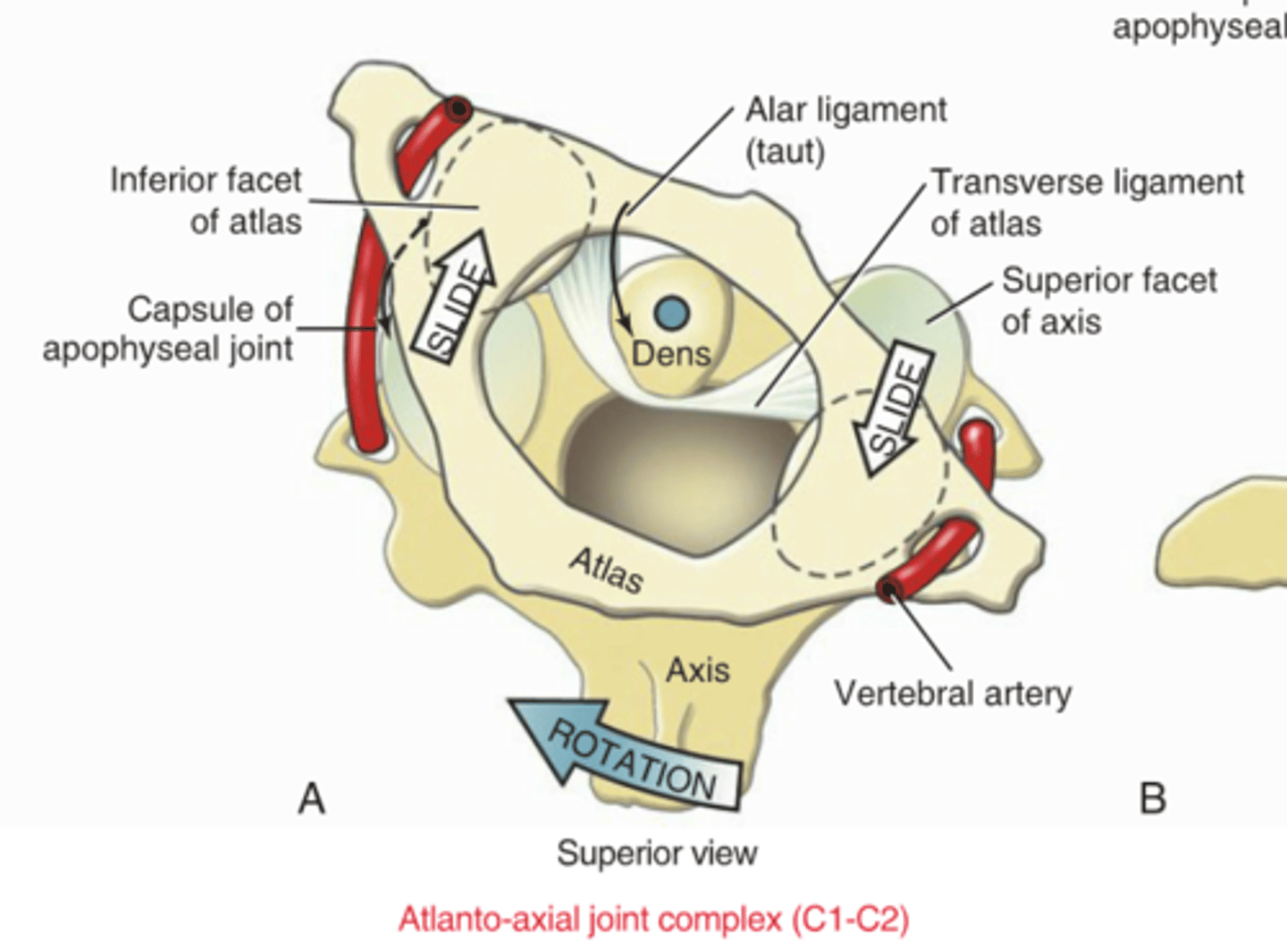
Atlanto-Axial Joint (AA)
3 synovial joints
– 1 pivot joint
- 2 facet joints
NO DISK.
Motion:
Rotation = 35-40°
but NOT coupled
+
Flexion/Extension is minimal (10-15°)
+
Lateral Flexion is even less
rotation atlas (C1)
on
stationary axis (C2)
Ex: Left rotation = atlas turns CC on axis.
IMPORTANT: Approximately 50% of the total rotation of the entire cervical region occurs at the AA before rotation occurs in the rest of the cervical region.
Which joint has the max rotation of c-spine
What is the ROM
Atlanto-axial (AA) Joint
35-40°
Describe the Progression of C-spine Rotation
Approx 50% of the total rotation occurs at the AA
BEFORE
any rotation occurs in the rest of the cervical region.
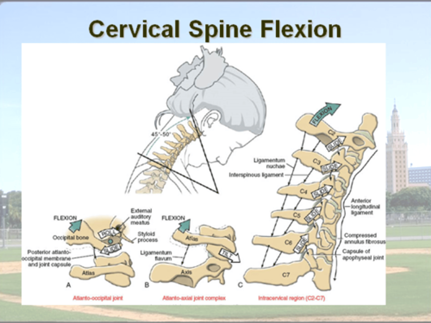
Lower Cervical Spine Motions (ROM) : Flexion / Extension
at what segments does the most movement occur?
C2-T1
max motion occurs at C4-5 & C5-6.
= these 2 segments are often the first to show signs of disk and joint degeneration.
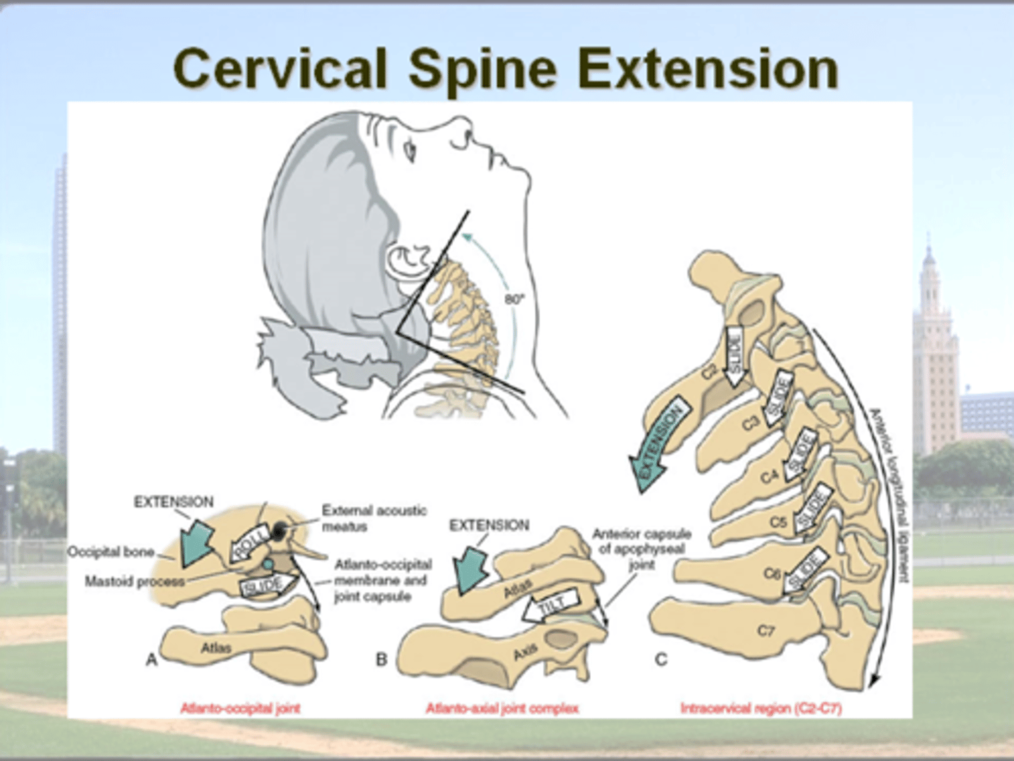
Lower Cervical Spine Motions (ROM) : Flexion/ Extension
describe the motions
at what segments does the most movement occur?
C2-T1
F = 45-60
E = 50 -65
max motion occurs at C4-5 & C5-6.
= these 2 segments are often the first to show signs of disk and joint degeneration.
C-Spine Segments that are often first to show disc + joint degeneration
Why?
C4-5 & C5-6.
because this is where max flexion + extension of c-spine occurs
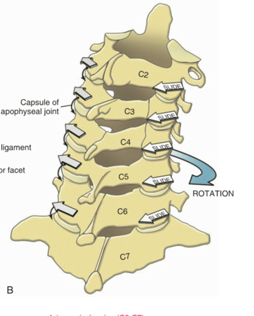
ex. Lower c-spine rotation to the right
- describe in terms of facet joints
right facet = closes
left facet = opens
Summary : Type I vs. Type II motion in the C-spine
- name the 3 segments and their respective motions
OA = Type I
AA - neither (rotation only)
C2-3 thru C7-T1 = Type II
Overall AROM at C-spine:
- what motions
- ranges
Flexion 45-60°
Extension 50-65°
Lateral Flex 40-45°
Rotation 70-80° each
(35-40 from AA prior to rest occuring)
ROM C-Spine Flexion
primarily occurs at what levels
45-60°
max motion occurs at C4-5 & C5-6.
ROM C-Spine Extension
primarily occurs at what levels
50-65°
max motion occurs at C4-5 & C5-6.

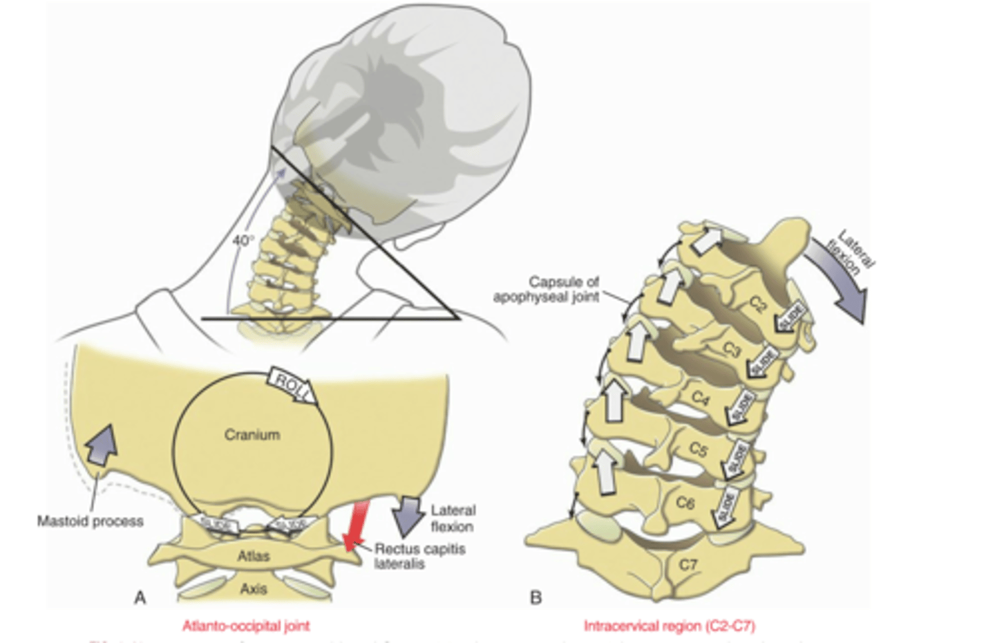
ROM C-Spine Lateral Flexion
primarily occurs at what levels
also coupled with what motion + type of coupling
40-45°
occurs at all levels C2-T4/5
Type II couple (same direction)
with rotation
ROM C-Spine Rotation
where does this occur at what levels
also coupled with what motion + type of coupling
70-80° TOTAL (each direction)
35-40 from AA BEFORE rest = Type I coupling (opposite directions)
35-40 from lower c-spine = Type 2 coupling (same directions)
Facet Joint Orientation at OA
- describe their orientation and how this impacts movement
O = convex
on
A = concave
CVX on CC = roll = opposite to slide
Facet Joint Orientation at AA
- describe their orientation and how this impacts movement
20° from transverse plane = ROTATION
Facet Joint orientation : lower c-spine
what segments
describe orientation
what motion does this favor?
C2 on C3 - C7 on T1 (6 segments)
= 45° from transverse (and frontal)
... transitions to ~30° at lower end (towards frontal)
= favors rotation + lateral flexion
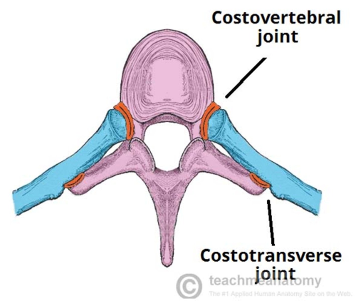
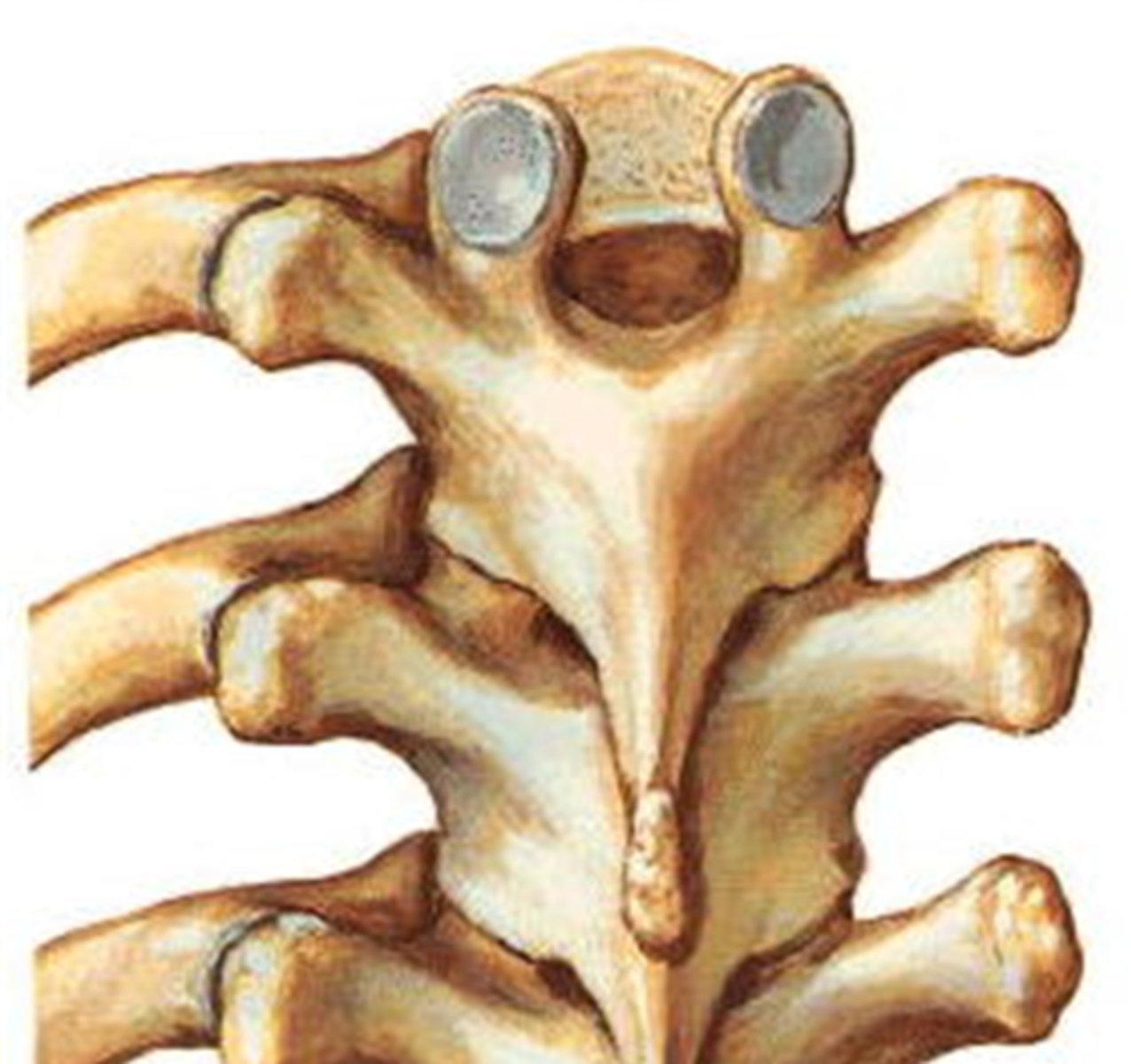
T- Spine : Unique Synovial Articulations (2)
what are they
classification of joints
are they mobile , well innervated?
Costovertebral Joints
Costotransverse joints
plane, synovial joints
mobile and well innervated
Motions of Thoracic Spine : Flexion / Extension
compare upper vs. lower
Possible throughout thoracic spine
extremely limited in upper T-Spine
= ROM increases caudally
More Flexion available overall
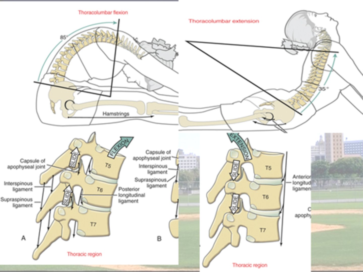
Motions available at T-spine
what are they
describe each in terms of rostral --> caudal
flex/ext = INCREASES caudally
very limited in upper t-spine
lateral flexion = INCREASES caudally
limited due to rib cage
rotation = DECREASES caudally
limited due to ribs + facet orientation
Facet Joint Orientation in the T-spine
- break it down by region
- how does this impact motion
T 1 - T 10 = Frontal Plane
vs.
T 10 - 12 = quick transition to Sagittal
= favors flexion & extension
Scoliousis Class Example
Upper thoracic vs. Lower thoracic / Upper Lumbar
- which side is the lateral flexion
- what happens to the ribs on each side
- which direction is the rotation
Upper Thoracic = L Lateral Flexion
ribs on R side
- elevated
- IC spaces more open
ribs on L side
- depressed
- IC compressed / shrink
Rotation to the Right
Posterior Rib Hump = RIGHT
(vs. rib flattening on Left)
VS.
Lower Thoracic / Upper Lumbar
= R Lateral Flexion
LEFT ROTATION
Posterior Rib Hump on LEFT
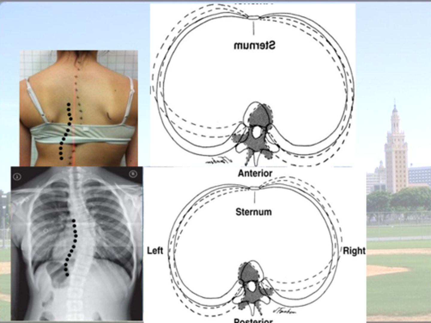
Scoliosis : Changes in Rib Position
Posterior Rib Hump
- Same side as rotation and thus opposite to side of lateral flexion
(be represents lower T-spine = type I coupling)
thus anterior hump = same side as lateral flexion
Contralateral to Lateral Flexion
- Ribs elevate
- Thoracic cage enlarged
- Intercostal spaces widen
vs.
Ipsilateral to Lateral Flexion
- Ribs depress
- Thoracic cage shrinks
- Intercostal spaces narrow
Any Spinal Curvature in the Frontal Plane =
Scoliosis
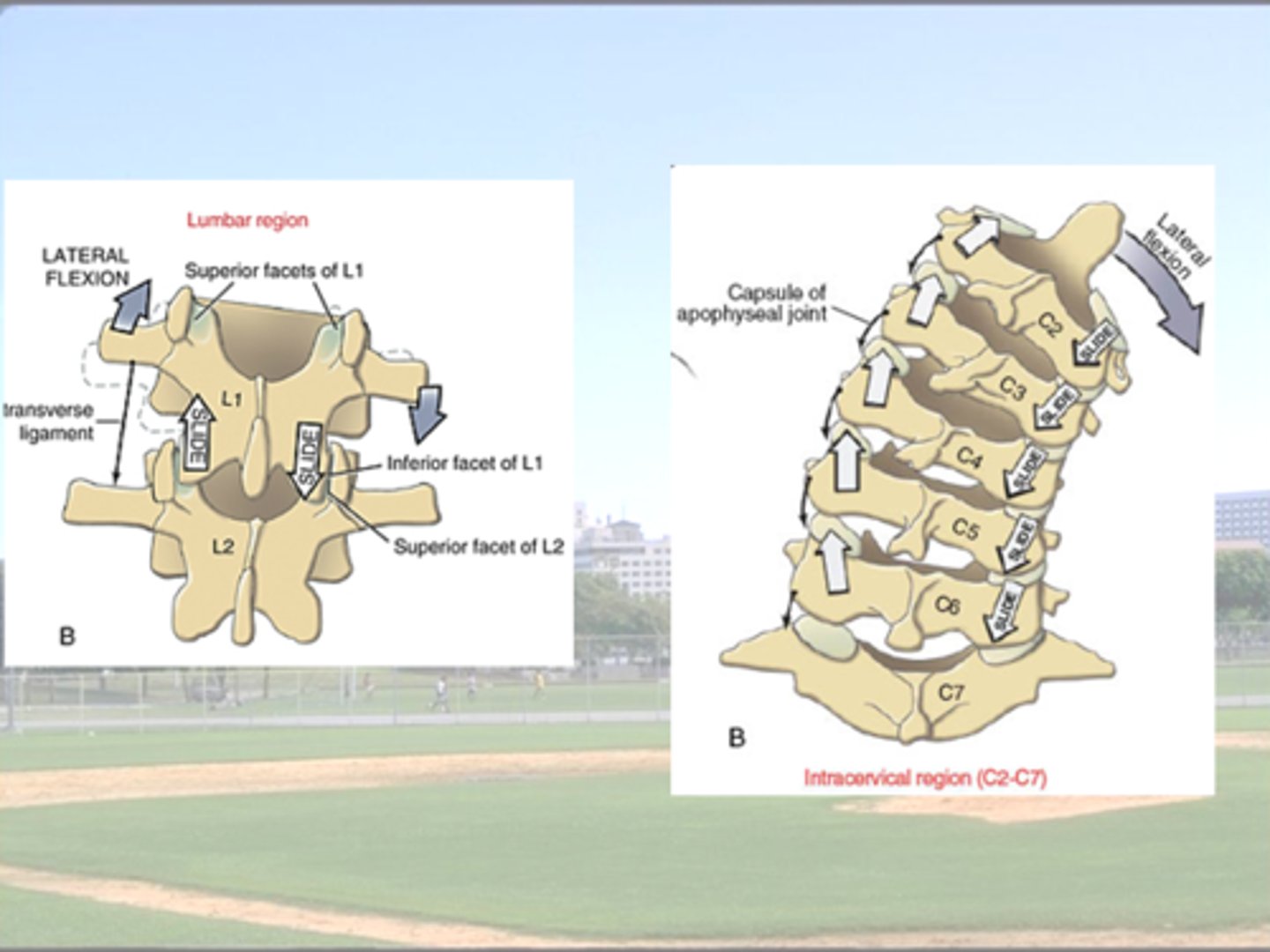
Lumbar Region : Facet joint Orientation
- overall orientation
- superior vs. inferior vertebrae for a given section
differentiate between 2 regions
L1-4 = Sagittal Plane (close to)
at each segment
- upper facet = faces laterally
vs.
- lower face = faces medially
L5-S1 = near the Frontal Plane
- facet surface of L5 faces anterior
- facet surface of S1 faces posterior
= resists anterior movement of L5 on S1 due to normally occurring anterior shear forces
(Just like T-Spine)
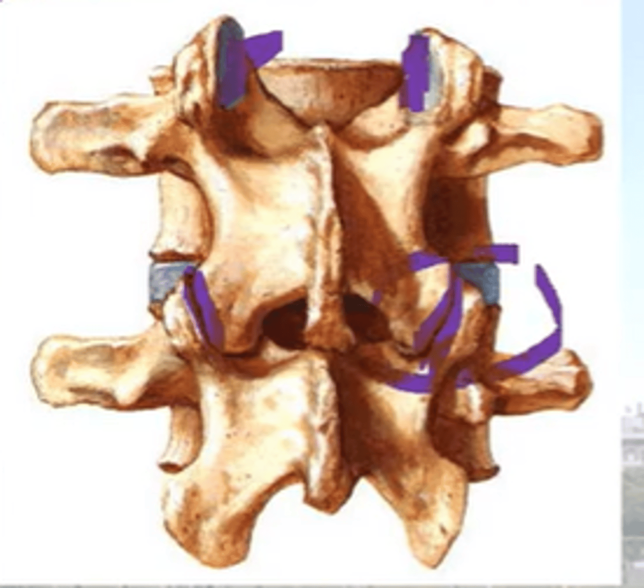
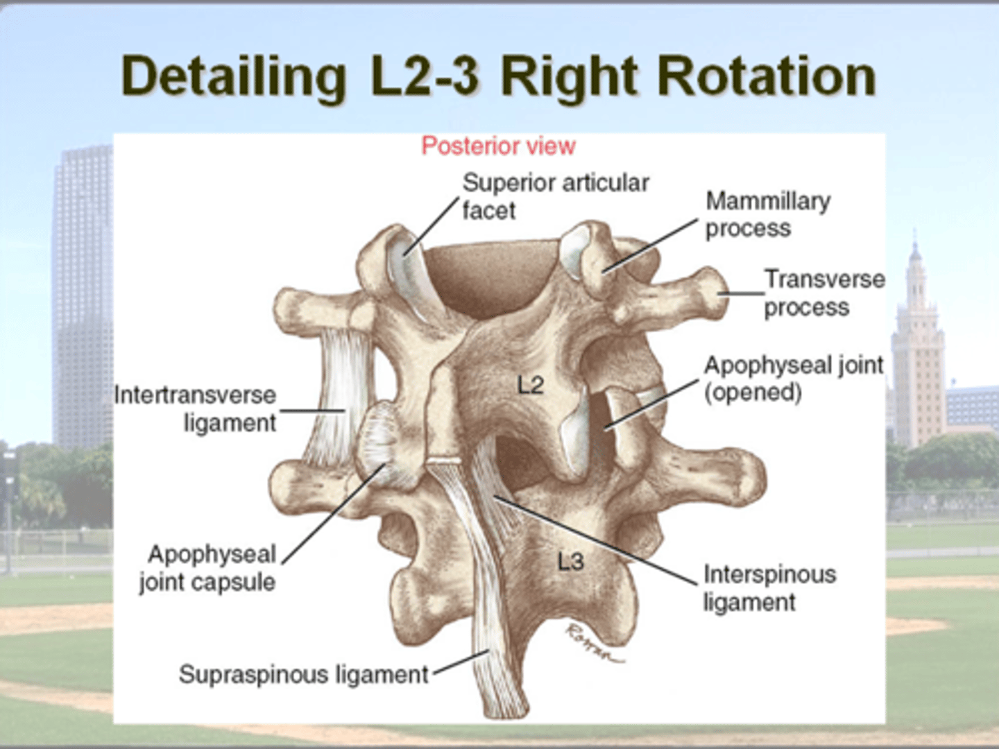
Motions : Lumbar Spine
primary motion(s)
coupling??
explain what happens to facet joints on each side
Primarily : Flexion & Extension
(due to facet orientation)
+
Lateral flexion + rotation
= limited due to orientation of facets.
Type I Coupling
( Lateral Flexion and Rotation )
= OPPOSITE DIRECTION
Ex.
R. lateral flexion = L. rotation (some)
Lateral flexion (pictured)
= Closing of ipsilateral facet
+ opening of contralateral facet
vs.
Rotation (pictured)
= opening of ipsilateral facet
+ closing of contralateral facet
Therefore, a person with a hypomobile lumbar facet joint on the RIGHT will be limited in right rotation and left lateral flexion (at that vertebral segment)
Q: How does this effect the entire lumbar spine?
A person with a hypomobile lumbar facet joint on the RIGHT will be limited in _____________
Explain
limited in:
- right rotation
+
- left lateral flexion
(at that vertebral segment)
bc TYPE I Coupled
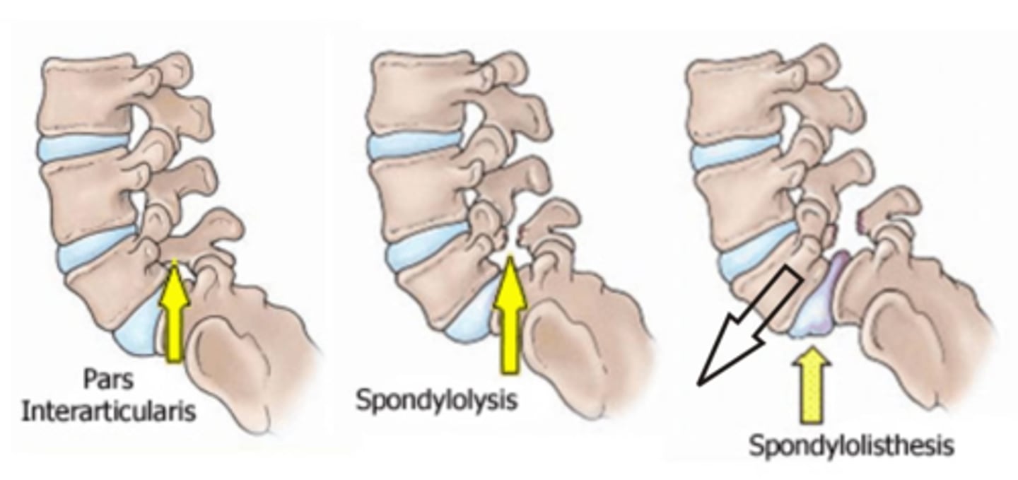
Spondylolisthesis
what is it
what structures support / protect against this?
Slippage of L5 on S1
- damage spinal nerves / cauda equina
limited by
facet orientation (frontal plane)
- L5 = anterior
vs.
- S1 = posterior
= BLOCKS L5 from slipping anteriorly
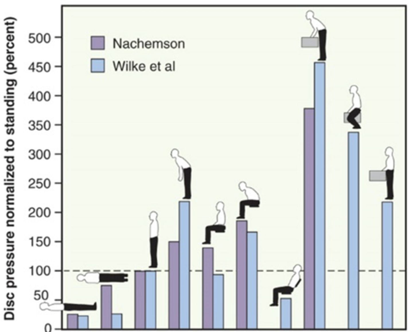
Intradiscal pressures in Lumbar Region
what is the baseline
describe trends
= what causes you to go above or below baseline
compression due to body weight & m contraction
standing = 100%
leaning forward = increases forces
supine = lowest
side
design programs to prevent lumbar disk NP herniation or limit any additional damage.
In order for the nucleus pulposus to escape the annulus fibrosis, the annulus must be weakened or torn.
Are SI joints strong/stable
why or why not
VERY STABLE
Extremely strong ligaments reinforce the joint on all sides.
Motion of Sacrum on Ilium:
how does this affect pelvic inlet/outlet
changes during pregnancy?
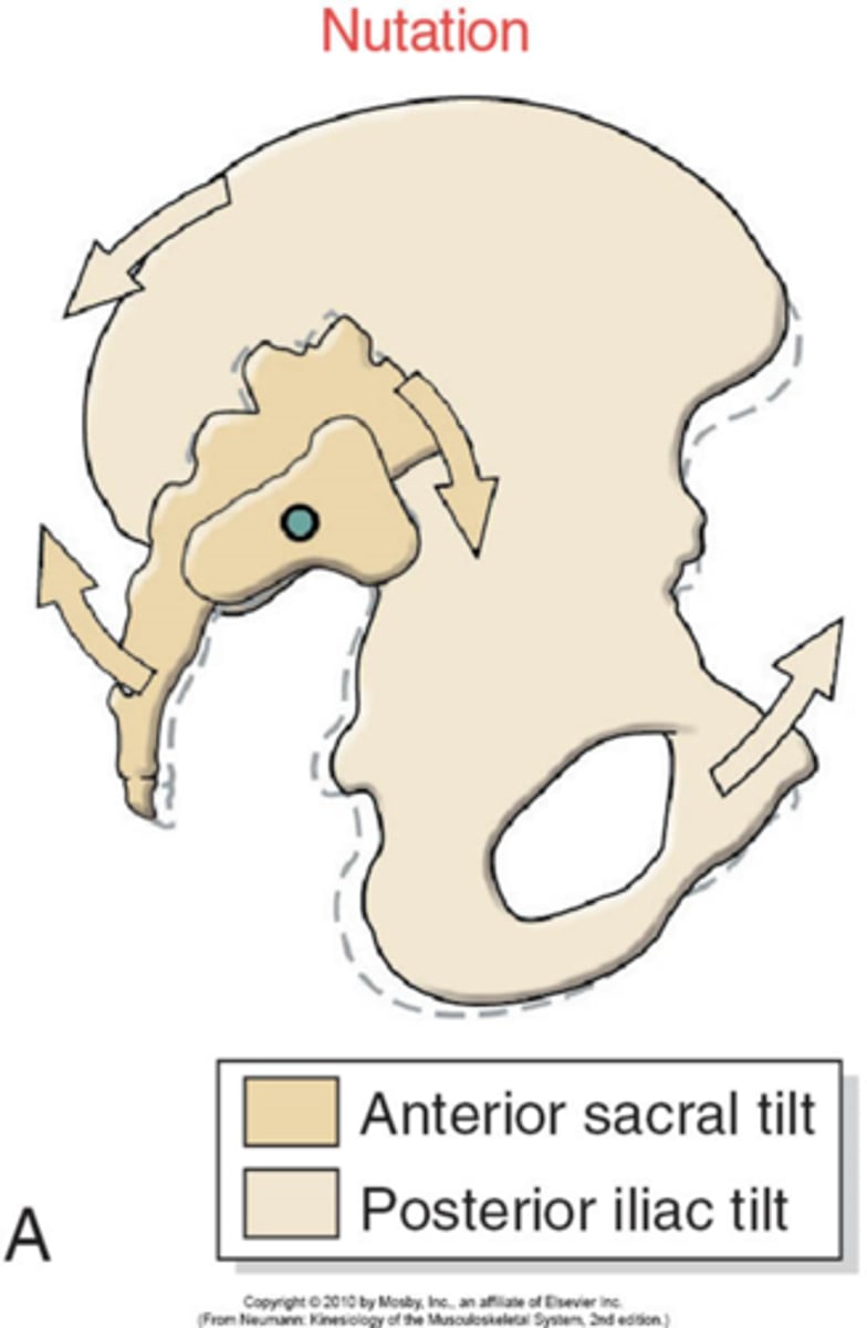
Primary motion of SI
Flexion (nutation) (nod)
– anterior tip of sacrum moves anteriorly + inferiorly
- coccyx moves posteriorly
vs.
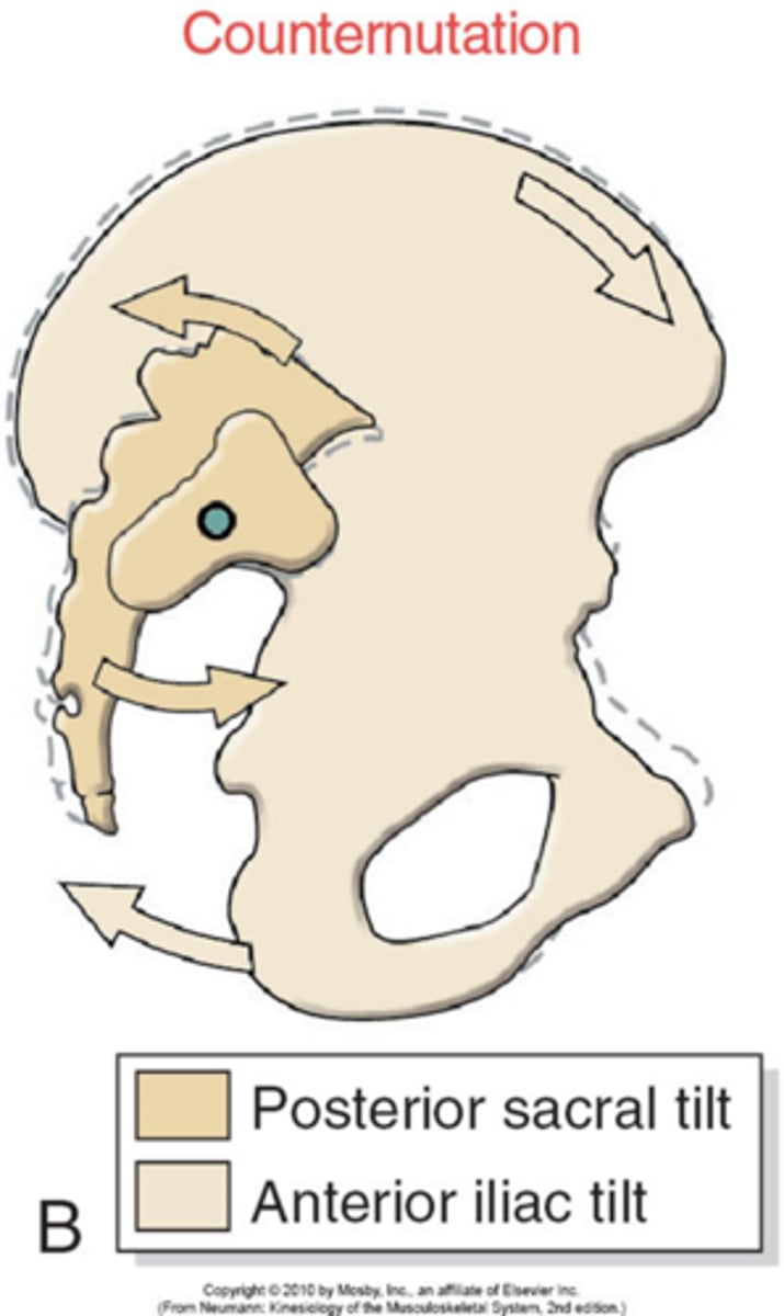
Extension (counternutation)
– anterior tip of sacrum moves posteriorly + superiorly
- coccyx moves anteriorly.
How do Nutation & Counternutation affect size of the Pelvic Inlet & Pelvic Outlet ??
pregnancy
- softening of ligaments
= increased joint mobility + decreased stability of SI joints
= likelihood of joint dysfunction and pain.
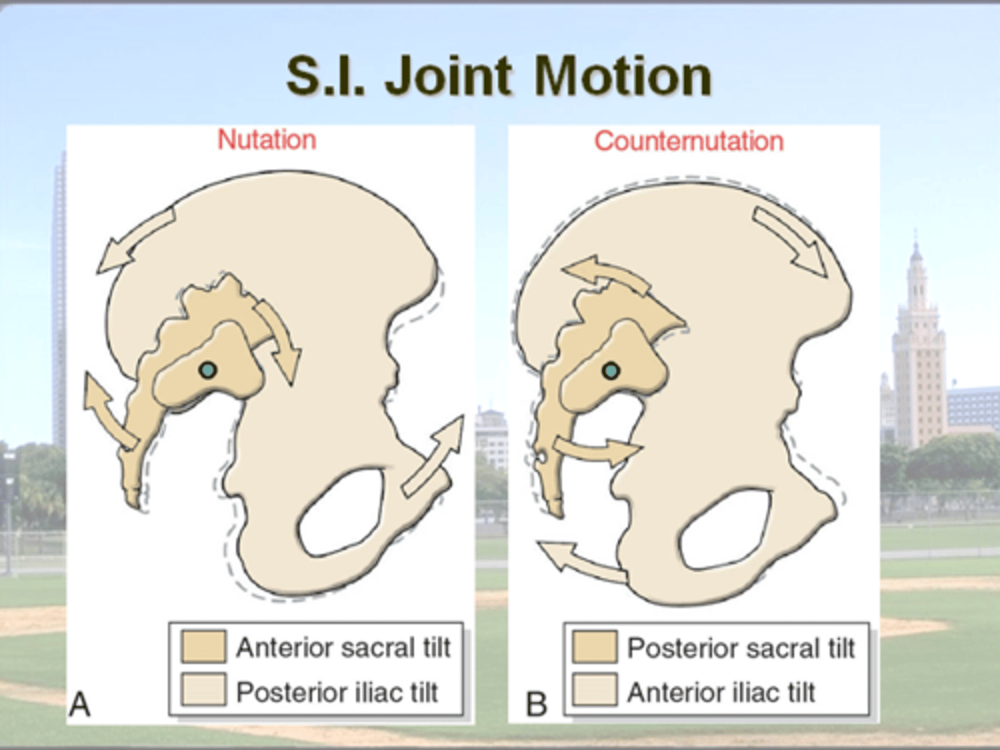
Nutation
what is it
2 possible ways for it to occur
SI "Flexion"
1. SI moving on stable Pelvis
2. Posterior Pelvic tilt
(stable sacrum)
Counternutation
SI "Extension"
1. SI moving on stable Pelvis
2. Posterior Pelvic tilt
(stable sacrum)
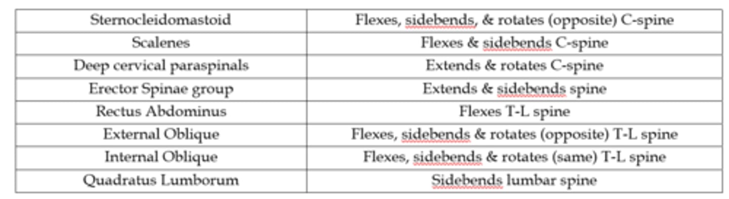
Muscles of the Spine/Trunk: Function to stabilize trunk & pelvis (control postures) and move the trunk/spine.
(TABLE)
External Oblique = SCM for Action
(ie Ipsi flexion + sidebend but opposite rotation)
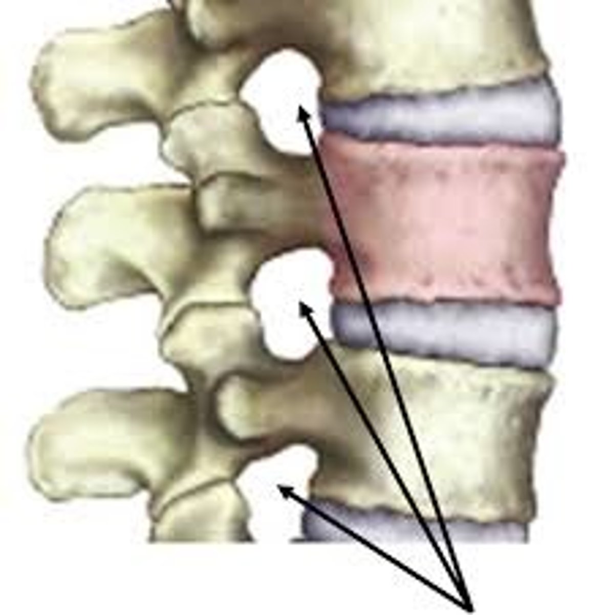
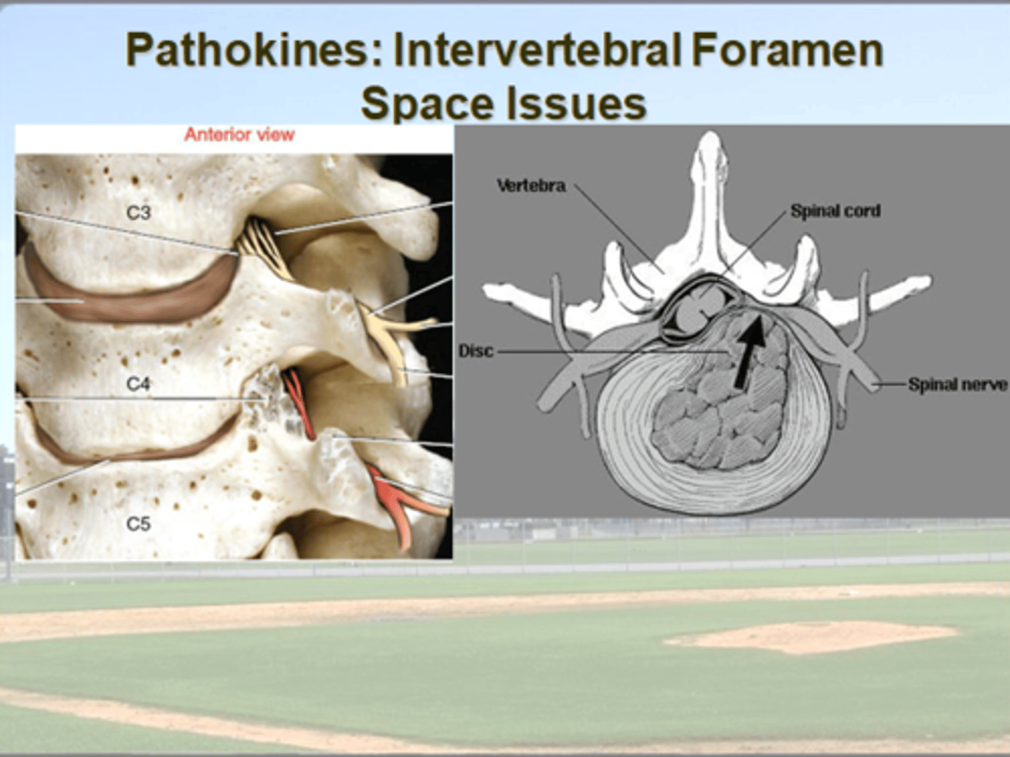
Intervertebral Foramen Space Issues:
describe structures in relation to IV foramen / spinal nerve
how does flexion / extension affect space
affect of disc height?
anterior = disk
posterior = facet joint
posteromedial
= ligamentum flavum
degeneration or dehydration of disc = narrows IV space
flexion = increases size
extension = decreases size
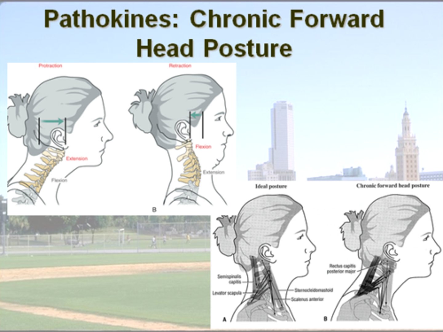
Protraction of the Head (Forward Head)
compare position of upper c-spine vs. mid to lower c-spine
what is the overall outcome of this?
upper C-spine = extension
vs.
Mid & Lower C-spine = flexion
increases stress on :
- levator scapulae
- semispinalis capitis
+
shortens SCMs