Functional Anatomy 1 - Lecture 14: Leg
 Call Kai
 Learn
Learn Practice Test
 Spaced Repetition
 Match
Match Flashcards
 Knowt Play
1/55

There's no tags or description
Looks like no tags are added yet.
Name | Mastery | Learn | Test | Matching | Spaced | Call with Kai | Chat |
|---|
No analytics yet
Send a link to your students to track their progress
56 Terms
leg
distal to knee and superior to the ankle
foot/ankle extrinsic muscles
muscle belly in the leg with tendon to the foot/ankle
osteology of the leg
tibia: larger, primary weight-bearing bone (90%)
fibula: smaller, less weight-bearing bone (10%)
tibial crest
anterior-medial surface: no muscle attachments
anterior-lateral surface: anterior compartment muscles
articulations with fibula
fibular facet (proximal)
fibular notch (distal)
soleal line
origin of soleus
reference for muscle attachments superior and inferior
inferior-medial direction
merges with medial border of tibia
proximal tibiofibular joint
synovial plane joint
two surfaces sliding on each other (tibia+fibia)
ligaments
anterior/posterior ligaments of fibular head provide stability
distal tibiofibular joint
fibrous joint (syndesmosis)
ligaments
interosseous membrane (IO)
anterior tibiofibular ligament
posterior tibiofibular ligament
distal tibiofibular joint
syndesmotic ankle sprain
high ankle sprain
forced dorsiflexion with rotation
damages anterior/posterior tibiofibular ligaments, possible IO membrane
forefoot
phalanges, metatarsal bones
midfoot
medial/intermediate/lateral cuneiform bones, cuboid bone, navicular bone
hindfoot
talus, calcaneus
talus
plantarflex ankle, palpate anterior/inferior to the malleoli
brings talar dome anteriorly
medial talar dome
plantarflex and evert the hindfoot
lateral talar dome
plantarflex and invert the hindfoot
calcaneal tuberosity
forms heel: Achille’s tendon attachment
sustentaculum tali
about 1 finger’s breadth distal to the tip of medial malleolus
navicular tuberosity
felt approximately 2 cm anterior to sustentaculum tali, prominent tubercle on medial aspect of foot
tibialis posterior attachment
tuberosity of 5th MT
prominence on lateral aspect of foot, lateral to base of 5th MT
peroneus brevis attachment
ankle movement
dorsiflexion/plantarflexion
sagittal plane (ML axis)
inversion/eversion
frontal plane (AP axis)
abduction/adduction
transverse plane (SI axis)
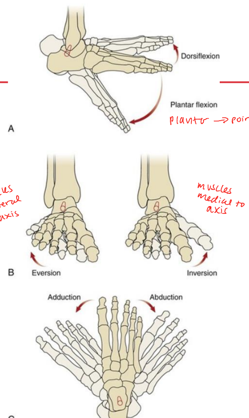
leg region innervation
sciatic (L4-S3)
superior angle of popliteal fossa
common peroneal (fibular) (L4-S2)
superficial peroneal (fibular) (L4-S2)
deep peroneal (fibular) (L4-S1)
tibial (L4-S3)
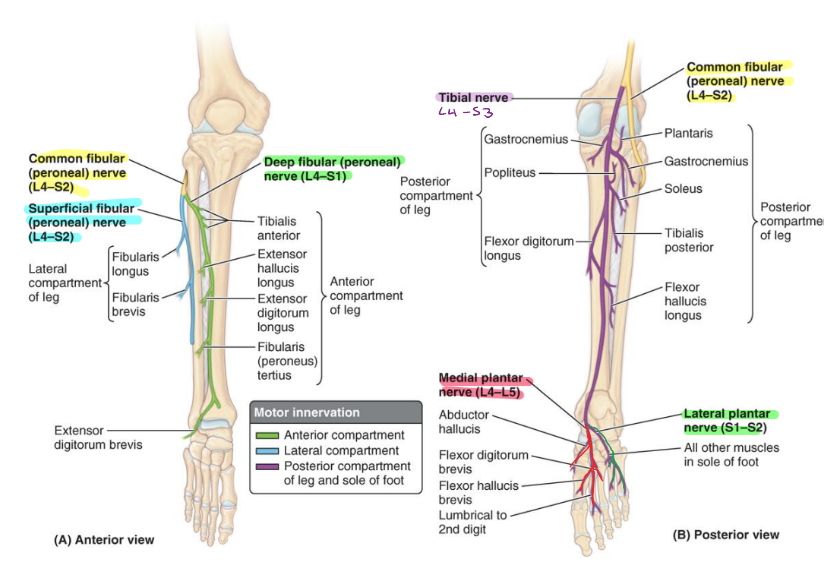
leg sensory innervation
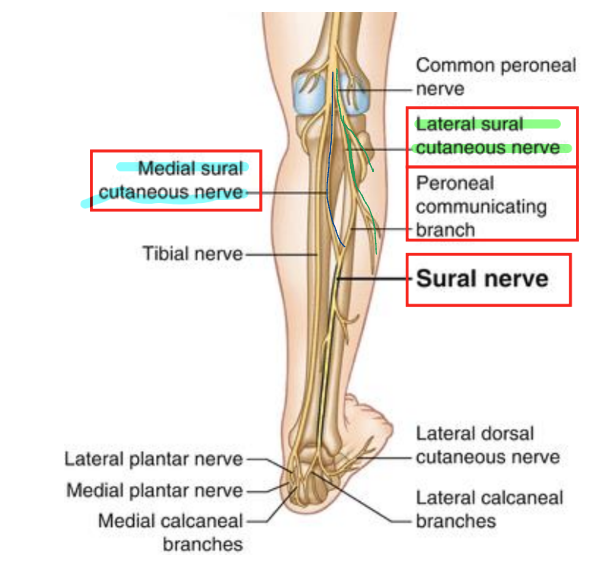
peripheral cutaneous innervation
medial knee and medial leg
saphenous n (cutaneous branch from femoral n)
lateral knee and leg
lateral sural cutaneous (branch from common peroneal n)
anterior lateral lower leg and some dorsal foot
superficial peroneal (fibular) n
posterior middle leg
sural cutaneous n (combined branches from tibial n and common peroneal n
webspace between 1st and 2nd digit
deep peroneal (fibular n)
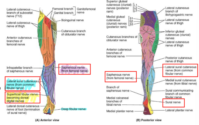
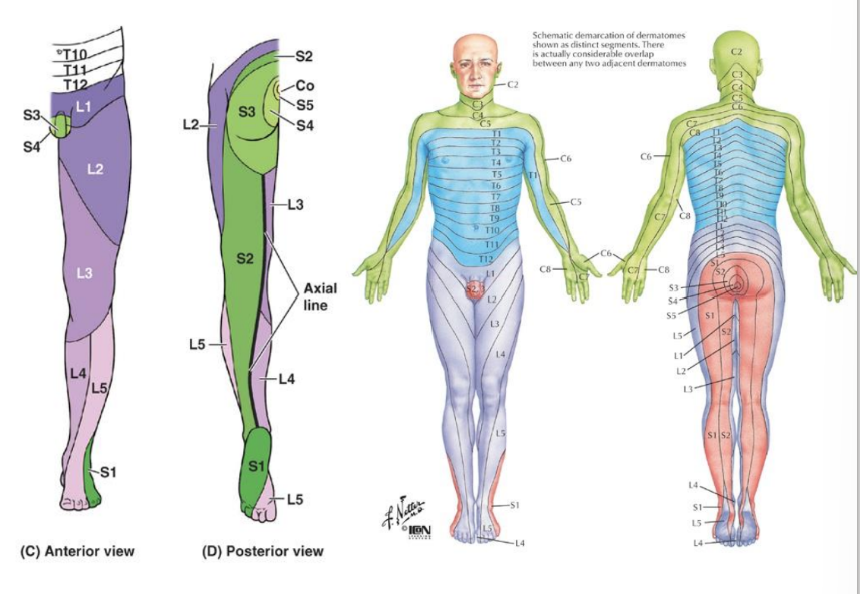
dermatomes sensory innervation of leg
L1: groin region near inguinal ligament
L2: superior-anterior thigh to lateral thigh
L3: distal anterior thigh to medial thigh
L4: medial lower leg
L5: lateral lower leg
S1: lateral foot/posterior medial thigh
S2: posterior lower leg/posterior medial thigh
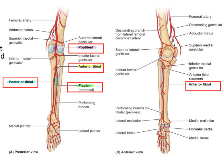
leg region arteries
popliteal a.
anterior tibial a.
anterior through gap in interosseous membrane
deep anterior compartment between tibialis anterior and EDL → dorsalis pedis a.
posterior tibial a.
deep posterior compartment
posterior to medial malleolus
peroneal (fibular a.)
supplies lateral compartment
descends near FHL
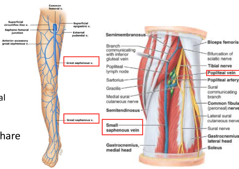
leg region veins
superficial veins
great saphenous v.
medial lower leg
to femoral v. in femoral triangle
small saphenous v.
lateral lower leg
to popliteal v. in popliteal space
deep veins
bundled with arteries, share a name
3 compartments of leg
anterior
lateral
posterior
superficial
deep
divided by:
interosseous membrane
anterior intermuscular septum
lateral intermuscular septum
anterior compartment
anterior to interosseous membrane lateral to tibial shaft and medial fibular shaft/anterior intermuscular septum
deep peroneal (fibular) n.
anterior tibial a.
ankle dorsiflexion (with eversion/inversion): digit extension
tibialis anterior
extensor hallucis
extensor digitorum longus
peroneus (fibularis) tertius
tibialis anterior
O: lateral tibial epicondyle and proximal 2/3 of lateral tibia and IO membrane
I: medial and inferior surfaces of medial cuneiform, base of 1st metatarsal
A: ankle dorsiflexion and inversion, assists with supporting medial longitudinal arch of foot during weight bearing positions
N: deep peroneal (fibular) n. (L4-L5)
B: anterior tibial artery
extensor hallucis longus
O: middle part of anterior surface fibula and IO membrane
I: dorsal base of distal phalanx of 1st digit
A: extension 1st digit, ankle dorsiflexion
N: deep peroneal (fibular) n. (L5-S1)
B: anterior tibial artery
extensor digitorum longus
O: lateral condyle of tibia, proximal 2/3 of anterior fibula, IO membrane
I: middle and distal phalanges of digits 2-5 (extensor hood)
A: extension of digits 2-5, ankle dorsiflexion
N: deep peroneal (fibular) n. (L5-S1)
B: anterior tibial artery
peroneus tertius
O: distal third of anterior surface of fibula and IO membrane
I: dorsal base of 5th metatarsal
A: ankle dorsiflexion and eversion
N: deep peroneal (fibular) n. (L5-S1)
B: anterior tibial a.
extensor retinaculum
needed to support line of pull of anterior compartment muscles and prevent bowstringing of tendons across the ankle
superior extensor retinaculum
between tibia and fibula
inferior extensor retinaculum
Y shaped from calcaneus to
medial malleolus
plantar fascia
lateral compartment of leg
lateral fibula to anterior and posterior intermuscular septum
ankle eversion
superficial peroneal n.
peroneal artery
peroneus (fibularis) longus
O: head of fibula and proximal 2/3 of lateral fibula
I: plantar base of 1st metatarsal and medial cuneiform
A: ankle eversion and plantarflexion, support arches of foot
N: superficial peroneal n. (L5, S1, S2)
B: peroneal artery
peroneus (fibularis) brevis
O: middle part of lateral fibula
I: tuberosity of 5th MT
A: ankle eversion and plantarflexion
N: superficial peroneal (fibular) n. (L5, S1, S2)
B: peroneal (fibular) a.
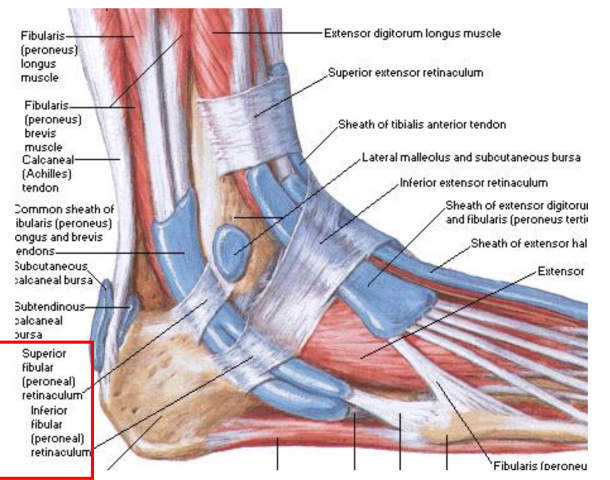
peroneal (fibular) retinaculum
supports stabilization of peroneal tendons as they cross lateral malleolus
superior: calcaneus to lateral malleolus
inferior: calcaneus to inferior extensor retinaculum
posterior compartment
posterior to interosseous membrane, tibia, fibular, and posterior intermuscular septum
further divided into superficial and deep by transverse intermuscular septum
ankle plantarflexion; digit flexion
tibial nerve
posterior tibial artery
superficial posterior compartment
gastrocnemius (medial and lateral heads)
plantaris
soleus
deep posterior compartment
flexor digitorum longus (medial)
tibialis posterior (middle)
flexor hallucis longus (lateral)
triceps surae
gastrocnemius, plantaris, soleus
common attachment to calcaneus via calcaneal tendon
generate up to 93% of plantarflexion force
superficial posterior compartment
gastrocnemius
O:
lateral head: lateral femoral condyle
medial head: medial femoral condyle and popliteal surface of femur
I: posterior calcaneus via Achilles tendon
A: ankle plantarflexion; knee flexion
N: tibial nerve (S1-S2)
B: posterior tibial artery
plantaris
O: inferior part of lateral supracondylar line femur
I: posterior calcaneus via Achilles tendon
A: weak ankle plantarflexion; knee flexion
N: tibial nerve (S1-S2)
B: posterior tibial artery
soleus
O: posterior head of fibula, superior posterior fibula, soleal line of tibia
I: posterior calcaneus via Achilles tendon
A: ankle plantarflexion
N: tibial nerve (S1-S2)
B: posterior tibial a.
tibialis posterior
O: IO membrane, posterior surface of tibia inferior to soleal line, posterior fibula
I: tuberosity of navicular, cuneiforms, cuboid, bases of MT 2-4
A: ankle plantarflexion and inversion
N: tibial nerve (L4-L5)
B: posterior tibial artery
flexor digitorum longus
O: medial posterior tibial inferior to soleal line
I: base of distal phalanges 2-5
A: flexion of MTPs and IPs 2-5, weak ankle plantarflexion and inversion
N: tibial nerve (S2-S3)
B: posterior tibial artery
flexor hallucis longus
O: distal 2/3 posterior fibula and part of IO membrane
I: base of distal phalanx of 1st digit
A: flexion of 1st digit MTP and IP; weak ankle plantarflexion and inversion
N: tibial nerve (S2-S3)
B: posterior tibial artery
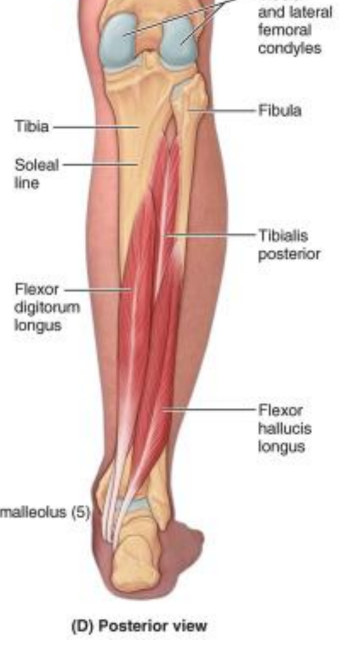
tibialis posterior
flexor digitorum longus
posterior tibial artery
tibial nerve
flexor hallucis longus
Tom, Dick, And Nervous Harry
flexor retinaculum
supports stabilization of deep posterior tendons as they cross medial malleolus
calcaneus to medial malleolus
tarsal tunnel
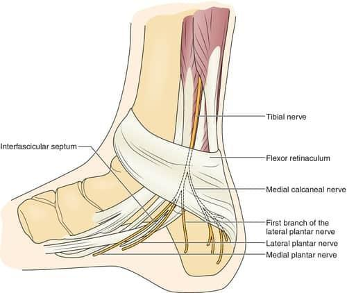
tarsal tunnel syndrome
narrowing of the tarsal tunnel, compression of the tibial nerve/posterior tibial artery
grooves
bony _____ for deep posterior muscle tendons
medial grooves
for tibialis posterior, flexor digitorum longus, flexor hallucis longus
lateral grooves
for peroneus longus/brevis
popliteus
O: lateral surface of lateral condyle of femur, lateral meniscus (intra-capsular)
I: posterior tibia superior to soleal line
A: internal rotation of the knee, flexion of the knee
N: tibial nerve (L4-S1)
B: posterior tibial artery