Central Ray Placement (Radiologic Sciences)
1/89
There's no tags or description
Looks like no tags are added yet.
Name | Mastery | Learn | Test | Matching | Spaced |
|---|
No study sessions yet.
90 Terms
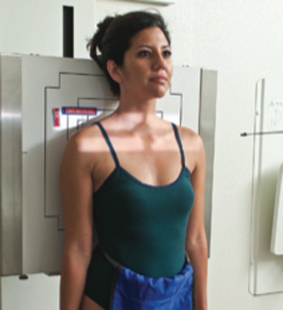
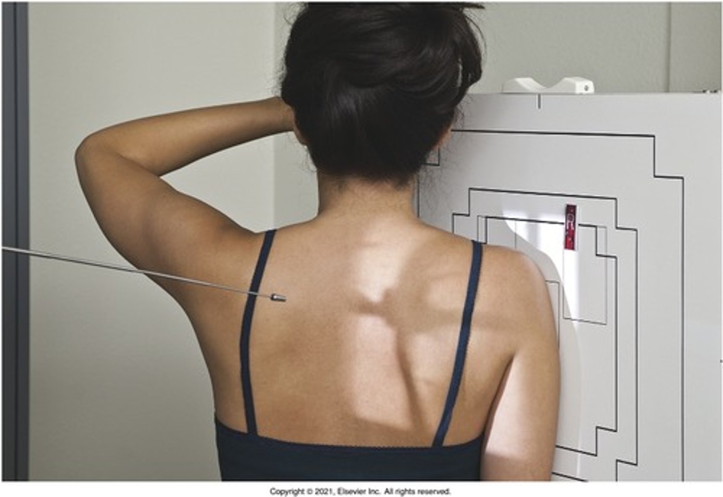
PA and Lateral Chest
perpendicular to T7

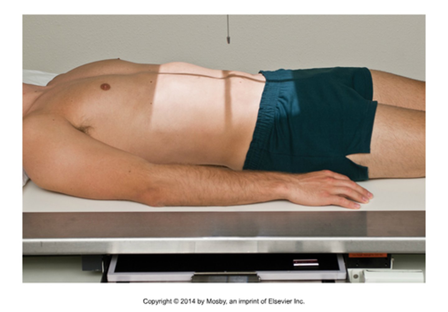
KUB
perpendicular to iliac crest (L4)
decubitus KUB
horizontal and directed 2 inches above iliac crest for air/fluid level

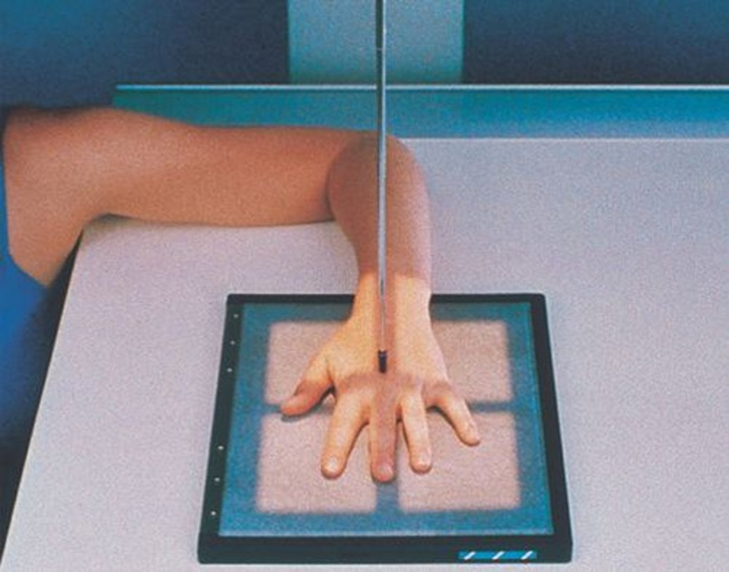
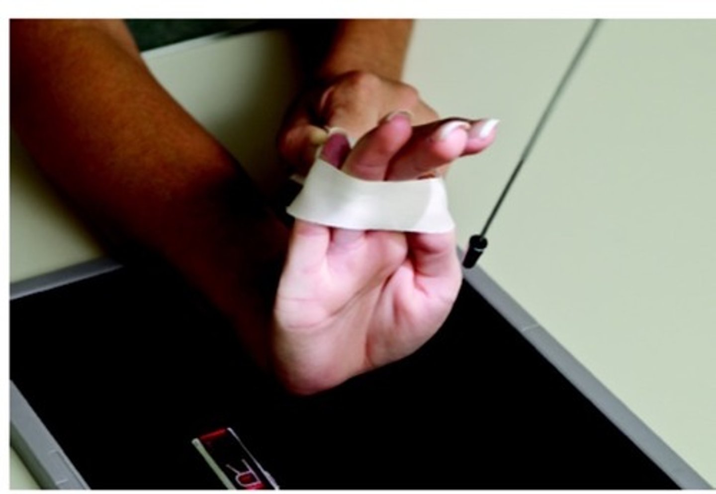
PA hand
perpendicular to 3rd MCP joint
oblique hand
perpendicular to 3rd MCP joint

fan lateral hand
perpendicular to 2nd MCP joint
lateral hand w/ extension
perpendicular to MCP joints

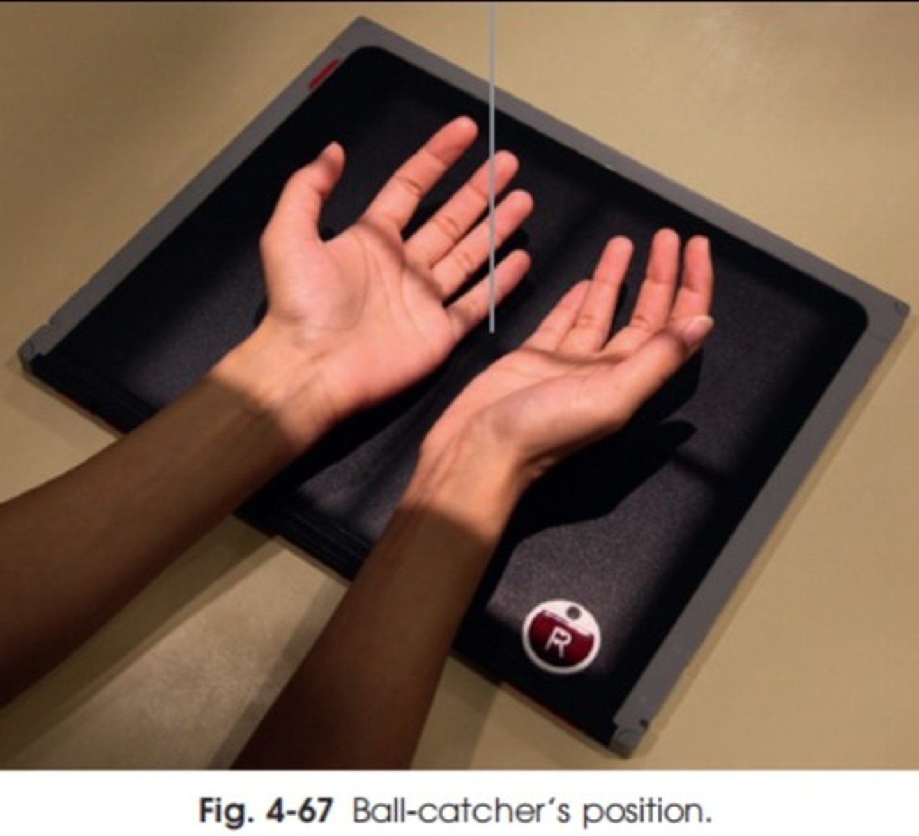
Norgaard Method/Ball Catcher hand
CR perpendicular, directed to midpoint between both hands at level of fifth MCP joints
PA finger
perpendicular to PIP joint

medial/lateral oblique finger
perpendicular to PIP joint
mediolateral/lateromedial finger
perpendicular to PIP joint
AP thumb
perpendicular to first MCP joint
PA oblique thumb
perpendicular to first MCP joint

lateral thumb
perpendicular to first MCP joint

modified robert's method (thumb)
CR directed 15 degrees proximal towards wrist entering first CMC joint
PA wrist
CR perpendicular to midcarpal area

PA oblique wrist
CR perpendicular to midcarpal area
Lateral wrist
perpendicular to midcarpal area
PA scaphoid wrist w/ ulnar devation
CR angled 10-15 degrees and enters scaphoid (2 cm distal and medial to radial styloid process)

modified stetcher method (wrist)
perpendicular to IR and directed to scaphoid

PA radial deviation (wrist)
CR perpendicular to midcarpal area
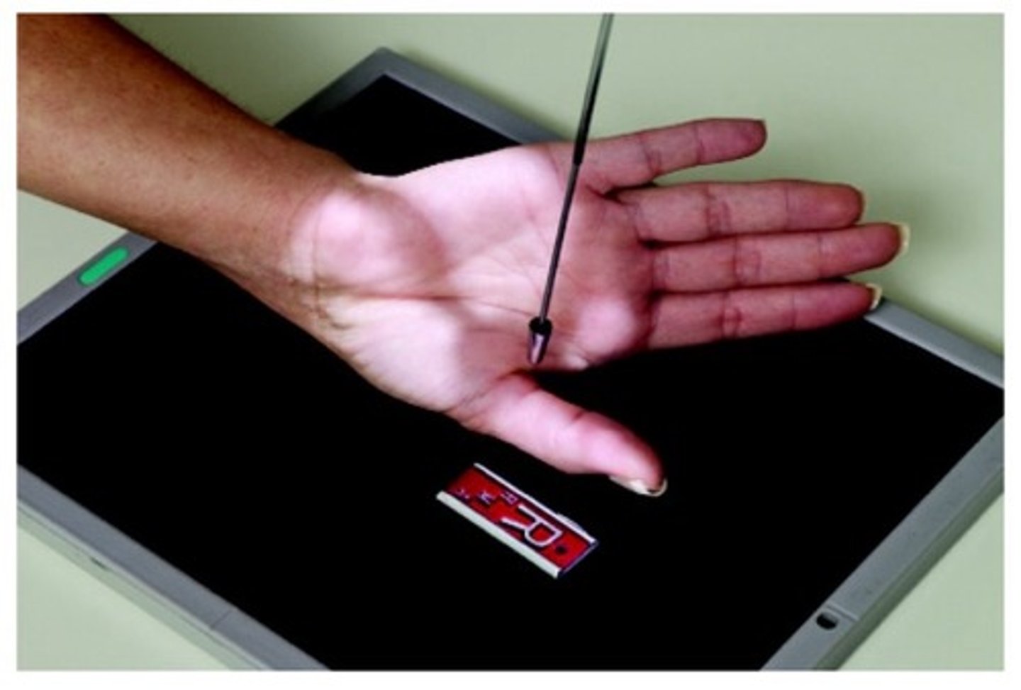
carpal canal tangential (gaynor-hart method)
angled 25-30 degrees, directed 1 inch distal to the base of the 3rd metacarpal (center of palm)
carpal bridge tangential (wrist)
CR angled 45 degrees directed to the midpoint of the distal forearm, about 4 cm proximal to the wrist joint

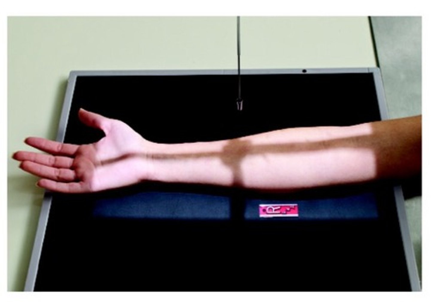
AP forearm
CR perpendicular to mid-forearm
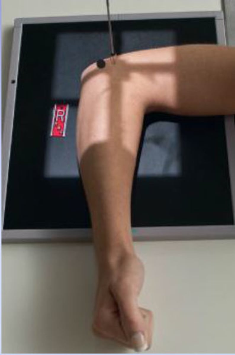
lateral forearm
CR perpendicular to mid-forearm

AP elbow
CR perpendicular to mid-elbow joint (2 cm distal to midpoint line between epicondyles)

AP external/internal oblique elbow
CR perpendicular to mid-elbow joint (2 cm distal to midpoint line between epicondyles)

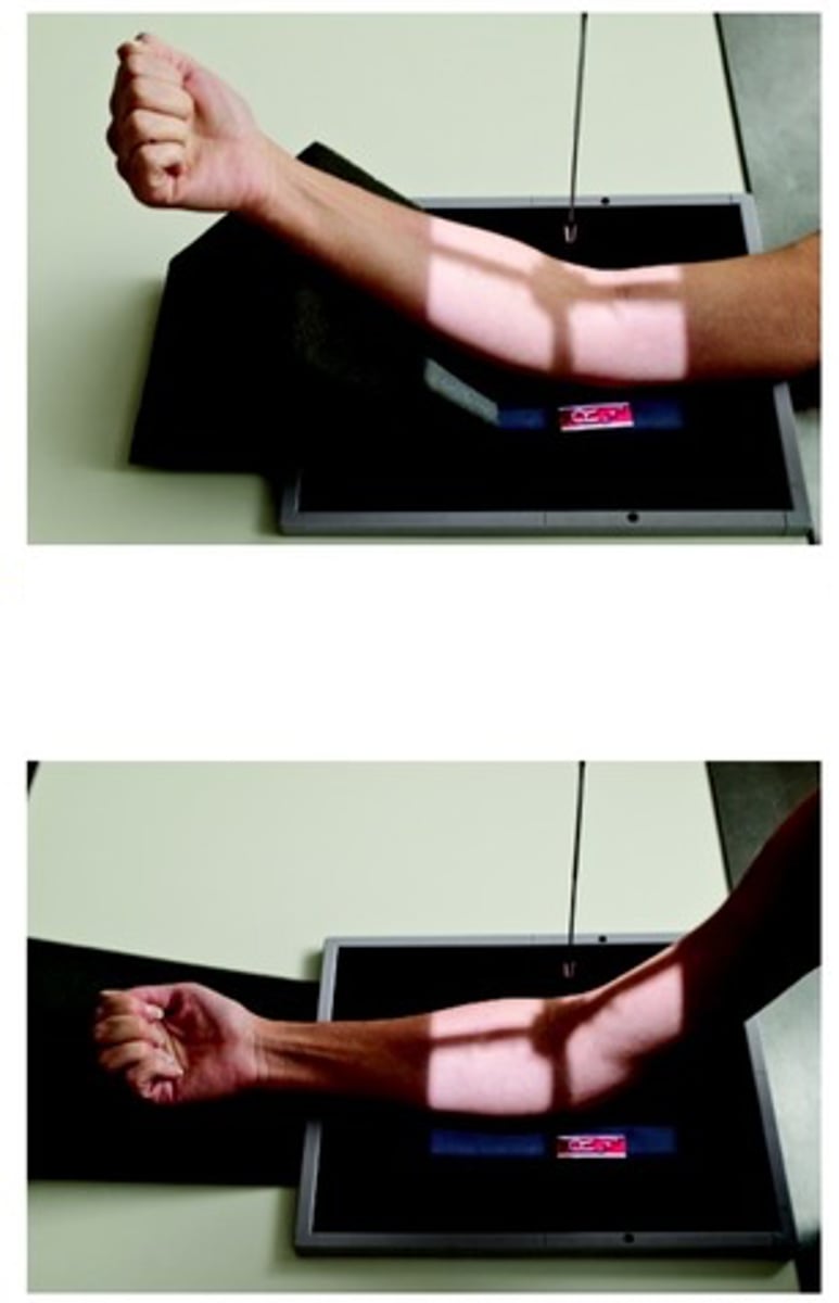
lateral elbow
CR perpendicular to mid elbow joint (4 cm medial to palpable posterior surface of olecranon process)
elbow acute flexion for distal humerus
CR perpendicular to midway between epicondyles

elbow acute flexion for proximal forearm
CR perpendicular to forearm (angling as needed) to a point 2 inches superior to olecranon process

AP elbow partial flexion
CR perpendicular to mid-elbow joint (2 cm distal to midpoint line between epicondyles)
coyle method for radial head (elbow)
CR angled 45 degrees towards should directed to mid-elbow joint

coyle method for coranoid process
angled 45 degrees away from shoulder directed to mid-elbow joint

AP humerus
perpendicular to midpoint of humerus
mediolateral/lateromedial humerus
perpendicular to midpoint of humerus

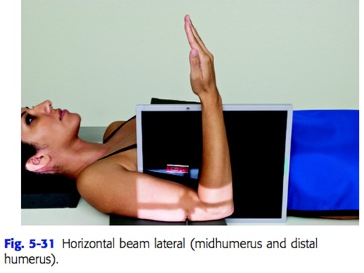
trauma horizontal beam lateromedial humerus
perpendicular to midpoint of distal two-thirds of humerus
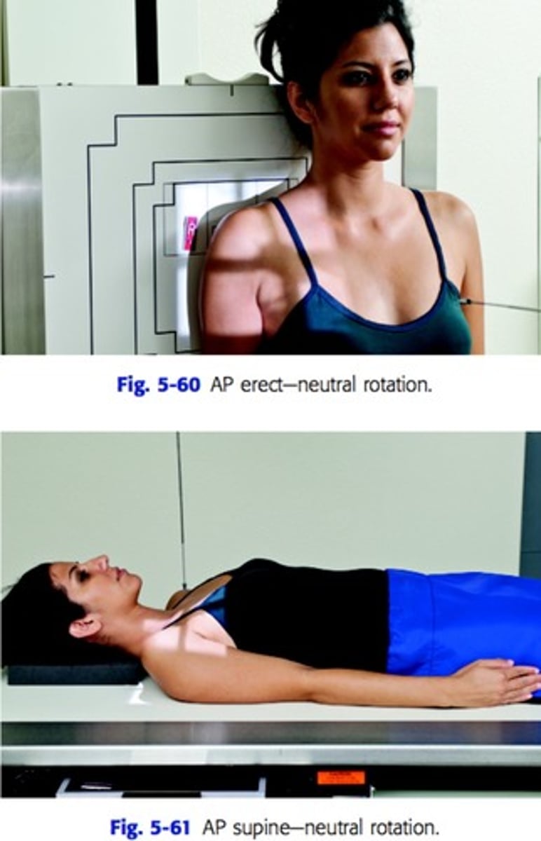
AP shoulder neutral rotation
CR perpendicular to mid-scapulohumeral joint
AP shoulder external rotation
perpendicular and directed 1 inch below coracoid process
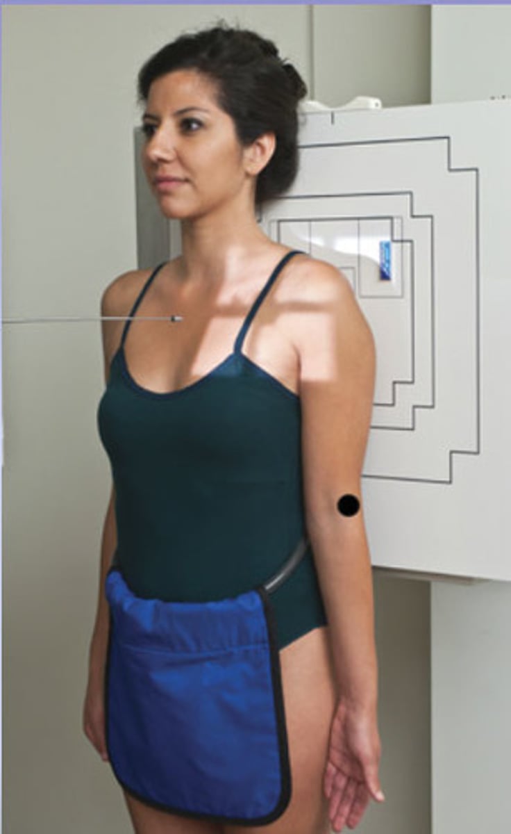
AP shoulder internal rotation
perpendicular and directed 1 inch below coracoid

inferosuperior axial projection/ lawrence method (shoulder)
CR angled 25-30 degrees medially to axilla and humeral head

grashey method shoulder
perpendicular to scapulohumeral joint (2 inches medial and inferior from superolateral border of shoulder)

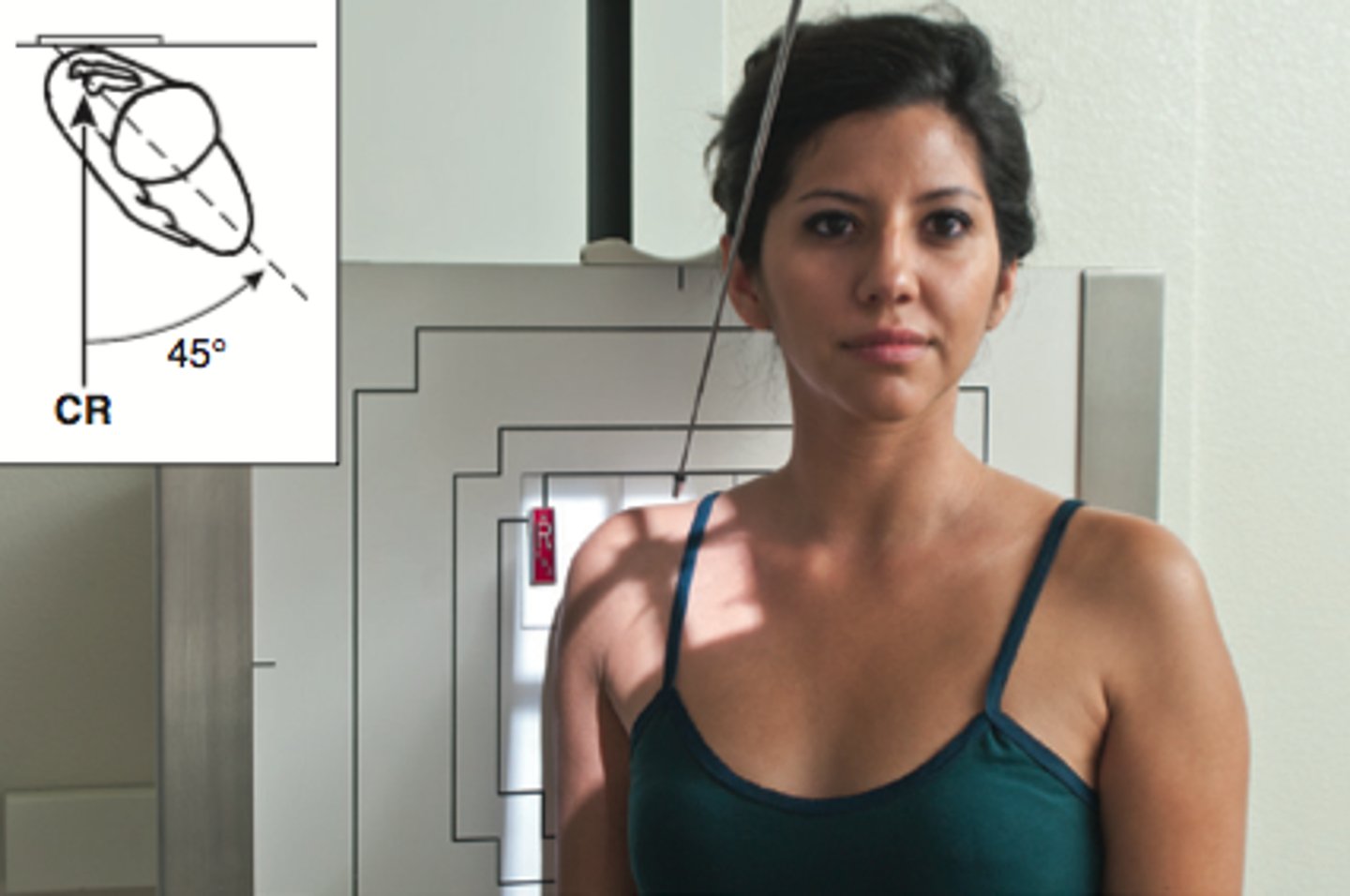
apical oblique axial/garth method (shoulder)
angled 45 degrees caudal centered to scapulohumeral joint
PA scapular y lateral
perpendicular and directed to scapulohumeral joint

AP clavicle
perpendicular to mid-clavicle
AP axial clavicle
angled 15-30 degrees cephalic to mid-clavicle
AP AC joints
perpendicular to midpoint between AC joints, 1 inch above jugular notch
AP scapula
perpendicular to midscapula (2 inches inferior to coracoid process or to level of axilla, and 2 inches medial from lateral border of patient)

neer method/tangential supraspinatous outlet
angled 10-15 degrees
lateral scapula
perpendicular to midvertebral border of scapula
AP toes
angled 10-15 degrees towards calcaneus to MTP joint (if a wedge is placed under the foot, the CR is perpendicular)

AP oblique toes
perpendicular to MTP joint of toe
lateral toe
perpendicular to IP joint for first digit and to PIP joint for second-fifth digits

prone lewis method tangential (foot-sesamoids)
perpendicular to first MTP joint
supine holly method (foot-sesamoids)
perpendicular to first MTP joint

AP foot
angled 10 degrees posterior (towards heel) to base of the third metatarsal
AP oblique foot
perpendicular to the base of the third metatarsal (midfoot)

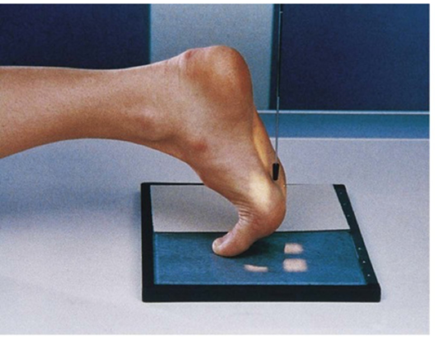
lateral foot
perpendicular to the base of the third metatarsal

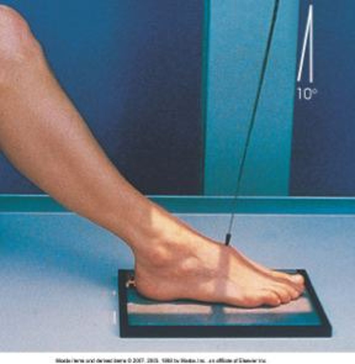
AP weight bearing foot
angled 15 degrees posterior to midpoint of feet at level of base of metatarsals
lateral weight bearing foot
perpendicular and directed to the base of the third metatarsal
plantodorsal axial calcaneus
angled 40 degrees cephalic directed to the base of the third metatarsal
lateral-mediolateral calcaneus
perpendicular directed to a point 1 inch inferior to medial malleolus
AP ankle
perpendicular and directed to a point midway between malleoli
AP ankle mortise
perpendicular and directed to a point midway between malleoli
AP 45 oblique ankle
perpendicular and directed to a point midway between malleoli
lateral ankle (mediolateral or lateromedial)
perpendicular to medial malleolus

AP tib/fib
perpendicular and directed to midpoint of lower leg
lateral tib/fib
perpendicular, directed to midpoint of lower leg
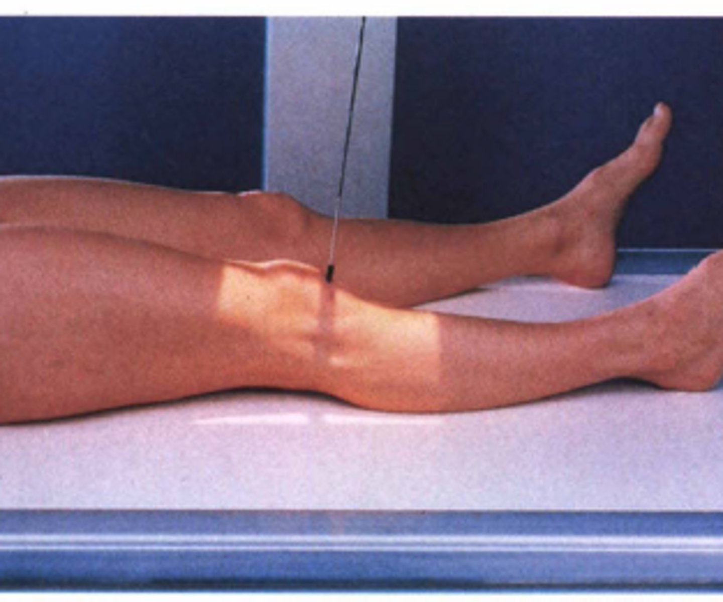
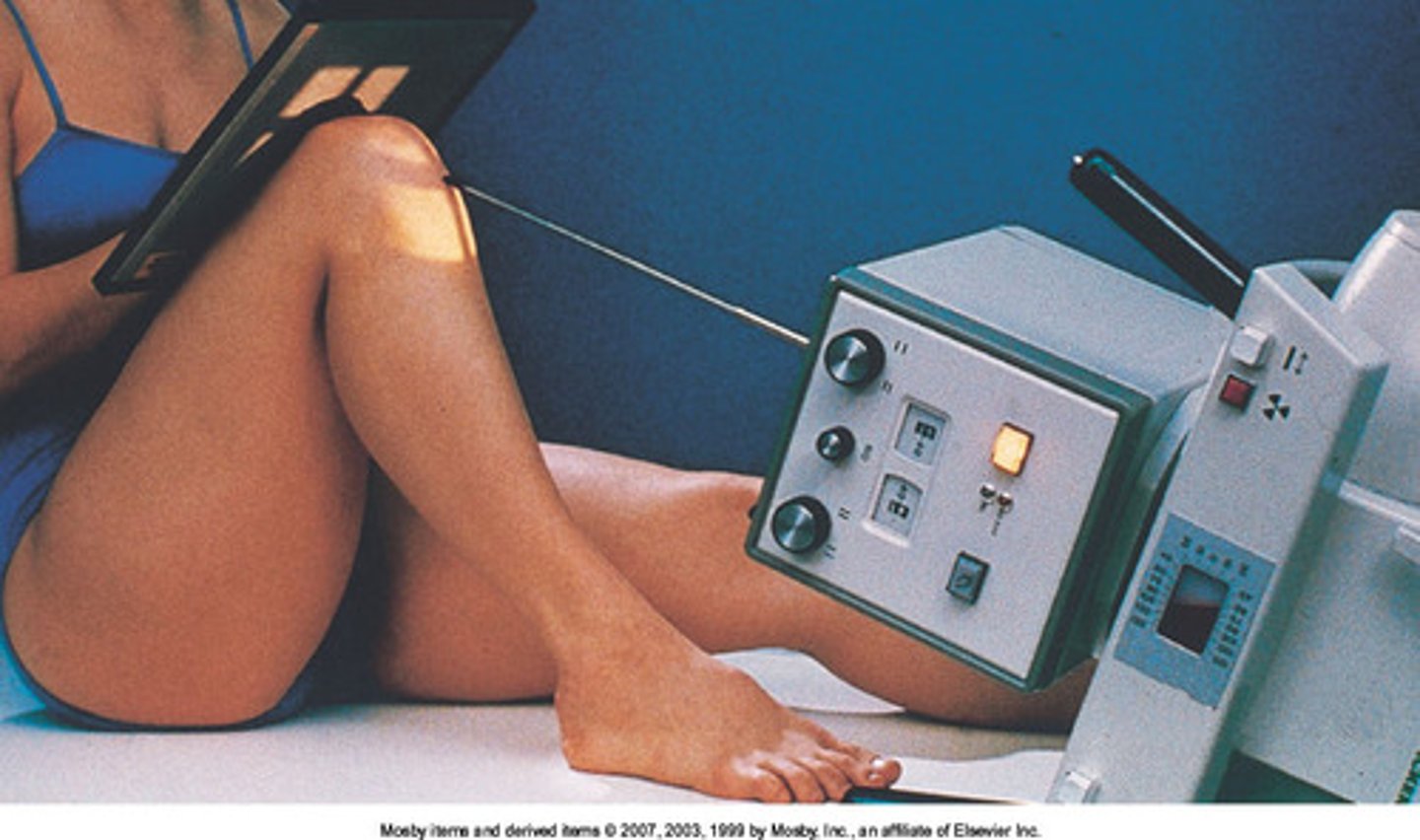
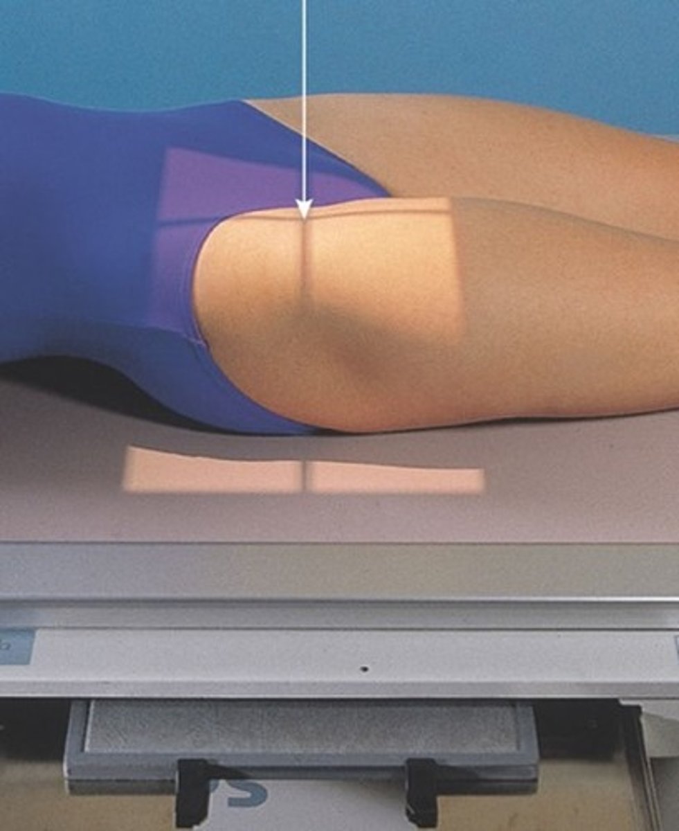
AP knee
perpendicular for average sized patients, 3-5 caudal angle for smaller patients, 3-5 cephalic angle for larger patients
directed to midpoint of knee at a level 1/2 inch distal to the apex of the patella
AP oblique knee (medial/lateral)
perpendicular for average sized patients, 3-5 caudal angle for smaller patients, 3-5 cephalic angle for larger patients
directed to midpoint of knee at a level 1/2 inch distal to the apex of the patella

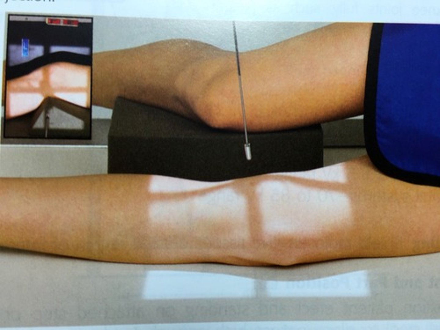
lateral knee
angled 5 to 7 degrees cephalic directed 1 inch distal to medial epicondyle
AP weight-bearing knee
perpendicular for average sized patients, 3-5 caudal angle for smaller patients, 3-5 cephalic angle for larger patients
directed to midpoint of knee at a level 1/2 inch distal to the apex of the patella
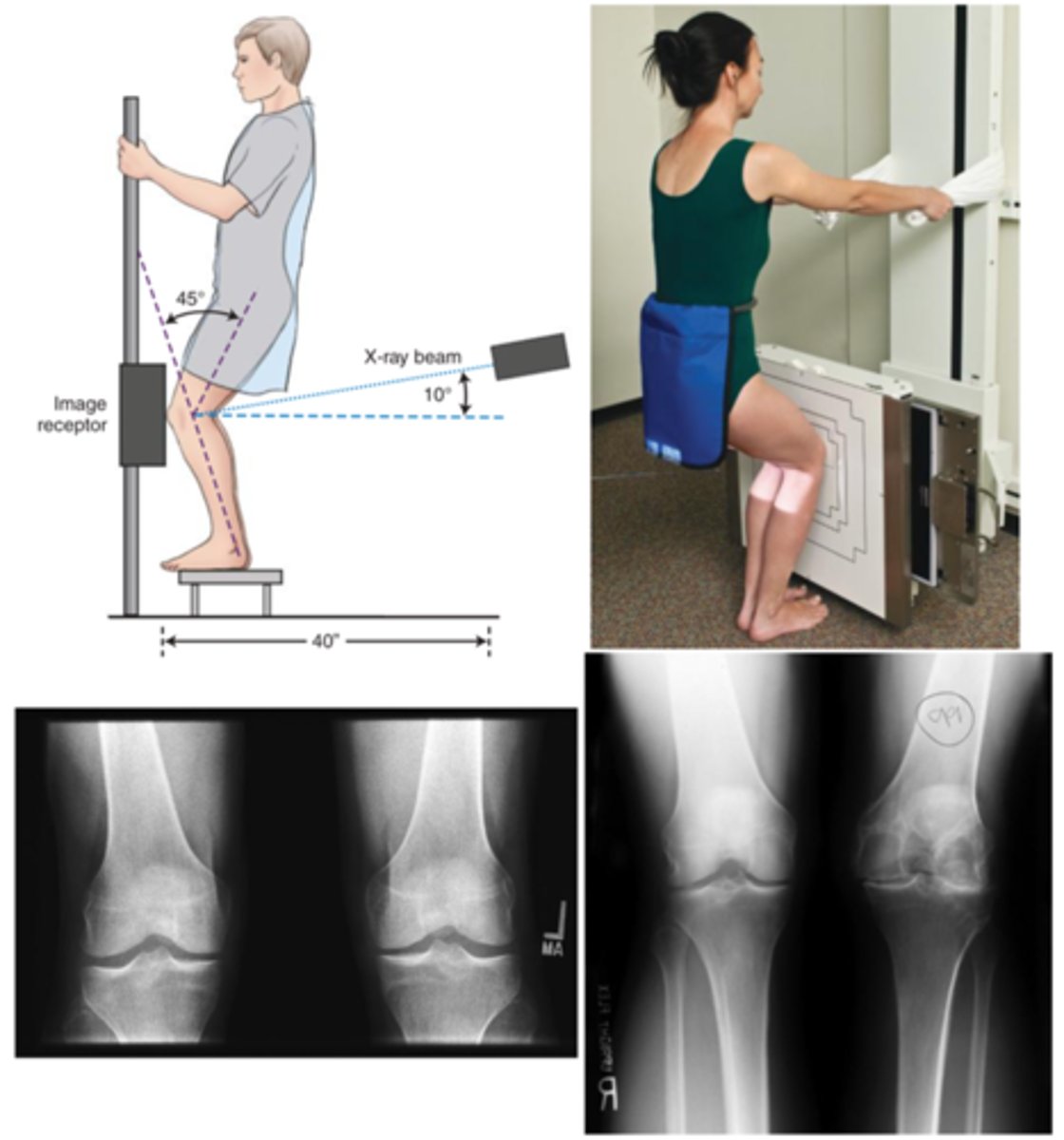
PA Axial Weight-Bearing Bilateral Knee Projection (Rosenberg Method)
CR angled 10 degrees caudal and centered to midpoint between knee joints at a level 1/2 below apex of the patellae
merchant bilateral method (knees)
CR angled 60 degrees caudal to a point midway between patellae
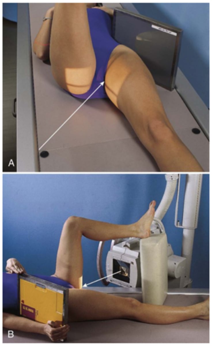
settegast method (knee)
angled 15-25 degrees cephalic and directed to patellofemoral joint space
hobbs modification (knee)
perpendicular to patellofemoral joint

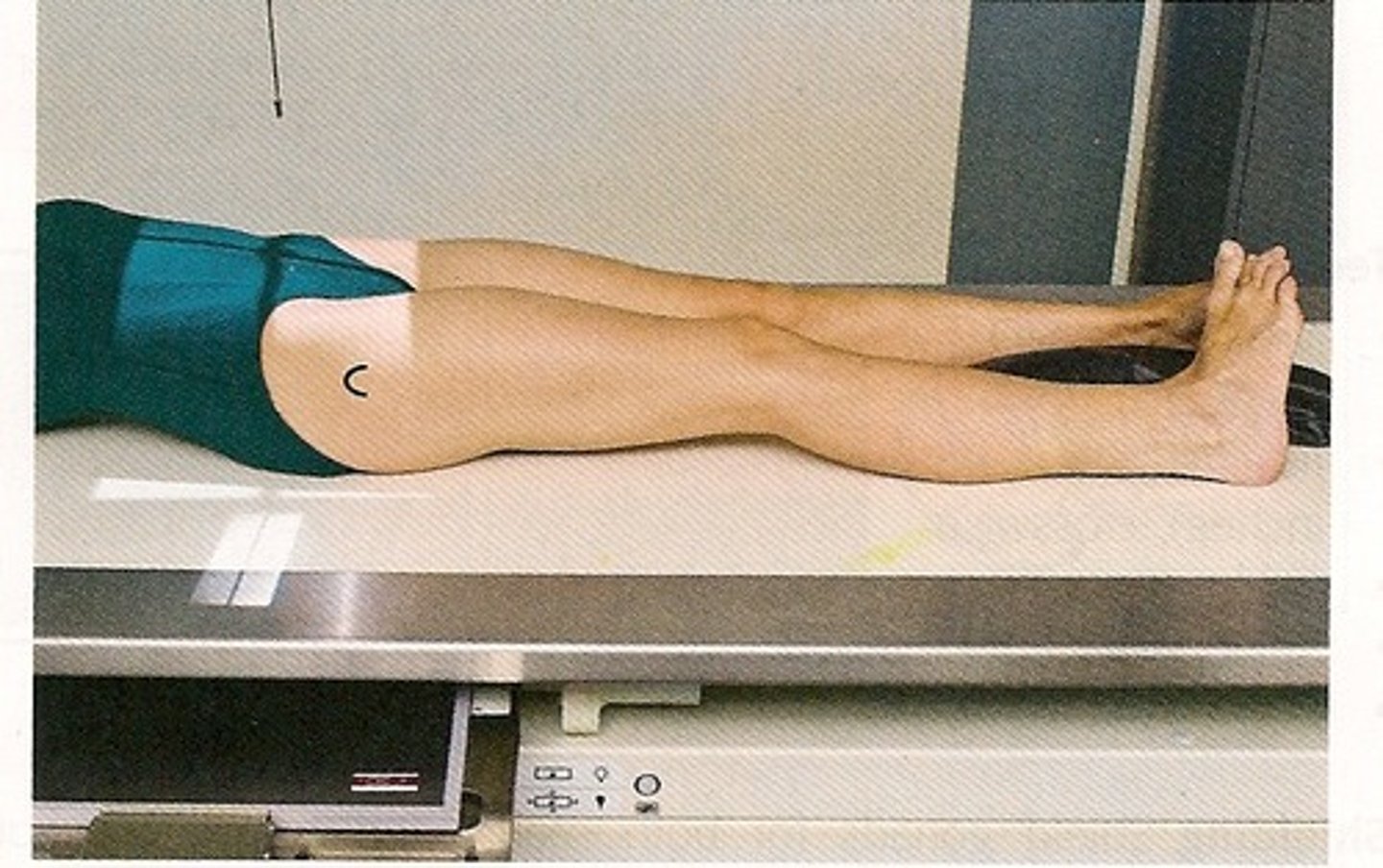
AP femur (mid and distal)
perpendicular and directed to midpoint of IR

AP pelvis (bilateral hips)
perpendicular and directed midway between ASIS and pubic symphysis
AP bilateral frog-leg pelvis (modified cleaves method)
perpendicular and directed 1 inch above pubic symphysis

AP axial outlet pelvis (taylor method)
angled 40 degrees cephalic directed 1-2 inches inferior to pubic symphysis

AP axial inlet pelvis (lilenfeld method)
angled 40 degrees caudal to ASIS

acetabulum iliac oblique (judet)
perpendicular and directed 2 inches medial and inferior to downside ASIS

acetabulum obturator oblique (judet)
perpendicular and directed 2 inches inferior to upside ASIS
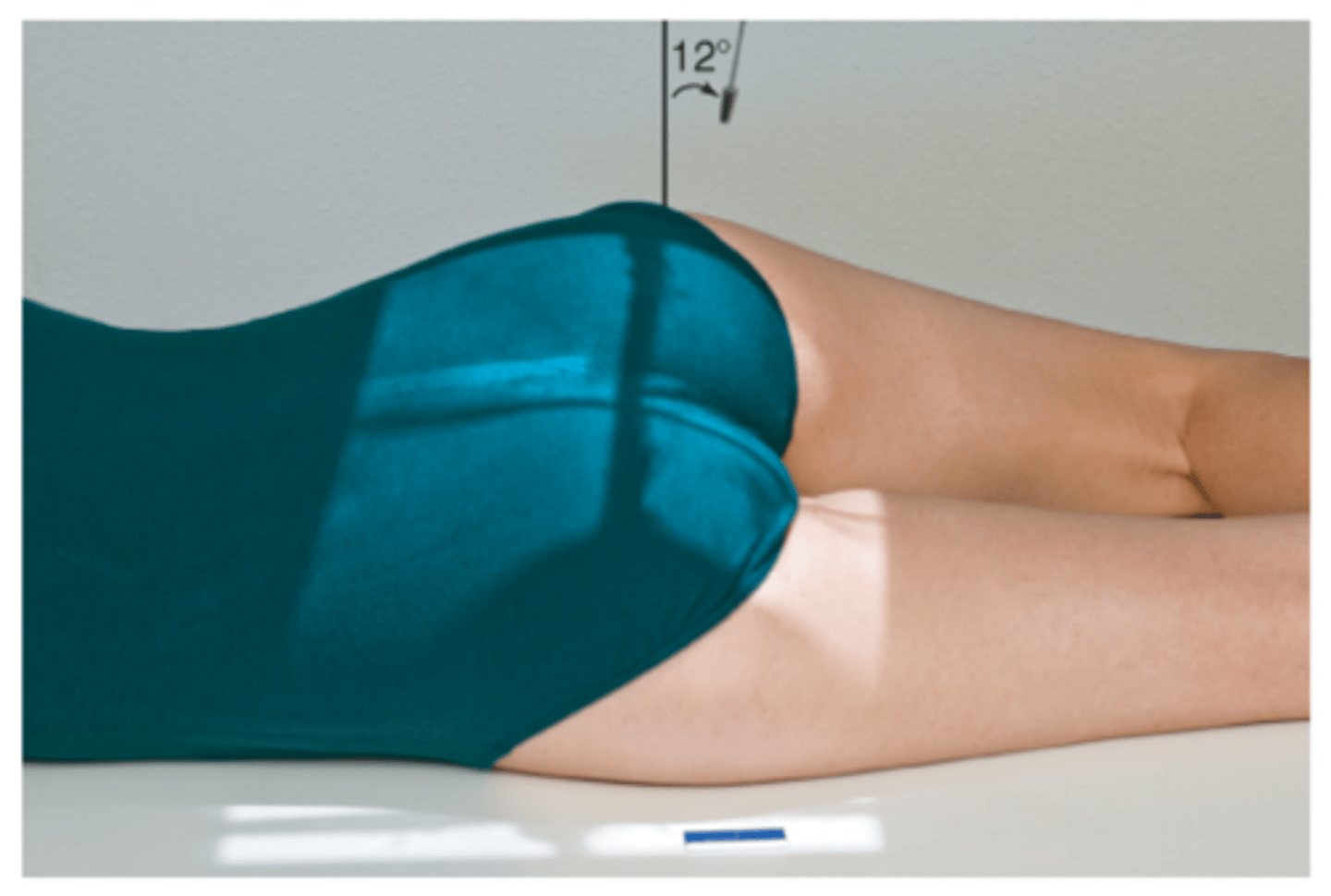
acetabulum PA axial oblique (teufel)
angled 12 degrees cephalic and directed 1 inch above greater trochanter/2 inches lateral to the midsagittal plane
AP hip
perpendicular to the femoral neck (1-2 inches medial and 3-4 inches distal to ASIS)
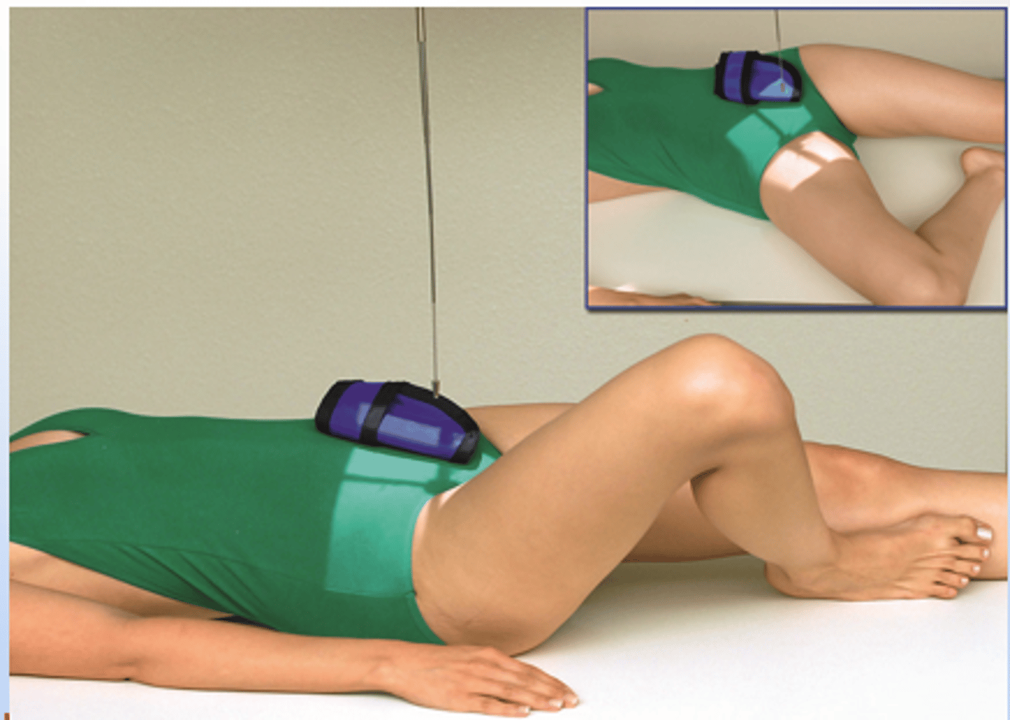
axiolateral inferosuperior hip (danelius-miller method)
horizontal and perpendicular to femoral neck and IR
AP unilateral frog leg hip
perpendicular to femoral neck
modified axiolateral hip (clements nakayama)
horizontal with a 15 degree posterior angle directed to femoral neck
AP axial sacrum
angled 15 degrees cephalic and directed 2 inches above pubic symphysis
AP axial coccyx
angled 10 degrees caudal 2 inches above symphysis