Brain Bee part 1
1/164
There's no tags or description
Looks like no tags are added yet.
Name | Mastery | Learn | Test | Matching | Spaced |
|---|
No study sessions yet.
165 Terms
How many operations per second can our brain handle?
1028
Is the brain capable of changing and adapting?
Yes
What allows neurons to release transmitter?
The fact that they change electric properties during an action potential
What are some examples of functions that the brain is responsible for?
Consciousness, thoughts, feelings, etc
What are the 3 main research designs to learn abt the brain?
Experimental design
Observational study
Case study
What is an experimental design?
Like a lab-step by step process-develop question, then hypothesis, then perform tests (experiment) to answer the question. -designing an experiment
Independent variable
The factor you choose to change
Dependent variable
Factor that is influenced by the independent variable
Confounding variable
Influence of external variables that can affect the results of the experiment
How can we eliminate confounding variables?
Thru a control group
Causality
Change in the independent variable directly causes a change in the dependent variable
Observational study
One type of an observational study is a quasiexperimental study
Have uncontrolled variables-therefore require 2 groups-experimental group and control group
Usually done when an experimental study is either impractical or unethical
Describe an experimental and control group in an observation study
Experimental-if the question we are trying to answer is if ppl with head injuries have worse hand eye coordination, the experimental group would be composed of ppl who are already diagnosed with a head injury
The control group would be composed of ppl that are demographically similar to the experimental group, but without head injuries
Then, we can observe the differences in the hand eye coordination between the 2 groups-usually points to more of a correlation rather than a causation tho
Case study
A detailed description of a single patient and their condition
Only show correlation not causation
Can help with development of hypothesis and later be tested experimentally
What are case studies usually used for?
Rare conditions-very specific injury/situation
Neurogenesis
Neuronal growth
Plasticity
The ability to change over time-brain can rewire/repair itself -figures out how to function without using damaged connections
Neurodegenerative
Symptoms get progressively worse over time-4 brain disorders
eg. Parkinson’s-ability to move and stuff decreases the more u have it, Alzheimer’s-memory deteriorates, etc.
Are there cures for neurodegenerative diseases?
Not currently-there also aren’t any treatments without risks/side effects yet
Is there strong evidence that the brain can recover from the destruction caused by a neurodegenerative disease?
No
Contralateral signaling in brain
Signaling pathways from left side of the brain cross over to communicate with the right side of the body and vice versa
Localization theory
Created by Paul Bronca-mid 1800s
Said that specific areas of the brain are responsible for carrying out specific functions
Distributive processing theory
Opposing theory to localization
Thought that behavioral functions require activation of cells across several areas of the brain-eg. complex behaviors like consciousness and emotion
Cognition
Generating knowledge thru a combo of senses, memories, and thoughts.
What is the most likely theory for how the brain works?
That some behaviors are more localized than others, but still rely on signals from across many other brain areas-basically a combo of localization and distributive theory
What did the invention of microscopes in the 1900s allow us to discover?
That different neurons have different shapes, and therefore carry out different functions-viewed and observed thru microscopes
This was our foundation for understanding cells that make up the nervous system
Electron microscope (include when made and what it was used for)
Made in 1954 + advancements in animal biology
Used to see space between neurons
fMRI (functional magnetic resonance imaging)-include when made, and what it allowed (include the spatial and temporal resolution qualities, advantages, disadvantages, what is it and application)
Made in 1991
Allowed us to visualize the brain activity when a person is doing smth-eg. making a decision, etc.-researchers use this to help correlate behaviour with activity patterns in specific parts of the brain
Good
Moderate-limited by speed of blood vessel dilation
Visualize real-time brain activity during complex behavioral tasks-can see which parts of brain are active and working thru blood flow change
Person can't have any metal (cause there’s a risk of injury or death due to the strong magnetic field if metal), test conditions make it difficult to assess children and those with anxiety/panic disorders (loud, small space, etc.) without endangering the patient, and data is hard to analyze + frequently has false positives (cause the change in BOLD signal is super small). Also, it assumes increased blood flow is directly correlated with the amnt. of neural activity which isn’t always the case
Study areas of the brain that are most responsive to specific tasks/conditions/emotions
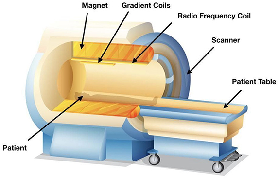
What is it?
Has 2 components-
A machine
As the person goes thru it, a powerful magnet revolves around their head (stronger the magnet better the spatial res. btw)
Circular tunnel
A device that emits radio waves
Revolves around head
Both components interact with protons-when protons enter a strong magnetic field, they align with or directly against the direction of the field. When it by radio waves, the protons lose their alignment going into a hi energy state. The protons will then fall back into a low energy state and return to alignment with the magnetic field.
The MRI detects this process
Since protons in oxygenated hemoglobin are sensitive to a magnetic field (diamagnetic) while in deoxygenated hemoglobin is not (paramagnetic), we are able to measure changes in oxygenation levels
MRI’s also rely on the fact that more metabolically active areas in the brain + body need more oxygen (like a PET scan with glucose)-they detect a change in blood flow (cause blood vessels dilate when need more oxygen)-This is called blood oxygenation lvl-dependent signal (BOLD signal)
CLARITY (include the spatial and temporal resolution qualities, advantages, disadvantages, what is it and application)
Very good
Poor
Microscopic level
Doesn't work on living matter-cause its destructive to tissue-all function is destroyed
Shows connectivity between cells
What is it?
A method to render the entire brain transparent-helps us map out the nature of the connections that span the NS thru the like scaffold thing
They flush the brain with chemicals and form a gel matrix that surrounds every cellular structure component. Then, it sues a chemical detergent to wash away the lipids while the gel stays unaffected-by washing away these light-affecting lipids, we can see where the connections were (cause of the “mold” we get)
Endogenous
Originating from within the body
Exogenous
Originating from outside the body
Action potential
All-or-nothing electrical output of neurons; temporary change in voltage caused by the movement of charged ions across the cell membrane.
It is a Short-lasting, temporary change in membrane potential that moves down the axon. All or nothing response: only a large change in potential will pass the signal forward.
Specific shape: depolarization, hyperpolarization, repolarization.
If intensity of stimulus increases the size of the action potential doesn’t become larger.
Myths abt neuroscience
We only use 10% of our brain
we use every part of our brain and most of our brain is active most of the time-just not at the same time-eg. does a traffic light only use 33% of tis lights? No, it uses all 3 lights at precise times. The brain also works like this. The activity of the brain is regulated by many mechanisms to prevent unusual activity-actually if many cells were active at the wrong times (if a traffic light shows both green and red) chaos would occur-one cause of seizures is actually excessive neural activity
Forming memories causes new neurons to be born
Memories aren’t represented by a new cell in our brain thru neurogenesis but rather they are probs stored at the synapse-the changes in ways neurons connect and communicate with each other is probs the mechanism behind how memories are formed and stored
The brain cannot repair itself
Plasticity allows the brain to learn how to rewire itself if a part of it is damaged-figures out how to still carry out functions without using the damaged connections. However, some conditions are neurodegenerative which means the brain can’t recover from the destruction of these diseases
If you are analytical, you are
left brain dominant, but if you
are creative, you are right brain
dominant
Nearly every function that the left half of the brain can do the right half can do just as well and vice versa.
WIFT
FWIT
What are the 2 diff forces that act on ions once an ion channel is open?
Electrical gradient
Chemical gradient
Electrical gradient
Refers to the electrical forces acting on charged molecules
Eg. pulling opposite charges together or pushing like charges apart
What are the 2 types of signals used by the nervous system to communicate?
Electrical (like an action potential) and chemical (like neurotransmitters) signals
What part of Phineas Gage’s brain was affected in his accident, and what did this reveal about that area’s function?
His frontal lobe was damaged after the accident and the changes in his personality revealed that one of the functions of this area is regulating our inhibitions
If an ischemic stroke were to cause damage to the right motor cortex of the brain, what kinds of effects would be observed?
Motor deficits on the left side of your body (cause of contralateral organization)
What is the benefit of model organisms for neuroscience research? What are some examples of model organisms?
Model organisms are non-human animals that share nervous system characteristics with humans, making them good alternatives for studies that cannot ethically be performed on humans. Some common model organisms are C. elegans, fruit flies, zebrafish, song birds, mice, rats, and macaque monkeys.
What is a common bias among selection of participants in neuroscience studies?
WEIRD subjects-Western, Educated, and from Industrialized, Rich and Democratic countries-this creates a bias since WERID ppl perform differently on behavioral tasks compared to others.
Most participants in neuroscience studies also tend to be undergraduate students that must participate in research for class credit. An acronym that summarizes the common biases
What parts of the body are part of the CNS (central nervous system)?
Brain + spinal cord
What parts of the body are part of the PNS (Peripheral nervous system)?
All other nerve cells in the body
Does info pass between CNS ad PNS or are they isolated from each other?
It does-pretty rapidly too
What is an incoming/ascending signal?
A signal that originates in the PNS and moves to the CNS
What is an outgoing/descending signal?
A signal that originates from the CNS that goes to the PNS
Afferent signal
Info that arrives into CNS
Efferent signal
Info leaving the CNS (like exits the CNS)
Brain (include how much energy it uses)
The main organ where movement, thoughts and consciousness originate
Uses up 1/5 of body/s total energy expenditure
Rostral
Parts of the brain that are more forward/in the front
Caudal
Opposite of rostral-structures more at the back of the brain
Anterior
Used interchangeably with rostral
Posterior
Used interchangeably with caudal
Dorsal or superior
Above/top-closer to the top of the head
Ventral or inferior
Below/bottom of head
Medial
Closer to the center (in the middle)
Lateral
Closer to the sides
Coronal slice
A projection of the brain that is sectioned to help with visualization and analysis of the brain
Its like literally vertical slices of the brain
Brain slices parallel to crown
Horizontal projection
A way to image the brain-displays brain from above
Brain slices that are parallel to the plane of the ground
Horizontal slice
Horizontal slices of the brain
Saggital
The slice that divides the brain into left and right hemispheres
Also called parasaggital
Brain slices parallel to the plane of the ground
Parasaggital
Slices parallel to the sagittal slice
These slices are never symmetrical tho-cause taken from one hemisphere of the brain at a time (from left to right)
White matter
Brain tissue that is almost white and pale
Allows for transfer of info form left and right hemispheres of the brain
Why does white matter appear white?
To allow neurons to send signals rapidly-they’re modified to add layers of fatty lipids (myelin)
This modification causes the light to reflect, making the tissue appear white to the eye
Decussation
When a white matter pathway crosses from one hem. to another
Corpus callosum
The main tract of white matter that allows the passage of info between the 2 hemispheres
Gray matter
Sections of brain tissue that have a darker pink/gray colour
Areas are usually dense with cell bodies
What 3 main germ layers are cells classified as when an embryo first starts to form?
Ectoderm
Mesoderm
Endoderm
Ectoderm
Usually develops into NS
Folds into itself-after merging at the surface, it creates the neural tube
Neural tube (include when it’s formed, and smth abt the 3 vesicle stage and the 5 vesicle stage)
Forms during 3-4th week of gestation
Cells becomes components of CNS
Undeveloped NS at this (the 3rd) stage is called the “3 vesicle stage”-because the neural tube has 3 distinct components early in development
A week later, parts of progenitor NS divide-becomes the “5 vesicle sage”
What are the 5 vesicles (from posterior to anterior)
Myelencephalon
Metencephalon
Mesencephalon (midbrain)
Diencephalon
Telencephalon (anterior)
The Rhombencephalon (hindbrain) divides into 1 and 2 by the 5 vesicle stage
The Prosencephalon (forebrain) divides into 4 and 5 by the 5 vesicle stage
Mesencephalon stays the same by the 5 vesicle stage-no change
Note: so at the 3 vesicle stage its just the hindbrain, midbrain and forebrain but then they divide into these 5
Rhombencephalon (hindbrain)
Oldest part of CNS-cause responsible for more basic survival things like motor control and unconscious functions of life,
Has 2 regions-Myelencephalon and Metencephalon
Myelencephalon
Develops into the medulla oblongata
The medulla oblongata is responsible 4 involuntary functions like breathing + can detect toxins in blood from dietary sources-triggers vomiting.
Metencephalon
Develops into Pons and Cerebellum
Pons: Contains areas that help us hear sounds + taste foods
Helps with involuntary functions
Cerebellum: Enables motor functions-balance, coordination, etc.
Mesencephalon (midbrain)
Structures don’t change from 3 to 5 vesicle stage
Contains structures like the red nucleus and substantia nigra-coordinate complex movements, or the tectum-helps us respond to visual stimuli
has a variety of increasingly complex functions related to coordination of more complex movements, reward and motivation processing, and more.
2nd most recent evolutionarily
Prosencephalon (forebrain)
Develops into the higher order brain regions-like the cerebral cortex
The most recently evolved region
Diencephalon
Contains thalamus + hypothalamus
Thalamus: Sensory info passes thru here-eg. taste, touch, etc.
Hypothalamus: Communication route to endocrine system
Telencephalon
Includes basal ganglia and cerebral cortex
Basal Ganglia: Used for motor and habit learning, emotional processing, and action selection
Cerebral cortex: Outermost layer of brain. Processes attention, memory and language
What brain structures are more basic functions usually controlled by? How about more complex ones?
Posterior brain structures
Anterior of the brain/brains structures
eg. brainstem in the hindbrain does stuff for basic survival (eg. respiration) while the forebrain does things involved with personality-much more complex
What is the nervous system?
a network of neurons connected that delivers information to and from the rest of the body. It is like a complex series of roads and highways that connect different cities, if the cities are the organs.
Design a study to answer the question “How do traumatic brain injuries affect short term memory?” What type of study would you do? How would you control the experiment? How do you select your participants? What are some weaknesses of your study design?
Observational (not experimental=not gold standard, but we can’t do an experiment because it is not ethical to give people head injuries)
Control for sex, age, environment, etc.
Selected based on rating of symptoms, diagnosis vs. no diagnosis, try to match for everything except TBI status, etc.
Medications, geographic, nature of head injury (no two are the same), etc.
How do you think the concept of emergence relates to localization theory and distributive processing theory? Which theory do you think is a more likely explanation for how the brain works?
Emergence and localization theory correspond to the same idea that each part of the brain does a specific function. The cases of Phineas Gage and Patient Tan both support this theory by providing insights about the effects of damage to very specific areas of the brain.
Emergence and distribution processing theory explains that the activation of cells across the whole brain is required to execute behaviour functions.
The most likely scenario is that both theories hold some truth: it is likely that some processes are highly localized in the brain, while still requiring input from other parts of the brain.
Note: emergence refers to the phenomenon where a complex system exhibits properties or behaviors that its individual parts do not possess on their own, arising only when those parts interact within a larger whole
Cortex
Bumpy outer surface of the brain
Made up of ridges (gyri) + grooved indentations (sulci-sometimes called fissure)
Longitudinal Fissure
Sulcus (singular of sulci)
Divides the 2 hemispheres (anterior-posterior axis)
Central Sulcus
Starts at the dorsal then runs abt half the length of the brain (vertically)
Lateral Fissure
Sulcus
Runs in anterior-posterior direction
Name the 4 major lobes the cortex is divided into
Occipital lobe
Temporal lobe
Parietal lobe
Frontal lobe
Are the lobes of the brain paired (one left on right) + roughly symmetrical?
Yes
Occipital Lobe
Smallest
Processes visual stimuli-V1-primary visual cortex of lobe
No borders
V1
Interprets signals into a visual representation of the world
Temporal lobe
Lateral fissure marks the border
Interprets sound waves thru auditory system-distinguishes btween talking, instruments, bark of dogs, etc. -this is due to the primary auditory cortex in the lobe (A1)
The ability to remember things (memory related processes) are also done here-thru hippocampus
Also has structures that help with language
Parietal Lobe
he front end (anterior) is is bordered by the central sulcus and the ventral border is the lateral fissure
Gives us with the ability to sense things with out body-eg. sense of touch- temp., pain, vibration, etc.
proprioception-ability to identify where parts of ur body are located is also processed here
All fo these functions are carried out by the primary somatosensory cortex-S1
Frontal lobe
Posterior border is the lateral sulcus
Largest of the 4 lobes
Contains the primary motor cortex (M1)
Carries out higher order functions-eg. Personality
Can also let us do mental math and suppress socially unacceptable actions
M1 (primary motor cortex)
Contains neurons to control movement of the body
Spinal cord (SC-not a scientific shortening of the word just my way of making things shorter)
Carries info towards the brain (upward) and towards body's other organs and muscles (downward)
Can also process sensations and form an appropriate motor response without brains input
Goes from the lvl of the neck down to the small of ur back
Housed with vertebral column-a series of bones
Is the diameter of the SC completely uniform?
No
How many iverluing vertebrae can the spinal cord be divides into?
A number and letter is used to identify each section of the spinal cord-letter is the vertebral section and number is the number of bones down from the previous section (smaller number= more anterior and larger=more posterior)
What 2 nerves branch off from each section or the spinal cord?
Afferent (incoming to CNS)
sensory nerve roots-sensations go to brain
Branch from dorsal side
Efferent (outgoing from CNS)
motor nerve roots-brain tells body to move
Branch from ventral side
These 2 branches meet and extend away from the spinal cord-after merging, they are called the spinal nerves-humans have 31 pairs of these
What are the 4 regions of the spinal cord?
Cervical
Thoracic
Lumbar
Sacral
Cervical
Upper 8 pairs of spinal nerves
Innervate muscles in neck, shoulder, arms and hands-SHAN
Sections C3-C5 innervate the diaphragm
Cervical areas of SC have the widest diameter