Parts of Spine
 Call Kai
 Learn
Learn Practice Test
 Spaced Repetition
 Match
Match Flashcards
 Knowt Play
1/14

There's no tags or description
Looks like no tags are added yet.
Name | Mastery | Learn | Test | Matching | Spaced | Call with Kai | Chat |
|---|
No analytics yet
Send a link to your students to track their progress
15 Terms

Spine
Parts of a spine:
Cervical Curve: 7 vertebrates (forms a convex curve)
Thoracic Curve: 12 vertebrates (forms a concave curve)
Lumbar Cuve: 5 vertebrates
Sacral Curve: 5 vertebrates
Kyphosis & Lordosis
Kyphosis refers to the natural curvature of the upper spine (primary curves in thoracic & sacrum)
Lordosis refers to the natural curvature of the lower spine (secondary curves in cervical & lumbar)
(Hyper in the prefix means extreme curvature of these)

Typical Vertebrae (Posterior/Anterior View)
Vertebral body: supports body weight and protects spinal cord
Pedicle: Connects to the lamina to make the vertebral foreman
Lamina: Connects to the pedicle to make the vertebral foreman
Vertebral foreman: the spinal cord goes in this hole
Transverse processes: attachment points for muscles and ligaments to stabilize spine
Spinal Processes: attachment point for muscles and ligaments to stabilize spine

Typical Vertebrae (Lateral View)
Superior articular process → Connects with another vertebrae’s inferior articular process, helps limit spinal movement from bending too back.
Superior articular facet → where a joint forms to connect other vertebrae
Superior articular notch → indentation that helps form the intervertebral foramen, where spinal nerves pass through.
Inferior articular process → Connects with another vertebrae’s superior articular process; helps stabilize movement.
Inferior articular facet → where a joint forms to connect other vertebrae
Inferior vertebral notch → indentation that helps form the intervertebral foramen for spinal nerves to pass through.
Vertebral body
Spinous process

Invertebrate Discs between vertebral body
Nucleus pulposus: soft, gel like centre of the disc that acts as a cushion for vertebral body
Annulus fibrosis: tough, fibrous ring that holds the nucleus in place, resists tensile & twisting forces
How does it work during movement?
Lying down:
Less body weight → disc is relatively thicker.
Standing:
Body weight compresses the disc → it becomes shorter/thinner
Lateral flexion:
One side of the disc is compressed, the opposite side is stretched

Spinal Ligaments
Anterior longitudinal ligament:
Covers the vertebral bodies and discs anterolaterally.
Extends from sacrum to occipital bone
Helps maintain stability of disc joints
Posterior longitudinal ligament: Extends from axis to sacrum. Helps prevent excessive flexion (bending forward).
Ligamentum flavum: Joins laminae of other vertebrae. Helps limit excessive flexion and helps the spine return toward its normal position after bending.
Supraspinous ligament: runs along the tips of the spinous processes. Helps limit excessive flexion.
Interspinous ligament → runs between the spinous processes. Also helps limit excessive flexion.
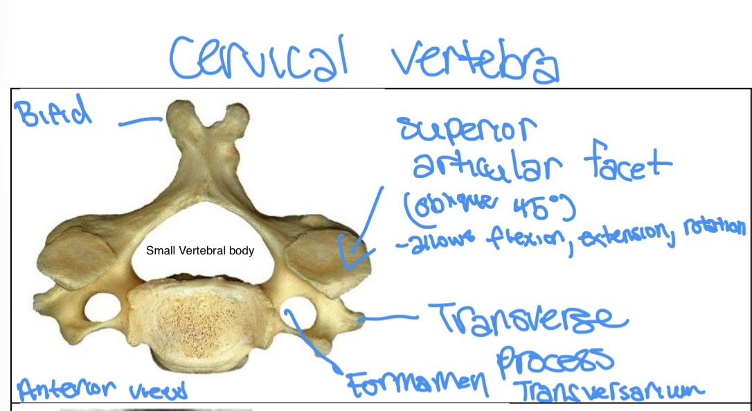
Cervical Vertebrae (anterior/posterior view)
Foramen transversarium → hole in the transverse process; vertebral arteries pass through it (mainly C1–C6).
Small vertebral body → smaller than the vertebral bodies of the thoracic and lumbar spine because they carry weight of head.
Bifid spinous process → spinous process is split into two parts at the tip, especially C2–C6. Attaches to ligaments and muscles
Articular facets: 45° oblique → connects to otherallows flexion, extension & rotation

C1 & C2
C1 (atlas):
Interior tubercle → Small bump at the front; provides attachment for muscles/ligaments.
Posterior tubercle → Small bump at the back; provides attachment for muscles/ligaments.
Large superior articular facets → Connect with the occipital joints of the skull → allow the head to nod up and down (“yes” motion).
Attachment point anteriorly for transverse ligament → The transverse ligament holds the dens of C2 against C1, keeping it stable.
C2 (axis):
Bifid spinous process → Split spinous process; provides attachment for muscles and ligaments that help move/stabilize the neck.
Odontoid process (dens) → A tissue that acts as a pivot for C1 and the skull → allows horizontal rotation of the head.
Minimal transverse processes → provide attachment points for muscles/ligaments.
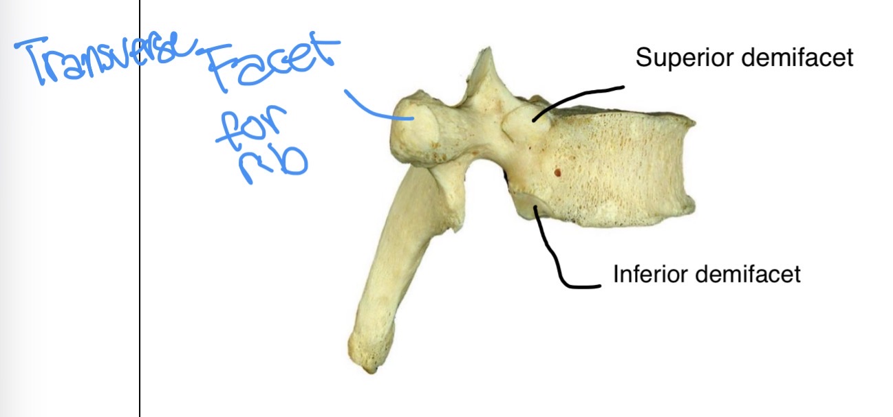
Thoracic Vertebrae (lateral view)
Superior demifacet: connects to the head of the rib above
Inferior demifacet: connects to the head of the rib below
Transverse facet for rib: connects to the tubercle of rib
Lumbar Spine (anterior/posterior view)


Sacrum & Coccyx
Base: the top part of the sacrum. Connects with L5.
Apex: the bottom point of the sacrum. Connects with the coccyx.
Anterior sacral foramen: openings on the front of the sacrum where sacral spinal nerves and blood vessels pass through.
Coccyx: small tailbone below the sacrum; provides attachment for muscles and ligaments and helps support the pelvic floor.
Auricular surface: Connects with the ilium of the pelvis (hip bone)

Parts of sternum
Manubrium: The superior part of the sternum and attaches to 1st rib. The central part is the suprasternal notch, the sides are the clavicular notches, where clavicles attach.
Sternum: sternal angle to xiphoid. attaches from 2nd-7th rib
Xiphoid: Bottom part of sternum. Attachment points for ligaments and muscles.

True, false, floating ribs
True (vertebrosternal) ribs: 1st-7th rib attached to sternum anteriorly through costal cartilages
False (vertebrochondral) ribs: 8th-10th ribs: have cartilages on their anterior ends that attach to the superior cartilage
Floating ribs: 11th-12th ribs that have small cartilages on their anterior ends that don’t connect
Typical ribs
Head:
Neck:
Costal Angle:
Tubercle:
Costal Groove:
Facets for Vertebrae:
Facet for costal cartilage:

Ribs & Sternum Joints
Sternocostal joints: costal cartilages attached to sternum (Most are synovial plane joints, expect 1st rib)
Costochondral joints: costal cartilages attached to ribs
Costalvertebral joints: head of the rib that connects to the vertebral body
Costotransverse joints: tubercle of the rib that connects to the transverse processes