Unit 7 Intrahepatic Biliary Neoplasms
1/45
There's no tags or description
Looks like no tags are added yet.
Name | Mastery | Learn | Test | Matching | Spaced |
|---|
No study sessions yet.
46 Terms
A Cystadenoma & Cystadenocarcinoma are:
Rare
Cystadenoma & Cystadenocarcinoma are most common in who?
Middle-aged women
What are the symptoms of Cystadenoma & Cystadenocarcinomas?
Abdominal pain, mass, or jaundice (if mass is near porta hepatis)
Cystadenoma & Cystadenocarcinoma US Findings: What do cystic mass have?
Multiple septa & papillary excrescences (lump)
Cystadenoma & Cystadenocarcinoma US Findings: Mass can show variations and can appear as what?
unilocular,
calcified,
or multiple
Cystadenoma & Cystadenocarcinoma US Findings: What can a lesion be associated with?
Dilation of intrahepatic ducts
What are the differential diagnosis for Cystadenoma & Cystadenocarcinomas?
Hemorrhagic cyst or infection
Echinococcal cyst
Abscess
Cystic metastasis
What are the 3 classifications of a Cholangiocarcinoma?
Intrahepatic
Hilar (Klatskin’s)
Distal
What is the second most common primary malignancy in liver?
Intrahepatic Cholangiocarcinoma
In a Intrahepatic Cholangiocarcinoma, incidence has risen and there is an increasing number patients with what diseases?
Cirrhosis and Hepatitis C
Intrahepatic Cholangiocarcinomas are often resectable or unresectable?
Unresectable
What is the outcome for a Intrahepatic Cholangiocarcinoma?
Poor prognosis
Intrahepatic Cholangiocarcinoma US findings: What is the size?
Large hepatic mass
Intrahepatic Cholangiocarcinoma US findings: What is the echogenicity?
Hypoechoic to hyperechoic
Intrahepatic Cholangiocarcinoma US findings: What is the texture?
•Heterogeneous texture or hypo vascular solid mass
Intrahepatic Cholangiocarcinoma US findings: What do 1/3 of the cases also have?
Biliary ductal dilation
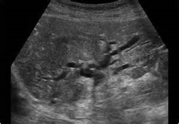
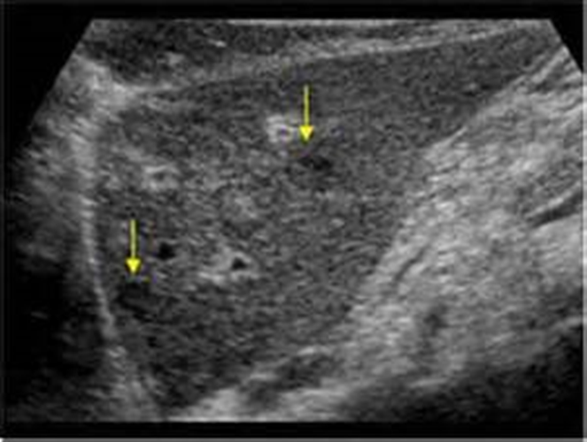
What is being shown in the image above?
Intrahepatic Cholangiocarcinoma
A Hilar Cholangiocarcinoma is also known as:
Klatskin’s tumor
Is Hilar Cholangiocarcinoma intrahepatic or extrahepatic?
Intrahepatic
Where is the Hilar Cholangiocarcinoma located?
At bifurcation of common hepatic duct
Hilar Cholangiocarcinomas are _____ _________
Slow growing
Hilar Cholangiocarcinoma can be _____ ____________
Late metastases
Clinically, what are the symptoms of a Hilar Cholangiocarcinoma?
Jaundice
Pruritus
Elevated cholestatic liver parameters
Klatskin’s Tumor US assessment: Where should the sonographer scan?
Porta hepatis
At level of obstruction
Klatskin’s Tumor US assessment: What should the sonographer assess?
Presence of a mass, lobar atrophy
Patency of MPV, RPV, LPV
Encasement of HA
Local or distant adenopathy & metastases
In a Klatskin’s Tumor if the ducts are dilated what should the sonographer do?
Follow course toward hilum to determine involved branches
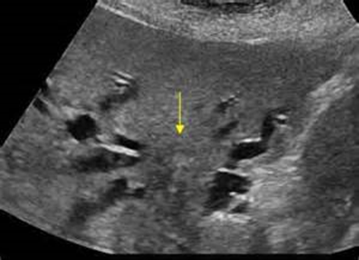
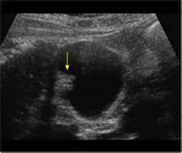
What is the arrow pointing to in the image above?
Hilar Cholangiocarcinoma/Klatskin’s Tumor
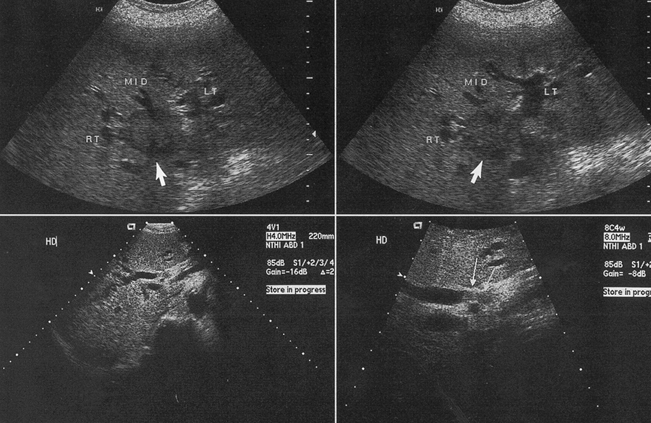
What are these images showing?
Klatskin’s Tumor
A distal cholangiocarcinoma is difficult to distinguish from what?
hilar cholangiocarcinoma
A main symptom of a distal cholangiocarcinoma is:
Progressive jaundice
A Distal Cholangiocarcinoma tumor can be either _____ or _______
Sclerosing or polypoid
With a Distal Cholangiocarcinoma what must be evaluated?
Tumor spread in superior ductal system; extrahepatic area; extension into adjacent lymph nodes
Distal Cholangiocarcinoma US findings: A sclerosing tumor is:
Nodular
Distal Cholangiocarcinoma US findings: What is shown with focal irregularities?
Ductal constriction & wall thickening
Distal Cholangiocarcinoma US findings: What is the echogenicity?
Hypoechoic & hypo vascular appearance
with poorly defined margins
With Distal Cholangiocarcinoma, a sclerosing tumor around the CBD is classified as what type of obstruction?
Extrinsic
With Distal Cholangiocarcinoma, a sclerosing tumor presents with what?
Nodular with focal irregular ductal constriction &
wall thickeningHypoechoic & hypovascular appearance with poorly
defined margins
With Distal Cholangiocarcinoma, a polypoid tumor within the CBD is classified as what type of obstruction?
Instrinsic
With Distal Cholangiocarcinoma, a polypoid tumor looks like what on ultrasound?
Hypovascular, well-defined mass
A sclerosing and polypoid tumor both dilate:
The entire biliary tree
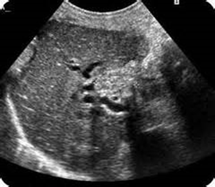
What is this image showing?
Distal Cholangiocarcinoma
Sclerosing Tumor
What are the most common tumor sites for metastases to the biliary tree?
Breast
Colon
Melanoma
What does metastases to the biliary tree affect?
Affects intrahepatic & extrahepatic ductal systems
What are the ultrasound findings for metastases to the biliary tree?
Similar to cholangiocarcinoma
Hyperechoic or hypoechoic
Hypovascular
Poorly defined margins
What is being shown in this image above?
Metastases to Biliary Tree
What is being shown in this image above?
Biliary Tree metastases