Microbio- perspectives, microscopy, and taxonomy+nomenclature
 Call Kai
 Learn
Learn Practice Test
 Spaced Repetition
 Match
Match Flashcards
 Knowt Play
1/67

There's no tags or description
Looks like no tags are added yet.
Name | Mastery | Learn | Test | Matching | Spaced | Call with Kai | Chat |
|---|
No analytics yet
Send a link to your students to track their progress
68 Terms
Microbiology (give example organisms)
The study of small (microscopic) organisms
Including: Bacteria(MAIN FOCUS! e.gProkaryotes), Archaea, Some Eukaryotes (Protists, Algae, and Fungi), and Viruses
Tree of Life
Shows the evolutionary relatedness of organisms, the one used by microbiologists has three sides of branches 1.Bacteria 2.Archaea 3.Eukarya
Almost entire tree is under microbiology except for small subsect of Eukaryotes (animals, plants fungi)
Bacteria Perspective: What are bacteria? How long have bacteria been around? What do they look like inside? What is it? What do bacteria do? Bacteria metabolism?
Bacteria are an evolutionary separate form of life, separate section of tree
They are very small, no organelles, they have nucleoids (DNA material but no membrane)
It is a prokaryote (meaning has nucleoid while eukaryotes have nucleous)
They have been around for billion of years
Bacteria consume(sugars;light;toxins;metal), and they grow (even in extreme conditions e.g. 80 degrees C hot springs or 4 degrees C arctic ocean. They can grow in strong acid, high in atmosphere, deep in the earth, etc, etc, etc)
some make alcohol, some breathe uranium, etc, etc, etc, can eat or breathe just about anything
Bacteria cycle the most oxygen, make the most carbon, fix nitrogen. Some bacteria make us sick, most make us healthy - primary decomposers. Bacteria build communities, cooperate with each other, and they prey on each other.
Bacteria have extremely diverse metabolism
What is unique about bacterial chromosomes?
Bacteria have circular chromosomes
Single, circular chromosomes rather than the multiple linear chromosomes of eukaryotes
Bacterial Ribosomes versus Eukaryotic ribosomes
Bacterial ribosomes are overall smaller (70S compared to Euk.80S), and their small and large compartments are divided into 50S and 30S while Eukaryotes divide into 60S and 40S
Bacterial Cell Wall
No cytoskeleton, then membrane, then cell wall composed of peptidoglycan
How many bacteria are there?
5×10^30 , a number so large it makes impossible things likely
Where are bacteria found?
water, soil, air, deep subsurface, high atmosphere, In/On all plants and animals…. everywhere, all the time!
Bacterial Visibility
Almost always invisible, but are sometimes visible e.g in a petri plate, acid mine drain(pH0), or rainbow pool in yellowstone national park
Visible sometimes due to the colonies of bacteria being very large e.g. milky seas which has a colony of bacteria the size of New Jersey
Vibrio Fischeri
This is a marine bacterium, it is a squid symbiont, luciferase(enzyme), produces light when it is at a high cell density, uses “quorum sensing”
Squid/Vibrio symbiosis —→ Counter-illumination
Chemical recognition between squid and the Vibrio Fischeri bacteria, squid feeds the bacteria which allows it to grow to a high density therefore emitting light
Counter-illumination: at night when the moonlight would cast a shadow highlighting the squids location to bigger fish below, the activated high colonies of Vibrio Fischeri light up from underneath the squid blocking its shadow from view
Each morning the squid expels the colony and starts over
The squid hide during the day
Symbiosis
Bacteria inside and on other organisms e.g. bacteria+animal, bacteria+plant, bacteria+fungi, bacteria+bacteria(biofilm)
Early Microscopes + Scientists
Antony van Leeuwenhoek - handheld, single lens microscopes, credited with discovering microorganisms which he called “Animacules” , 1676
Even earlier than 1676- Robert Hooke’s microscope (looks normal on one side with eyepiece, barrel, objective, and specimen holder but has water flask and oil lamp off to the side for light reflection) , he had simplified drawings of Myobacterium tuberculosis
Voynich Manuscript
1620, Yale secret library, included weird drawings and an unknown language, pictures depict imagery similar to 17th century microscopes; imagery similar to cells (first microbiology textbook?)
Potential Author: Cornelius Drebbel bc designed early microscopes
Microscopy (what is it needed for, and what are the requirements)
required to see individual bacteria
Magnification
Resolution
Light Quality
Contrast
Magnification
relative increase in image size, increase in apparent size of image, increasing the focal length (length between the objective and the slide) decreases the magnification, decreasing the focal length (moving in closer to slide) increases magnification
*empty magnification: bigger isnt always better, have to increase resolution so the image doesnt appear fuzzy closer in
Resolution
the ability to distinguish two points that are close together, higher numerical aperture (angle of light / closer means wider angle) means higher resolution.
resolution is ultimately limited by light quality because objects that are smaller than the wavelength of light cannot be detected red-orange-yellow-green-blue-violet (lowest to highest resolution bc reds wavelength is the biggest, cant detect anything smaller)
Light Quality
sets limit of resolution
Contrast
the ability to detect objects against a background
Refraction
light bends when it passes from one medium to another (straw as it passes from the air into the water)
bending occurs because light travels more slowly in higher density mediums
Lenses (measurment of it?)
lenses refract visible light to focus all the beams of light on a single point
*refractive index is a measurement of how much a lens will bend light
Stains
help with contrast, fixes(kills) cells
Bright Field Microscopes
typical microscope with eyepiece and objective
Phase contrast microscopy
uses diffraction and interference to generate contrast —> no staining necessary , cells stay alive
phase plate with gaps for the diffracted light , makes a sort of halo/white glow come from behind the bacterium to help it stand out
Transmitted Light Microscopy
when light passes through the specimen
Fluorescent/light Microscopy
stains different parts of a eukaryotic cell with different colors of light
light does NOT pass through the specimen, a mirror is used to reflect light (rainbow of hues) to the specimen, light excites fluorescent molecules which then emit light themselves
depending on strength of light, or stains used can kill cells
Green Fluorescent Protein (GFP)
protein from aqueoria victoria aka jellyfish
gene fusions to GFP causes a membrane stain, MinJ-GFP, fuses two gene sequences together to make a hybrid protein , molecular genetic cloning
protein location informs proteins function
2 different forms of light emission
fluorescence: excitation causes emission of light, so something that emits light after photo excitation , example: GFP
Luminescence: cell emits light from its own chemical reaction, example Luciferase
Luciferase
also useful for microscopy, when the reaction occurs you can see the yellow portions locations looking at the slide
Electron Microscopy (2 kinds)
uses a beam of electrons with a very short wavelength allowing for a very high resolution, much clearer and more zoomed in scale available then light microscopy
transmission- cells stained and killed, electron source coming from underneath, through the specimen, through magnet, to detector
scanning- cells will be strained and dead, electron source comes from top down onto specimen, electron source through a magnet → specimen → detector
Electron Cryotomography (Transmission EM)
Freeze ice in sample, take TEM pictures and tilt the stage, gets an image of each tilt added together to build a 3D image, computer reassembles the image
cells stay alive
As wavelength goes down…
resolution goes up
Atomic Force Microscopy (AFM)
a type of microscopy that does not use light (kind of)
an ultra fine probe taps over objects and light detects probe displacement
touches down (emitter), emits light to detector
does NOT require fixation or staining so the cells are still alive although the probe may damage soft objects
Microbial fossils ex.
Old remnants of single cell life on earth
E.G Stromatolites ancient, fossilized microbial mats in western Australia, look like those weird wavey rocks, aprox. 3 billion years old - can ee the structure of the cell life
The question “How many kinds of bacteria are there” depends on:
How you name them! E.g. taxonomy versus nomenclature
Taxonomy
a system of naming
placing organisms in groups
Nomenclature
a system of naming:
Kingdom
Phylum
Class
Order
Family
Genus
Species
E.G. Homo(Genus) Sapiens(Species) = human ; Escherichia(Genus) Coli(Species) = bacterium
Species
an interbreeding population that is reproductively isolated
HOWEVER! Bacteria do not sexually reproduce meaning the species concept fails therefore there are separate naming strategies for bacteria
Classification strategies for bacteria
Numerical Taxonomy (traits)
DNA-DNA hybridization (genome comparison)
Phylogenetic (molecular chronometer)
Polyphasic approaches (combination)
Naming by disease
Numerical Taxonomy
method of naming bacteria based on trait similarity: the more similar the traits, the more similar the organisms
take 100 traits and convert them to yes or no qualifies (e.g. if you have three bacteria make a table, list the traits, and say yes or no as to if each bacteria has the trait)
make pairwise comparisons (see the percentage of similar yes or nos between the bacteria) with the similarity coefficient
create similarity coefficient, bacteria then get ordered based on similarity

Similarity Coefficient
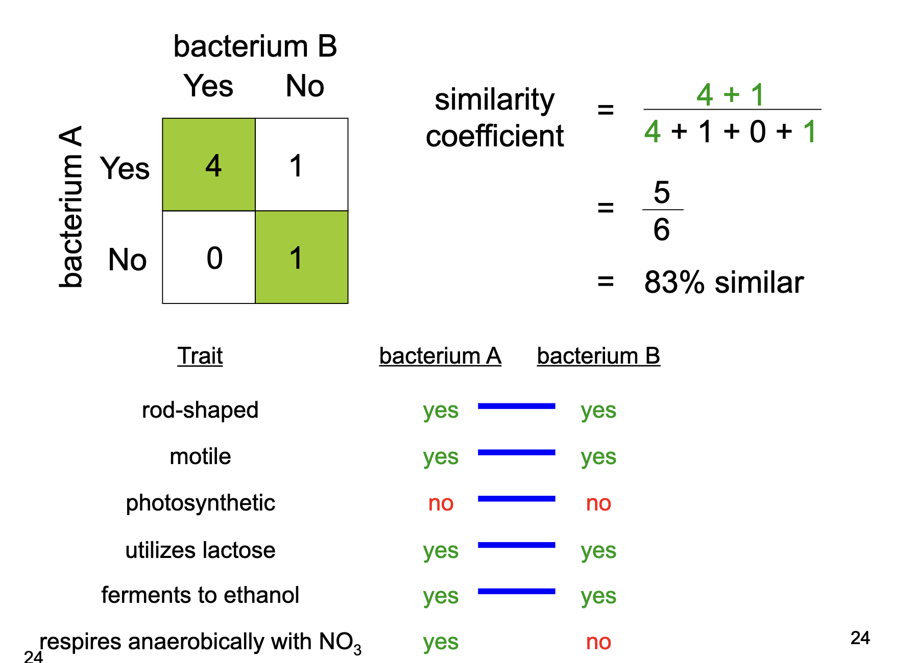
Part of determining numerical taxonomy (trait comparison)
Problems with numerical taxonomy / similarity coefficient naming
the idea that if two strains share enough important traits then they are the same species
advantages: the trait information is very useful and meaningful
disadvantages: trait choice is arbitrary, all traits are weighed equally while some traits are simple and others are very complex, traits are not necessarily related to one another, the same trait may arise through different mechanisms
shape! two organisms can have wildly similar shapes while one is a bacteria and another is an archaea
all traits are encoded by DNA, to improve tree use genetic information
DNA-DNA hybridization
compare entire genomes
grow one bacteria strain (A) in N15 and the other (B) in N14
heat up to denature bonds creating single stranded DNA molecules
cool them to reanneal—> bacteria A’s DNA may fully re-from into double stranded while bacteria B may still have some single stranded = hybrid
CsCl gradient ultracentrifugation
Detect UV absorbance
treat with ssDNA nuclease to enhance
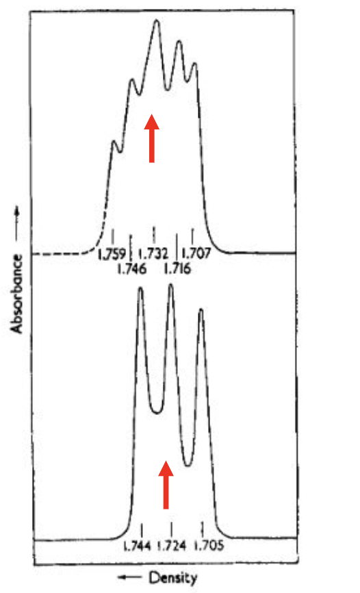
**graph peaks=hybrid
** >70% DNA/DNA hybridization = same species
DNA-DNA hybridization advantages versus disadvantages
advantages: extremely accurate and relevant, the only formal definition of bacterial species today is >70% DNA-DNA hybridization
disadvantages: the two organisms being compared must be very genetically similar for the technique to be meaningful, technically cumbersome and impractical
*only very close comparisons work
Phylogenetics
classification based on evolutionary relatedness by sequence similarity
this means that if two strains share enough important traits then their genomes would have a similar sequence, so a molecule (specifically chosen protein or gene sequence) called a molecular chronometer , as time passes sequences evolve —> organisms with fewer changes in sequence are more closely related
molecular chronometer: must be a molecule found in all representatives of the groups studies, function of it must be the same in all representatives as well, there must be sufficient similarity between molecules so the sequences can be aligned, BUT there also must be enough differences so that each sequence has its own signature
Example: these are all RecA protein sequences, they all function for homologous recombination, but they all have slightly different amino acid sequences so they can be distinguished with their signatures:

Ribosomes
structural component in a cell that makes proteins, in bacteria the ribosome is 70 and has two subunits: 30S (small subunit, contains 16S rRNA and 21 proteins) and 50S (large subunit, contains 23S rRNA + 5S rRNA + 34 proteins)
all living things have ribosomes so they all have rRNA
16S rRNA phylogeny
a specific nucleaic acid sequence found in the 30 subunit of the ribosome, found in all living things (everything can be compared using this), it is a part of the ribosome (same function), relatively consistent regions (anything can be aligned, can compare even ancient relative strains), has highly variable regions (signature, compare recent relatives)
GREAT molecular chronometer for phylogenetics
essentially “numerical taxonomy” with base pairs: sequence and compare two organism’s 16 rRNA —> each nucleotide position is a trait
more similar 16S rRNA sequence=more similar bacteria
BIG results of 16S rRNA phylogeny (mention 6 things)
completely revised the tree of life!! bacteria now parallel to eukaryotes, no longer under, now we have three parallel branches
revised bacterial branch: used to think important traits like photosynthesis were narrowly distributed meaning all right next to each other (old bacterial branch = numerical taxonomy tree —> NEW bacterial branch = 16S rRNA tree showing photosynthesis as widely distributed throughout organisms
also showed that the few unusual bacteria on the tree were actually archaea —> have new archaea domain on tree
molecular microbial ecology - quickly assess microbial members of an environment
can figure out properties of the environment by the bacterial groups thriving within it
microbiome- 16s rRNA tech can help determine all organisms in a region of the body - bacterial population profile associated with health and disease
endosymbiotic theory- mitochondria and chloroplasts have bacterial 16S rRNA
molecular microbial ecology
a way to quickly figure out what microbial organisms are in an enviornment by harvesting and sequencing environmental mixture of 16s rRNA
endosymbiotic theory
idea that eukaryotic organelles were once bacteria / have bacteria 16S rRNA inside of them
Phylogenetics by 16S rRNA sequencing (basic one sentence of what it is, advantages, and disadvantages)
Phylogenetics classification strategy using 16S rRNA subunit of bacterial ribosomes as the molecular chronometer
advantages: easy in the “sequencing age” (modern tech has made DNA sequencing super fast and cheap ; massive data sets accumulating because of fast and easy sequencing so easy to cross check what organism it is ; fantastic for higher order comparisons
disadvantages: poor at species designation meaning two different bacteria could have practically identical 16S rRNA sequences, no direct train information as its part of the ribosome meaning this method of classification can only tell you who this microbe is related to not what it does bc it is part of the ribosome (protein making sub unit)
Why can bacteria not be organized by traditional species concepts?
They do not sexually reproduce
Bacterial Classification Strategy Number 1
Numerical Taxonomy
comparing organisms based on similar traits
**informative but too arbitrary meaning personal choice
Bacterial Classification Strategy Number 2
DNA/DNA Hybridization
compare organisms based on total genome similarity
**precise but too specific
Bacterial Classification Strategy Number 3
Phylogenetic - 16S rRNA sequencing
compare organisms based on evolutionary relatedness
**universal but does not tell you about the properties of the organism
Bacterial Classification Strategy Number 4
Polyphasic Approach
combination
1-phylogenetic 16S rRNA sequencing
2-numerical taxonomy
3-DNA/DNA hybridization
Bacterial Classification Strategy Number 5
Naming by disease
Polyphasic approach
naming by a combination of approaches
steps: isolate and grow a new bacterium
1-sequence 16S rRNA and compare to database to find closest relative aka.phylogenics
2-then compare physical traits to closest type strain aka.numerial taxonomy
3-then compare genome to the type strain to determine if it is the same or different species aka.DNA/DNA hybridization
Bacterial “species” in terms of polyphasic approach (type strain)
bacterial species is a collection of strains that share important traits and 70% DNA/DNA genome hybridization with a type strain (an arbitrarily selected bacterial strain that has all of the hallmark traits of the species to which it belongs)
*compare traits of a new isolate to the chosen type strain , confirm in same bacterial species with >70% DNA/DNA hybridization
How do you know which type strain to compare?
establish the neighborhood or section of DNA to look at first before jumping to the expensive, intensive DNA/DNA hybridization
this is why you complete 16S rRNA sequencing first in polyphasic approach, get a more narrowed down view
ex process of naming a new bacteria
new isolate discovered!
1-16S rRNA sequencing , check data base for any close relatives, this allows you to identify close relatives and choose relevant type strain for trait comparison
if no close relatives, it is in a new family
2-compare traits to closest type strain—>numerical taxonomy
if similar not similar to type strain, probably a new genus
3-DNA/DNA hybridization of genomes
<70% similar then it is a brand new species
>70% similar then it is the same species
Organism of the Week- Epulopiscium Fishelsoni
bacteria found in gut of surgeon fish
1 million times larger than e.coli
visible to the naked eye
thought to be eukaryotic cell: large and looked like it had “organelles”
until 16S rRNA proved it was an enormous bacteria!
cannot be cultured in the laboratory
very unusual cell division
new daughter cell grows inside mother cell, mother cell is killed when daughter is released
Naming by disease
pathogen species are named based on the disease they cause
e.g. tuberculosis —> mycobacterium tuberculosis
complications to naming bacteria
characterization can be a long and difficult process
some important traits can be hard to observe
there are more bacteria than microbiologists
some bacteria can not be grown in the lab
we are not looking in the right places for new bacteria
naming is dynamic
the more we learn, the more names change
Organism of the Week- E.Coli (2 types)
E. coli “K12”: the most studied organism ever, no pili (finger like projections), powerful genetic system, helpful to human gut microbe, instrumental in molecular biology **harmless symbiot
E.coli “O157:H7”: recently emerged pathogen, has toxin+pili from a phage infection! caused “Jack in the Box” outbreak and contamination of spinach in California **dangerous pathogen
Identical 16S rRNA but chromosomes only 75% similar for comparison Humans and Chimp’s genomes are 98% similar
Phages
viruses that infect bacteria
viruses are comprised of proteins and nucleic acids
phage infection in E.coli “O157:H7” causes pili and adds a toxin
they have no cytoplasm, no way to make energy, no way to self replicate
Lysogeny
phage inserts its genome into the genome of E.coli and goes dormant, E.coli replicates the phage and expresses its genes like its own as it grows
“prophage” introduces toxin and pilus
E.coli now secretes Shiga toxin (StxAB) that damages human cells, benefitting both bacterium and phage
Naming strains by “Serotype”
“strains” are variants within a species
“serotypes” name pathogens based on the antigen (unique structures or patterns on outer surface) variation , antibodies bind to + destroy specific antigens
if two variants of a virus have the exact same surface antigens (same antibodies can recognize and destroy them), then they belong in the same serotype