Amaya's Radio exam 1 (chap. 1-8 & 18-19)
 Call Kai
 Learn
Learn Practice Test
 Spaced Repetition
 Match
Match Flashcards
 Knowt Play
1/93

There's no tags or description
Looks like no tags are added yet.
Name | Mastery | Learn | Test | Matching | Spaced | Call with Kai | Chat |
|---|
No analytics yet
Send a link to your students to track their progress
94 Terms
Who invented/discovered the x ray
Roentegen
Radiation
A form of energy carried by waves or a stream of particles
Fluorescence
a glow that results when a fluorescent substance is struck by light, cathode rays, or x-rays
Radiograph
an image or picture produced on a receptor (radiation-sensitive film, phosphor plate, or digital sensor) by exposure to ionizing radiation; a two-dimensional representation of a three-dimensional object
Dental radiograph
A photographic image produced on a receptor by the passage of x-rays through teeth and related structures
Radiography
The art and science of making radiographs by the exposure of a receptor to x-rays
Dental radiography
The production of radiographs of the teeth and adjacent structures by the exposure of an image receptor to x-rays
Radiology
The science or study of radiation as used in medicine; a branch of medical science that deals with the therapeutic use of x-rays, radioactive substances, and other forms of radiant energy
X radiation
a high-energy radiation produced by the collision of a beam of electrons with a metal target in an x-ray tube
X ray
A beam of energy that has the power to penetrate substances and record image shadows on photographic recepotors
Who exposed the first dental radiograph
Walkhoff
Who exposed the first dental radiograph in the U.S. (living patient)?
Kells
What charge does a proton have
positive
What charge does a neutron have
neutral (no charge)
What charge does an electron have
negative
Radioactivity
the process by which certain unable atoms or elements undergo spontaneous disintegration/ decay in effort to attain a more balanced nuclear sate
Ionizing radiation
Radiation capable of producing ions by removing or adding an electron to an atom
Labeling of the X ray tubehead

What parts of the x ray tube head contain lead?
Lead collimator
Metal housing
Tube head seal
Position indicating device
Leaded glass housing
What are the periods for radiation exposure?
Latent period
Period of injury
Recovery period
Cumulative effects
Latent period
the time that elapses between exposure to ionizing radiation and the appearance of observable clinical signs
Period of injury
After latent period, cellular injuries occurs such as
cell death
changes cell in function
breaking or clumping of chromosomes
formation of giant cells
cessation of mitotic activity
Abnormal mitotic activity
Recovery period
Last event in sequence
Cell damage followed by repair
Cumulative effects
Repeated radiation exposure can lead to health problems
Radiosensitive Tissues / Organs
small lymphocyte
bone marrow
reproductive cells
intestinal mucosa
skin
lens of eye
oral mucosa
Radioresistant tissues/organs
Muscle tissue
nerve tissue
mature bone
cartilage
salivary gland
thyroid gland
kidney
liver
Somatic radiation effects
Injury in somatic cells that produce poor health and iridate the individual
Genetic radiation effects
Effects not seen in the irradiated person but are passed to future generations
Collimation
Restricts size and shape of x ray beam and reduce patient exposure
Soamtic cells
Cells in the body except reproductive
Genetic cells
The reproductive cells (Ova and sperm)
Inherent filtration
When primary beam passes through X ray Tube ,insulating, oil, and tube head seal with .5 to 1.0 mm of filtration
Added filtration
Aluminum disks in path of X ray beam, between collimator and x ray seal to filter out longer wavelengths and low energy x rays
Total filtration
Inherent + added
Machines at or below 70 kV need a minimum of 1.5 mm of aluminum filtration Above 70 kV you need 2.5 or above
What beam is wanted for an x ray?
A high-energy, penetrating, and hardened x-ray beam
What does maximal permissible dose mean?
Maximum does the body can endure with little or no injury
What is the maximal permissible dose?
50 mSv / year
.05 Sv/ year
5.0 rem/year
ALARA Concept
As low as reasonably achievable
Ways to protect the patient
Proper prescribing of dental images
Proper equipment (aluminum filter, collimator, PID)
Thyroid collar
Lead apron
Digital sensors/fast film
Beam alignment devices
Exposure factor selection
Proper technique when taking images
Proper film processing and receptor handling
X ray beam quality definition
Describe the mean energy or penetrating ability of the x ray beam
What is the quality of a beam controlled by?
Kilovoltage because it regulates the speed/energy of electrons and determines penetrating ability
What is x ray beam density?
Overall darkness/blackness of an image
What happens to density when kilovoltage is increased?
Image has more density and is darker
What happens to density when kilovoltage is decreased?
Images has less density and is lighter
What is contrast?
how sharply dark and light areas are differentiated or separated on image
What happens to contrast when kilovoltage is decreased?
High contrast
Many black and white areas with few shades of gray
Used for carries
What happens to contrast when kilovoltage is increased?
Low contrast
Many shades of gray
Use for periapical disease
What is exposure time?
Interval of time during which x rays are produced
What is the relationship between exposure time and kilovoltage?
Inverse
more exposure = more x rays = darker image
What is quantity of a beam controlled by?
Milliamperage
What is x ray beam quantity?
Number of x rays produced in the dental x ray unit
What happens to density when milliamperage is increased?
Higher density and a darker image
What happens to density when milliamperage is decreased?
Lower density and a lighter image
What happens to exposure time when milliamperage is increased?
Exposure time was decreased
What happens to exposure time when milliamperage is decreased?
Exposure time was increased
What is x ray beam intensity?
Both quality and quantity as a product
X ray beam intensity and Kilovoltage relationship
Higher kV = Higher intensity because more energy and shorter wavelengths
X ray beam intensity and Milliamperage relationship
Higher mA = higher intensity because of more electrons/ x rays produced
X ray beam intensity nd exposure time
Higher exposure time = more intense
Target surface distance
The distance from the source of radiation (tungsten target in anode) to the patients skin

Target object distance
The distance from the source of radiation to the tooth
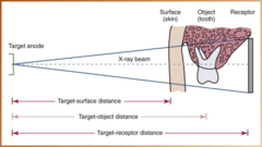
Target receptor distance
The distance from the source of radiation to the receptor

Inverse square law
the intensity of radiation is inversely proportional to the square of the distance from the source of radiation
Half value layer (HVL)
the thickness of aluminum , when placed in the path of the x-ray beam, reduces the exposure rate by one-half.
Parts of X ray machine?
control panel, extension arm, tube head
What is kilovoltage used for?
The peak voltage of an alternating current
What is milliamperage used for?
Regulates the temperature of the cathode filament
Radiolucent
Portion of an image that is dark or black
Radiopaque
Portion of image that appears light or white
What is a step wedge used for ?
To check contrast
Sharpness
aka detail, definition or resolution
Capability of receptor to reproduce the distinct outlines of an object
What factors influence sharpness?
Focal spot size, film composition, movement
Penumbra
Unsharpness, blurry, fuzzy, unclear area
Magnification
Radiographic image that appears larger than actual size of the object it represents
What factors influence magnification?
Target-receptor distance
Object-receptor distance
Distortion
variation in the true size and shape of the object being radiographed
What factors influence distortion?
object-receptor alignment
x-ray beam angulation
Intraoral X ray machine parts
tube head, extension arm, control panel
Extra oral x ray machine parts
Position indicating devices
Panoramic
Cephalometric
Skull imaging
Cone beam computer tomography
What are the types of dental receptor holders
Bite block
Endoray
Snap a Ray
XCP
What devices are used for digital imaging
X ray unit
Sensor
Computer
Direct digital imaging
Method of obtaining a digital image, in which an intraoral sensor is exposed to x-radiation to capture a radiographic image that can be viewed on a computer monitor
Indirect digital imaging
A sensor after exposure is scanned and converted toa digital form that can be viewed on a computer monitor
Advantages of digital imaging
-Superior gray-scale resolution
-Reduced exposure to x-radiation
-Increased speed of image viewing
-Lower equipment and film cost
-Increased efficiency
-Enhancement of diagnostic image
-Effective patient education tool
Periapical examination purpose
used to examine the entire tooth (crown and root) and supporting bone

Interproximal examination purpose
To examine the crowns of both the maxillary and mandibular teeth on a single image

occlusal examination purpose
Used to examine large areas of the maxilla or mandible on one image

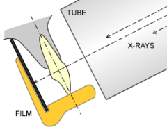
Parallel technique
1) Long axis of tooth and the receptor are parallel to each other
2) The central ray is directed perpendicular to the long axis of the tooth and film
3) The beam alignment device must be used to keep the receptor parallel to the tooth
Relationship between a focal spot and x ray image
smaller focal spot = Sharper image
Relationship between PID length and target receptor distance
A longer PID has less magnification
Relationship between distortion and object receptor distance
More distortion the further you are from an image
Relationship between receptor image and distortion
If the receptor is not parallel with the object the more likely distortion will occur
What happens when vertical angulation is steep (+70 degrees)
Foreshortening
What happens when the vertical angulation is flat (+20 degrees)
Elongation