BIOL 2401 Lab Exam
 Call Kai
 Learn
Learn Practice Test
 Spaced Repetition
 Match
Match Flashcards
 Knowt Play
1/18

There's no tags or description
Looks like no tags are added yet.
Name | Mastery | Learn | Test | Matching | Spaced | Call with Kai | Chat |
|---|
No analytics yet
Send a link to your students to track their progress
19 Terms
When should the inoculating loop be flamed when streaking agar plates?
Before streaking the first 1/3
Before streaking second 1/3
After streaking the last 1/3
Supplies needed for catalase test
Microscope slide
Toothpick (to transfer isolated colonies)
Hydrogen peroxide
Appearance of positive catalase test
Bubbles after adding hydrogen peroxide
Supplies needed for coagulase test
Reaction card with two circles (one for control, one for test)
Control reagent
Test reagent
Agar plate with cultures
Appearance of positive coagulase test
Clumping (agglutination)
Components of colony morphology (SSPMET)
Size, Shape, Pigmentation, Margin, Elevation, Texture
Examples of shape (colony morphology)
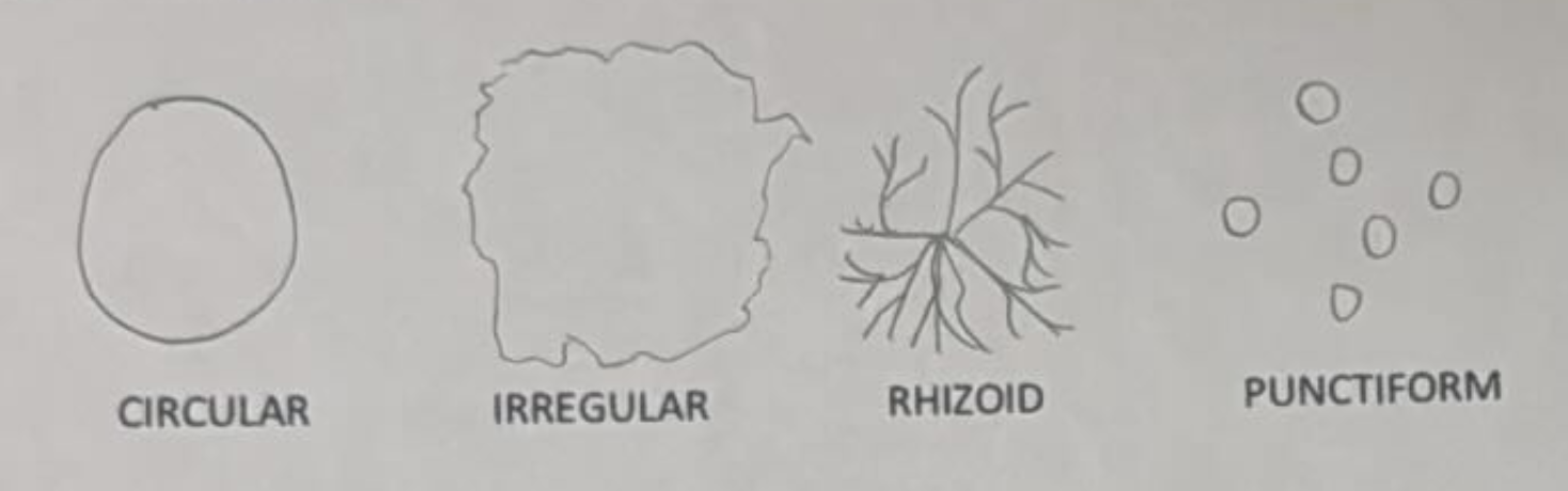
Circular
Irregular
Rhizoid
Punctiform
Examples of margin (colony morphology)
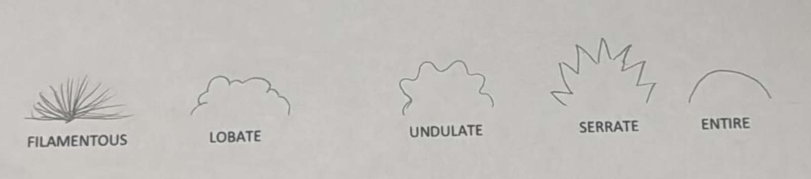
Filamentous
Lobate
Undulate
Serrate
Entire
Examples of elevation (colony morphology)

Raised
Convex
Flat
Umbonate
Crateriform
When should a slide be heat-fixed (gram staining)?
After the bacterial smear has air-dried and before adding any gram stain reagents
Steps of gram staining (CIDS)
Crystal violet → 60s → rinse
Iodine → 60s → rinse
Decolorizer drop-by-drop → rinse
Safranin → 60s → rinse, gently blot or air dry
Components of cell morphology (GCC)
Gram reaction, cell shape, cell size
Examples of cell shape (cell morphology)
Coccus
Coccobacillus
Bacillus
Vibrio
Spirillum
Spirochete
Steps to adjust microscope for Köhler Illumination (set up at 40x)
Close aperture diaphragm (limits light)
Close field diaphragm (makes hexagon)
Raise condenser fully, adjust (makes sharp hexagon)
Adjust screw (centers hexagon)
Open field diaphragm
Open aperture diaphragm
Steps to prepare a slide before gram staining
1, Sterilize loop
Add loopful of water to slide
Sterilize loop
Smear part of isolated colony on slide
Let fully air dry
Heat fix
Positive mannitol fermentation result

Red agar turns yellow
Beta hemolysis
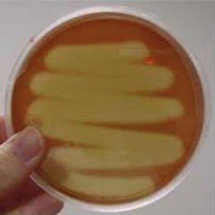
Complete hemolysis → turns clear
Alpha hemolysis
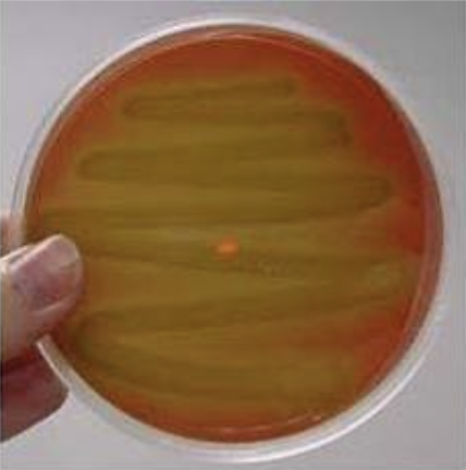
Partial hemolysis → turns green
Gamma hemolysis
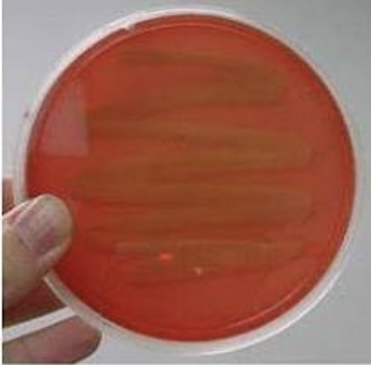
No hemolysis → stays red