Looks like no one added any tags here yet for you.
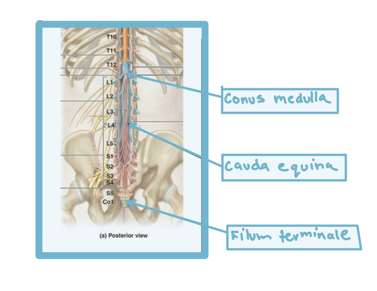
List the three major components of the gross anatomy of the spinal cord
Conus medullaris
Cauda equina
Filum terminale
Conus medullaris
Is the terminal spinal cord at L1
Stops age 4
Cauda equina
Cauda = tail
equina = horse
Filum terminale
Terminal thread
Study
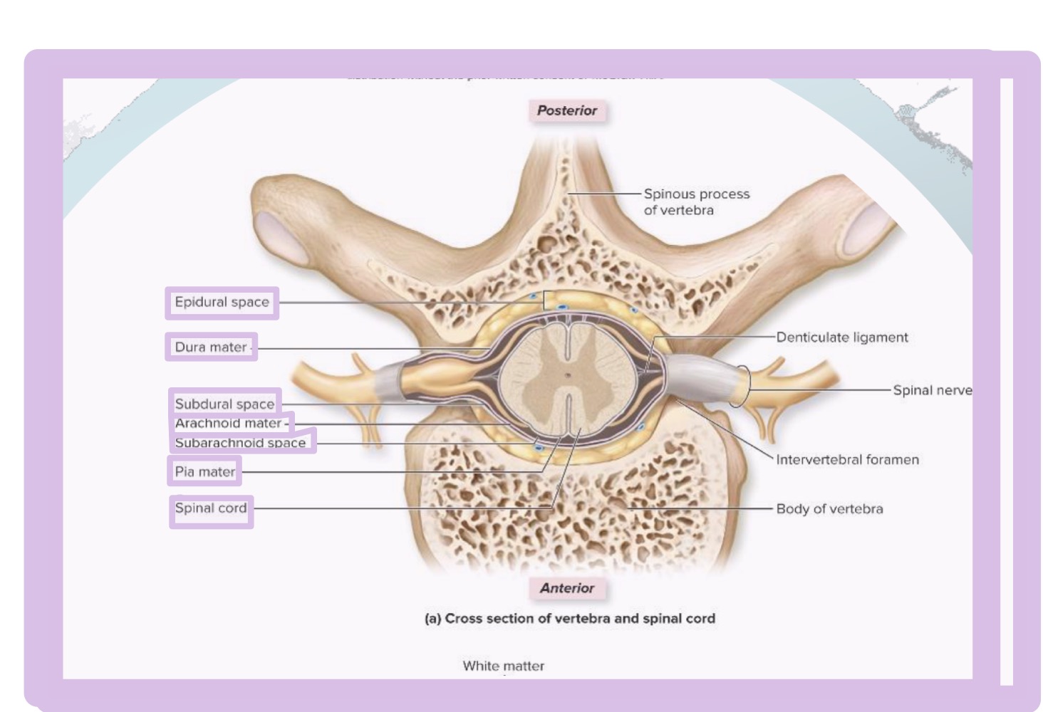
What is in the spinal cord meninges and place in order from superficial to deep
Superficial
Epidural space
Dura mater
Subdural space
Arachnoid mater
Subarachnoid space
Pia mater
Deep
Epidural space
Above
Real
Subdural space
“Potential” space
Subarachnoid space
Contains CSF
Reason why it is unique!
What are the three meninges and place them in order from superficial to deep
Superficial
Dura mater
Arachnoid mater
Pia mater
Deep
Dura Mater
Tough outer layer
Thickest layer
Directly under skull and vertebral column
Provides protective covering for brain and spinal cord
Arachnoid mater
Middle layer
Web-like structure
Beneath dura mater
Contains subarachnoid space
Filled with cerebrospinal fluid that cushions the brain and spinal cord
Pia mater
Delicate inner layer
Thinnest layer
Directly adhering to the surface of the brain and spinal cord
It follows the contours of the brain, including the sulci (grooves) and gyri (ridges)
Study
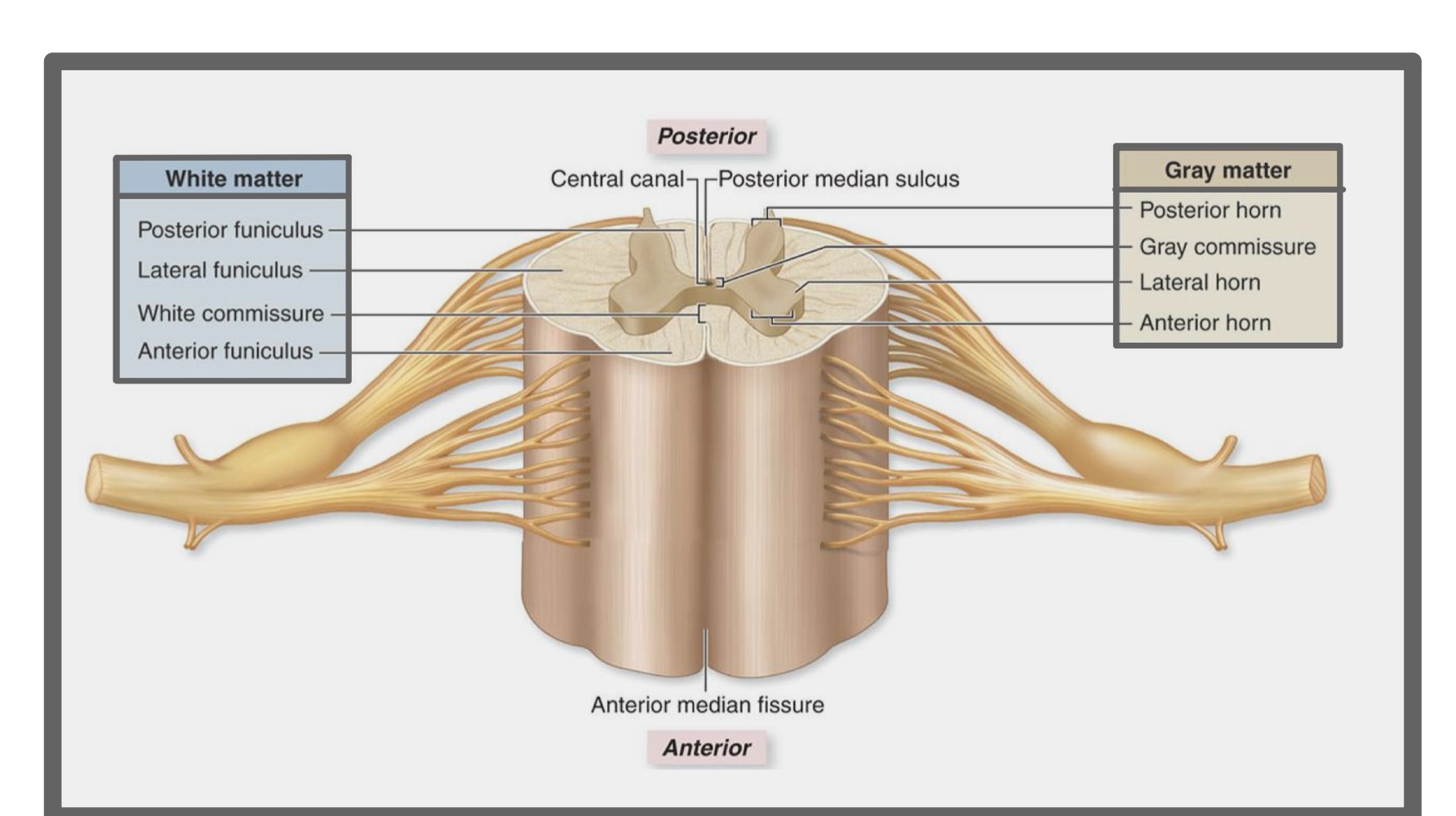
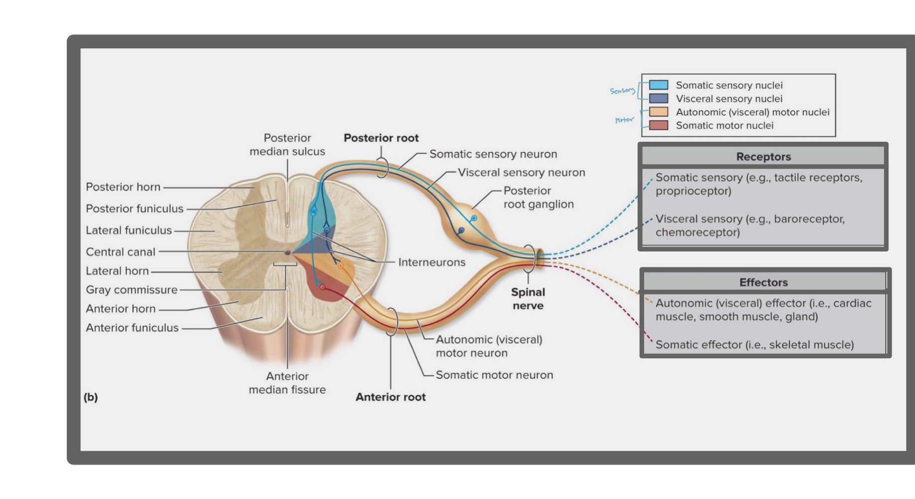
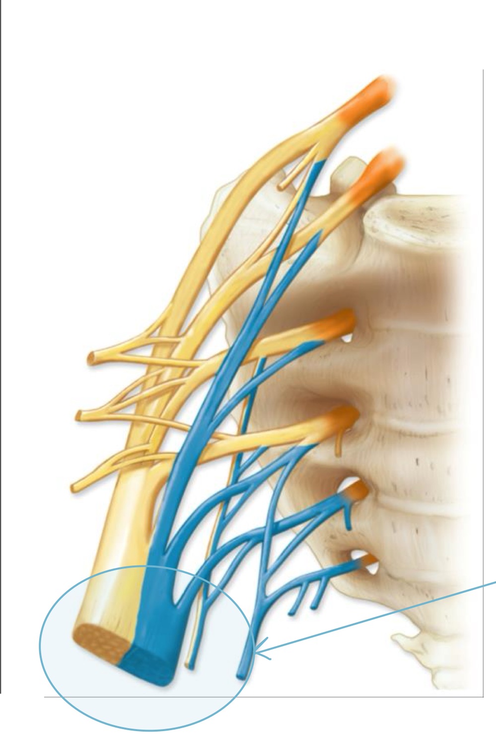
List what is located in the nuclei of gray mater in the spinal cord?
Posterior horn
Gray commissure
Lateral horn
Anterior horn
Posterior horn
Afferent
Sensory
Axons of sensory neurons
Cell bodies of interneurons
unipolar
Function of posterior horn
Receives sensory information from the body, including pain, temperature, touch, and relays it to the brain
Structural classification of neuron in posterior horn
Somatic sensory neuron (receptor)
Visceral sensory neuron (receptor)
Posterior root ganglion
Somatic sensory
Pain and pressure receptors in skin
Visceral sensory
Stretch receptors in smooth muscle (like organs) from viscera
Posterior root ganglion
Collection of cell bodies of sensory neurons
Anterior horn
Efferent
Motor
Cell bodies of somatic motor neurons that innervate skeletal muscle
Multipolar
Function of anterior horn
Sends signals from motor neurons to skeletal muscles, enabling movement and motor control
Structural classification of neuron in anterior horn
Autonomic (visceral) motor neuron (effectors)
Somatic motor neuron (effectors)
Autonomic (visceral) motor neuron
Innervates and controls smooth muscle, cardiac muscle, and gland tissue
Somatic motor neuron
Innervates skeletal muscle, enabling voluntary movements and muscle contractions
Lateral horn
Efferent
Motor
Cell bodies of autonomic (visceral) motor neurons
Multipolar
Function of lateral horn
Fuses spinal cords, controlling the autonomic nervous system
Autonomic (visceral) motor neurons
Innervates smooth muscle, cardiac muscle, and glands
Study
Study
What are the four nerve plexuses?
Cervical nerve plexus
Brachial nerve plexus
Intercostal nerve - No thoracic plexus
Lumbar nerve plexus
Sacral nerve plexus
Cervical Nerve plexus
Innervates the muscles of the neck
Has phrenic nerve
Innervates diaphragm
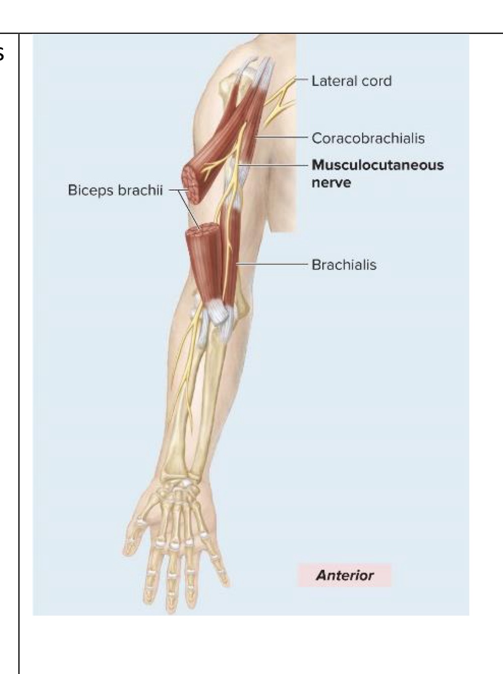
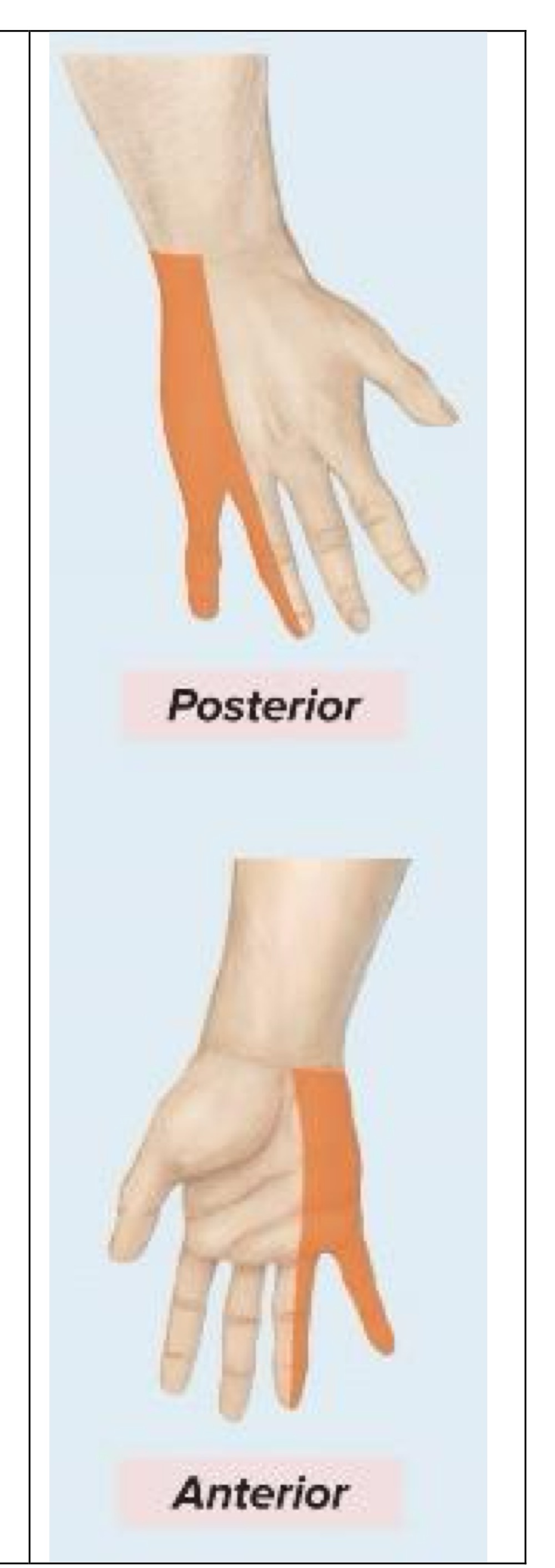
Brachial nerve plexus
Supplies upper limbs
What are the five major Brachial plexus terminal branches that innervate the arm?
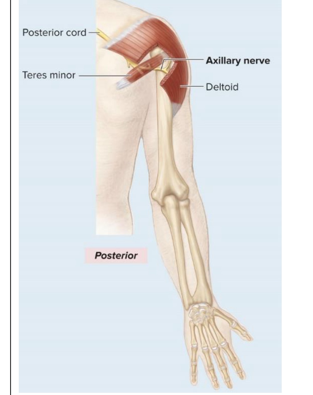
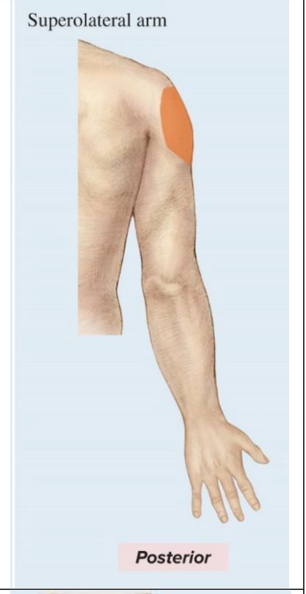
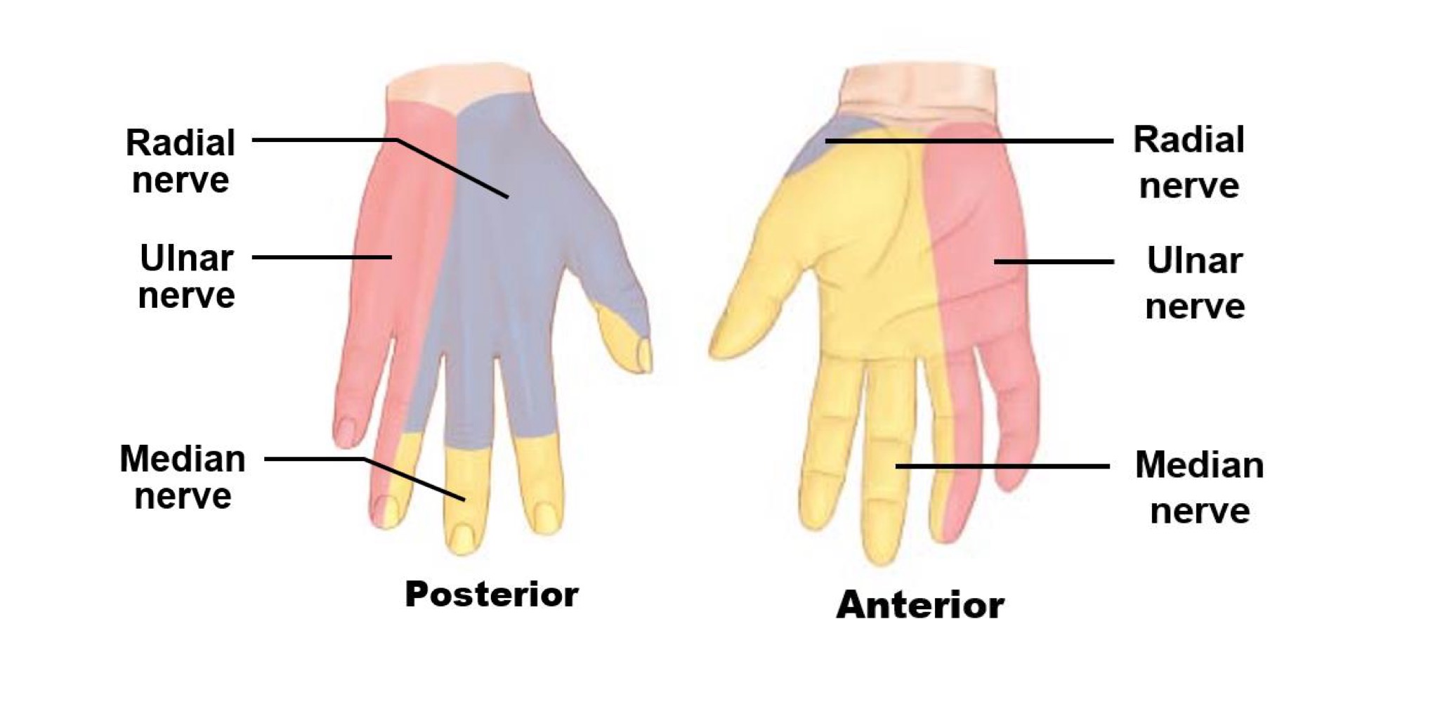
Axillary nerve
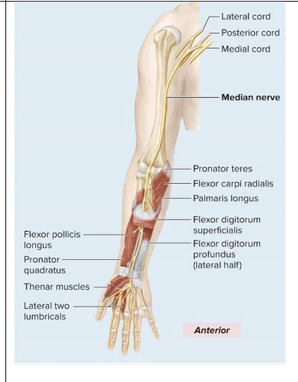
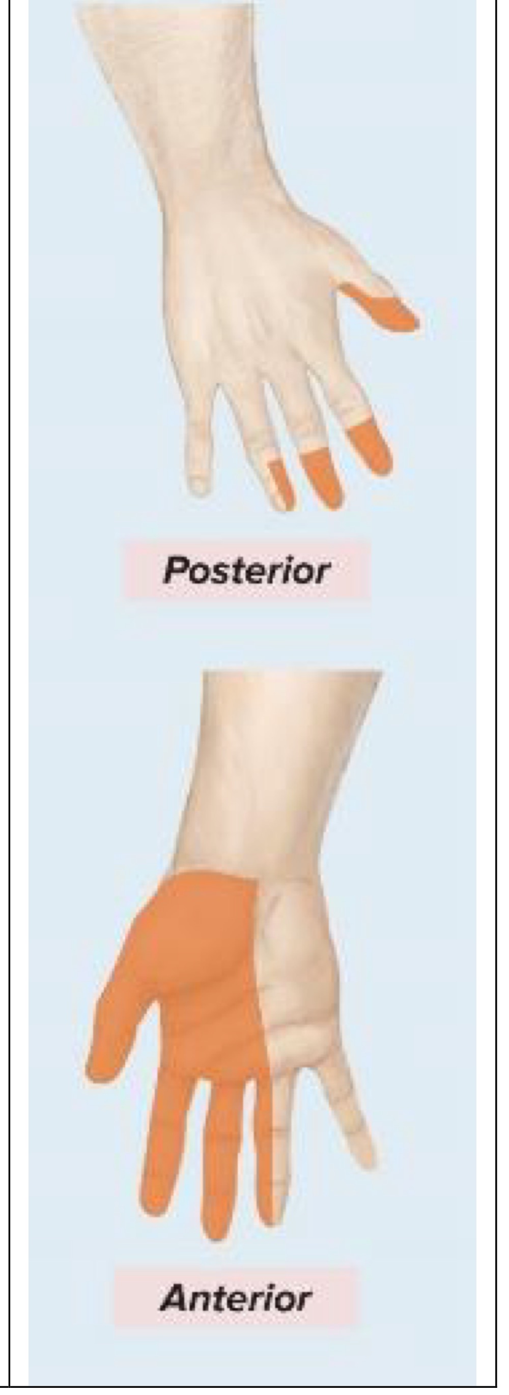
Median nerve
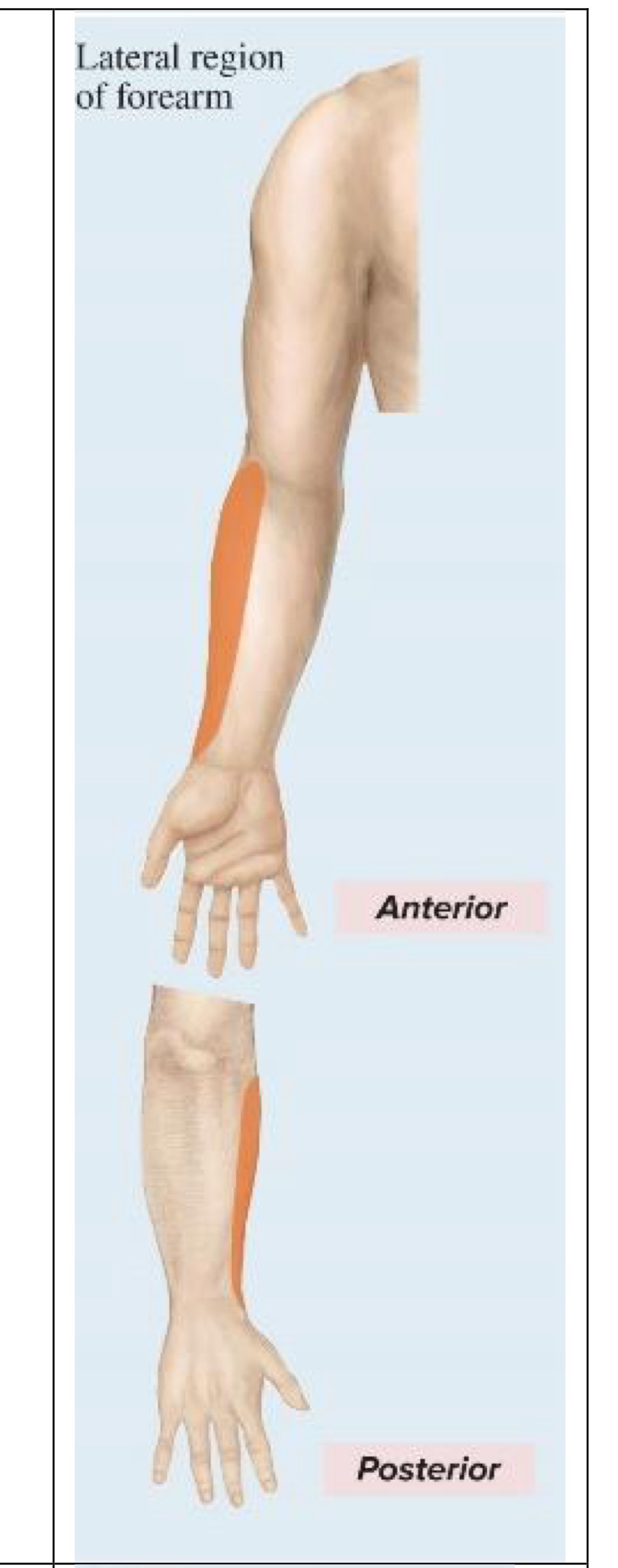
Musculocutaneous nerve
Radial nerve
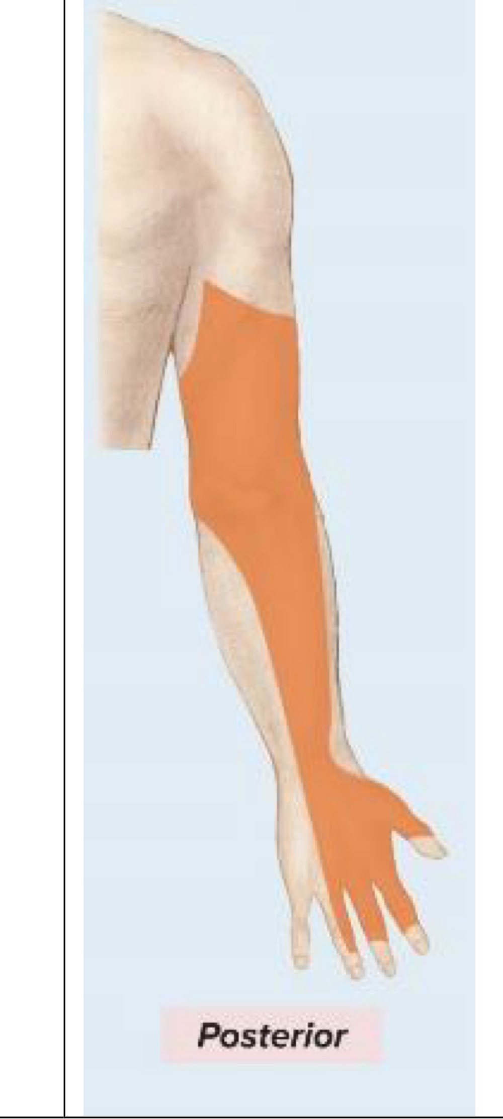
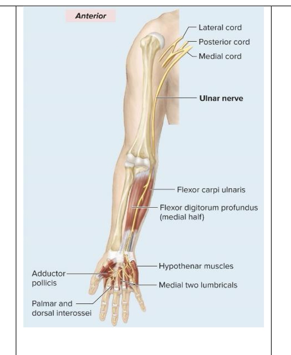
Ulnar nerve
Motor innervation of axillary nerve
Arm abduction
Sensory innervation of axillary nerve
Superolateral arm
Motor innervation of median nerve
Wrist, digit flexors
Sensory innervation of median nerve
Palmar aspects and dorsal tips of lateral 3 ½ digits
Thumb
Index finger
Middle finger
½ ring finger
Motor innervation of musculocutaneous nerve
Forearm flexors
Sensory innervation of musculocutaneous nerve
Lateral region of forearm
Motor innervation of radial nerve
Forearm, wrist, digit extensors
Sensory innervation of radial nerve
Posterior region of arm
Posterior region of forearm
Dorsal aspect of lateral 3 digits
Except their distal tips
Motor innervation of ulnar nerve
Wrist, digit flexors
Sensory innervation of ulnar nerve
Dorsal and palmar aspects of medial 1 ½ digits
Little finger
medial aspect of ring finger
Study
Lumbar nerve plexus
Innervates abdomen, some external genitalia, anterior and medial thigh
Sacral nerve plexus
Innervates:
Pelvis
Posterior thigh
Leg
Foot
The main nerve is the sciatic nerve
Largest and longest nerve in the body
Main nerve (sciatic nerve)
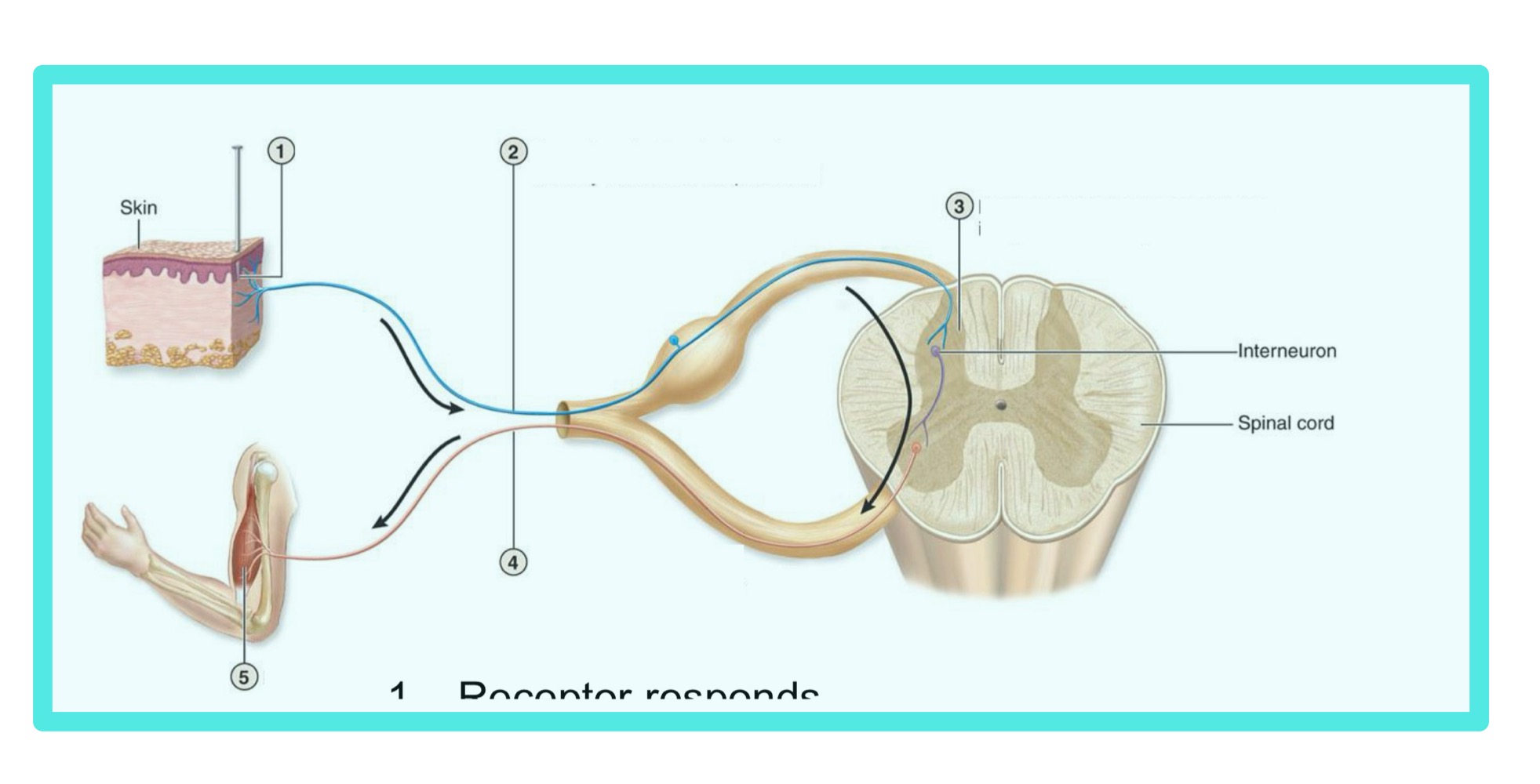
Reflexes
Rapid, automatic, involuntary reactions of muscles or glands to a stimulus
What are the characteristics of reflexes?
Stimulus
Rapid response
Automatic response
Involuntary response
Reflex arc
Neutral wiring of a single reflex
Receptor in PNS → CNS
What are the steps of a simple reflex arc?
Receptor responds
Sensory neuron to spinal cord
Integration - sometimes by interneuron (monosynaptic and polysynaptic)
Motor neuron to effector
Effector responds
Study how steps look on diagram
Classification of Ipsilateral
Receptor and effector on the same side
Classification of Contralateral
Receptor and effector on opposite sides
Classification of Monosynaptic
Does not involve interneuron
Ex: Knee-jerk (stretch reflex)
Classification of Polysynaptic
Involves interneuron
Ex: Withdrawal reflex