Orbital Nerves: Cranial Nerves II, III, IV and VI
 View linked note
 Call Kai
 Learn
Learn Practice Test
 Spaced Repetition
 Match
Match Flashcards
 Knowt Play
1/72

There's no tags or description
Looks like no tags are added yet.
Name | Mastery | Learn | Test | Matching | Spaced | Call with Kai |
|---|
No analytics yet
Send a link to your students to track their progress
73 Terms
List the extrinsic eye muscles
Levator palpebrae superioris m., superior oblique m., Lateral rectus m., Superior rectus m., Inferior oblique m., Inferior rectus m., Medial rectus m.
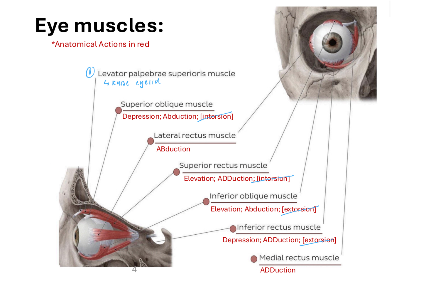
Which extrinsic eye muscles originate from the common tendinous ring?
Superior, Inferior, Lateral, Medial rectus
Major action of levator palpebrae superioris
Raise eyelid (assisted by superior tarsal m.)
Major action of superior oblique m.
Depression, ABduction
Major action of lateral rectus m.
ABduction
Major action of superior rectus m.
Elevation, ADduction
Major action of inferior oblique m.
Elevation, ABduction
Major action of inferior rectus m.
Depression, ADduction
Major action of medial rectus m.
ADduction
Where are intrinsic muscles located?
Within the eyeball
What is the role of intrinsic muscles?
Control lens shape and pupil size
CN II is also known as…
Optic n.
CN III is also known as…
Oculomotor n.
CN IV is also known as…
Trochlear n.
CN V1 is also known as…
Ophthalmic division of trigeminal nerve
CN VI is also known as…
Abducent n.
List the orbital nerves
CN II, CN III, CN IV, CN V1, CN VI
CN II fiber type
SSA
CN III fiber type
SM (GSE) and Parasympathetics (GVE)
CN IV fiber type
Somatomotor (GSE)
CN V1 fiber type
Somatosensory (GSA)
CN VI fiber type
SM (GSE)
CN II function/innervation
Special sense of vision
CN III function/innervation
GSE: Levator palpebrae suprerioris, Superior rectus, inferior rectus, medial rectus, inferior oblique
GVE: Sphincter pupillae, ciliary m.
CN IV function/innervation
Superior oblique
CN V1 function/innervation
Skin around the eye, eyelid, lacrimal gland, conjunctiva
CN VI function/innervation
Lateral rectus
List the orbital nerves that run along the lateral wall of the cavernous sinus (inferior → superior)
CN V1, CN IV, CN III
Which orbital n. passes through the cavernous? sinus (not along the wall)
CN VI
Path of sensory information from retina to visual cortex
Retina, L and R optic n., Optic canal, R and L optic n. join at optic chiasm in chiasmatic groove, Optic tract, Midbrain (postsynaptic), Visual cortex (in occipital lobe)
What covers CN II?
Pia, arachnoid, and dura mater
What a. travels along CN II?
Central retinal a.
Pupillary light reflex
Changes how much light enters eyes (dilate and constrict pupils)
Accommodation reflex
Changes focus with distance (change thickness of lens)
Where do L and R optic nerves cross each other?
Optic chiasm
Each eye has what 2 visual fields?
Nasal and temporal
Path of light from L nasal field
Light captured by R side of R eyeball, R optic nerve, R lateral geniculate body, R optic tract, Occipital lobe
Path of light from R nasal field
Light captured by L side of L eyeball, L optic nerve, L lateral geniculate body, L optic tract, Occipital lobe
Path of light from L temporal field
Light captured by R side of L eyeball, L optic nerve, Optic chiasm cross to R side, R lateral geniculate body, R optic tract, Occipital lobe
Path of light from R temporal field
Light captured by L side of R eyeball, R optic nerve, Optic chiasm cross to L side, L lateral geniculate body, L optic tract, Occipital lobe
If someone loses vision in 1 eye, what kind of lesion is this?
Prechiasmatic lesion (damage to retina or optic n.)
If someone loses temporal visual field, what kind of lesion is this?
Chiasmatic lesion
If someone loses input from the contralateral visual field from both eyes, what kind of lesion is this?
Postchiasmatic lesion
What are the main orbital branches of V1 (medial to lateral)
Nasociliary n., Frontal n., Lacrimal n.
What does the upper division of CN III innervate?
Levator palpebrae superioris and superior rectus
What does the lower division of CN III innervate?
Inferior rectus and inferior oblique
Which ganglia along the sympathetic trunk do sympathetics to the head and neck synapse at?
Superior cervical ganglia
What plexus carries post-synaptic sympathetics into the head?
Carotid plexus (ICA)
2 sympathetics pathways to vessels within the eye
ICA, Nasociliary n./Plexus branch, Ciliary ganglion, Short ciliary nn.
Path of sympathetics to dilator pupillae m.
ICA, Nasociliary n., Long ciliary n., Sclera, Dilator pupillae
Within the eye, what do parasympathetics innervate?
Sphincter pupillae (pupillary light reflex)
What do ciliary m. do?
Controls lens thickness for accommodation reflex
Shape of lens when ciliary m. are relaxed
Flattened
Shape of lens when ciliary m. are contracting
Thickened
What fibers attach lens to ciliary m.
Zonular fibers
Afferent limn of pupillary light reflex
CN II from stimulation to retinal ganglion cells
Efferent limb of pupillary light reflex
CN III parasympathetics via short ciliary nn. to sphincter pupillae
How to test for pupillary light reflex?
Shine bright light and observe if pupils change size
Afferent limb of accommodation reflex
CN II from stimulation of retinal ganglion cells
Efferent limb of accommodation reflex
CN III parasympathetics via short ciliary nerves to ciliary body and sphincter pupillae
How to test accommodation reflex
Ask patient to watch object (like a pen) as it gets closer (convergence, pupil constriction, lens thickening)
Describe Horner’s syndrome
Neurologic disorder that affects the sympathetics (especially to the head)
Where are lesions in Horner’s syndrome located?
Sympathetic ganglion/sympathetic chain
Clinical manifestations of Horner’s syndrome
Ptosis, miosis, and anhidrosis
Why would you observe ptosis in a patient with Horner’s syndrome?
Loss of control of superior tarsal muscle
Why would you observe miosis in patients with Horner’s sundrome
Loss of control of pupillary dilator m.
Why would you observe anhidrosis in patients with Horner’s syndrome?
Loss of control of smooth muscle within the aa. of the forehead
Goal of the H Test
Isolate actions to isolate specific nerves
Testing superior oblique m. (CN IV)
Look medially (ADduct) and downwards
Testing superior rectus (CN III)
Look laterally and upwards
Testing inferior rectus m. (CN III)
Look laterally and downwards
Testing medial rectus m. (CN III)
Look medially
Testing inferior oblique m. (CN III)
Look medially and upwards